1Thara .N 2Sai Kumar .N 3Vinod Babu .K 4Akshata Akalwadi
CORRESPONDING AUTHOR
2SAI KUMAR .N, MPT
Professor & Principal,
K.T.G. College of Physiotherapy and KTG Multi Speciality Hospital, Bangalore-560 091, India.
Int J Physiother. Vol 2(5), 824-833, October (2015) ISSN: 2348 - 8336
ABSTRACT
Background: Task specific training of upper limb may result in learning of new motor task through
transfer after repeated practice. Mirror therapy and motor imagery are effective emerging techniques used as an adjunct in rehabilitation of upper limb function in hemiplegia. The purpose of the study is to find comparative effects of task specific motor imagery with mental practice over task specific mirror therapy on upper limb functional activities for subjects with sub acute hemiplegia.
Method: An experimental study design with two groups conducted on 30 subjects with sub-acute
hemiplegic. Thirty subjects randomised, 15 subjects into group A and 15 into group B. Group A subjects received task specific motor imagery with mental practice thrice a week for 10 weeks and Group B received task specific mirror therapy thrice a week for 10 weeks. In both groups, each session consisted of 60 minutes. The outcome measure such as Action Research Arm Test (ARAT) was measured before and after 10 weeks of intervention.
Results: Comparison of post intervention means of ARAT using Independent t test and Mann-Whitney
Test showed that there is no statistically significant difference in grasp and gross movement between the groups and there is a statistically significant difference in grip, pinch and total score between the groups.
Conclusion: The present study concludes that 10 weeks of task specific motor imagery with mental
practice and task specific mirror therapy both shown significant effect on improvement of upper extremity function. However, greater percentage of improvement was found using task specific motor imagery with mental practice in hand function when compared to task specific mirror therapy.
Keywords: Motor imagery, mental practice, mirror therapy, task specific exercises, sub-acute
hemiplegia, hand function, action research arm test
DOI: 10.15621/ijphy/2015/v2i5/78241
1Physiotherapist,
2Professor & Principal,
3,4Associate Professor,
K.T.G. College of Physiotherapy and KTG Multi Speciality Hospital. Bangalore. India. Affiliated to Rajiv Gandhi University of Health Sciences, Karnataka. India.
Received 20th August 2015, revised 17th September 2015, accepted 03rd October 2015
INTRODUCTION
The World Health Organization has defined stroke as “rapidly developing clinical signs of focal (at times global) disturbance of cerebral function, lasting more than 24 hours or leading to death with no apparent cause other than that of vascular
origin.”1,2 In India, the stroke contributed to 41% of
deaths and 72% of disability adjusted life years amongst the non-communicable diseases (ICMR
2004).3 The prevalence rate of stroke in India is in
the range of 84-262/100,000 in rural and 334-424/
100,000 in urban area.4
A large number of new rehabilitative techniques potentially capable of stimulating cerebral plasticity among these techniques, large interest is devoted to treatment approaches aimed to improve motor functions, including constraint-induced movement therapy, mental practice, mirror therapy, virtual reality, robotics, and brain
stimulation techniques, bilateral training,
functional electrical stimulation, repetitive task
training, electromyographic biofeedback.1,5
The concept of Mirror therapy (MT) and Motor imagery (MI) is based on mirror neuron system, that have been explained in several neuroimaging studies revealing that the adult human brain consists of mirror neurons, a combination of sensory and motor properties in a single, simple unit. They have genetically inherited mechanism that unifies action perception and action execution. MI with Mental practice (MP) is a non-invasive technique in which physical tasks, scenarios, or both are imagined and cognitively rehearsed, usually without voluntary physical movements.It is a dynamic state during which the representation of a specific motor action is internally activated
without any motor output.6,7 Inspite of the brain
being damaged by stroke, its ability to train using MP seems to be retained, especially when combined with functional training, since it
reinforces cerebral reorganization.8, 9
Relatively many studies have been done where sub-acute stroke patients were benefited with Task Specific Mirror therapy and Motor Imagery with Mental Practice. Previous studies in stroke such as that of Marian Michielsen et al., suggested that mirror therapy using a mirror reflection can facilitate motor learning and may be beneficial for
motor functional recovery in the paretic hand.10
Whereas, it can be observed from studies done by Sjoerd de Vries, et al., that motor imagery training resulted in significant changes in task performance and creating a neural level reorganization similar
to the one related to normal (physical) training.11
Since motor imagery with mental practice and mirror therapy are relatively new techniques, experiment on short term treatment and guidance on more specific intervention is required. Therefore, the Study with research question, Whether the task specific motor imagery with mental practice and task specific mirror therapy does have a difference on improving upper extremity function for subjects with sub acute hemiplegia? The purpose of this study to find the comparative effects of task specific motor imagery with mental practice over task specific mirror therapy on upper limb functional activities for subjects with sub acute hemiplegia. It was null hypothesized that there will be no significant difference between task specific Motor imagery with mental practice versus task specific Mirror therapy on improving upper limb functions for subjects with sub-acute hemiplegia.
METHODOLOGY
An experimental study design with two groups, Group A: Motor imagery with mental practice group, Group B: Task specific Mirror Therapy Group. As this study involved human subjects the Ethical Clearance was obtained from the Ethical Committee of KTG College of Physiotherapy and K.T.G. Hospital, Bangalore as per the ethical guidelines of Bio-medical research on human subjects. This study was registered under Rajiv Gandhi University of Health Sciences for subject for registration for dissertation with registration number 09_T031_47082. Subjects included in the study were unilateral hemiplegic stroke right or
left,5,8 Subjects with sub acute hemiplegia between
2 to 6 months post stroke, 8,12 Ischemic stroke,13 age
above 45 years and below 80 years,both male and
female subjects, Brunstrom stage of motor
recovery of 3 to 5,5,14 Modified Ashworth scale score
<2.15 Subjects were excluded with wrist and/or
Procedure for intervention for Group A: 16-18
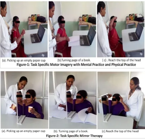
In Group A, subjects were treated with task specific motor imagery with mental practice incorporating physical practice with videotape consisting of 5 tasks for duration of 60 minutes for 3 days a week for 10 weeks.
The subject was made to sit comfortably in a chair in front of the table containing task related materials (videotape displayed on the computer screen, empty paper cup without handle, book, phone and pen). The affected limb and unaffected limb was placed on the table. The subject was first asked to observe the videotape of motor tasks and was instructed to physically practice each task for ten times. Then the subject was asked to mentally practice each task ten times with an interval of five minutes between them. The subject was instructed to indicate the beginning and ending of each task
saying “GO”.15 The total duration of motor tasks was
for 60 minutes per session.13 The motor tasks that
were given were, picking up an empty paper cup without handle and bringing it to the mouth in order to touch it, and then returning the cup to its initial position, turning page of a book, reaching the top of the head, picking up a phone and proper
holding of a pen to write. 19, 20
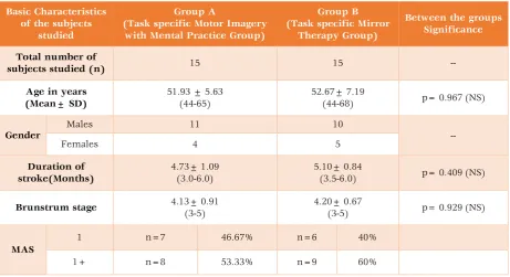
Procedure for intervention for Group B: 21,22
In Group B, subjects were treated with task specific mirror therapy consisting of 5 tasks for duration of 60 minutes for 3 days a week for 10 weeks.
The subject was positioned on a height adjustable table comfortably with the mirror accommodated between the affected and unaffected limbs. The affected limb was placed behind the mirror and unaffected limb in front of the mirror. The mirror was positioned in front of the patient’s midline, so that the affected limb was fully covered by the mirror and the reflection of the unaffected limb was completely visible. The subject was first demonstrated to perceive the limb differently to how it actually is. Next, subject was instructed to observe the mirror reflection for one to two minutes, trying to visualize the mirror image as the affected limb. Once the subject got engaged with the mirrored limb they were asked to perform slow, easy to achieve bilateral movements (perceived bilateral movements) while continuing
to look at the reflected image.23 The motor tasks
that were given were, picking up an empty paper cup without handle and bringing it to the mouth in order to touch it, and then returning the cup to its initial position, turning page of a book, reaching the top of the head, picking up a phone and proper holding of a pen to write. The total duration of intervention was given for 60 minutes per
session.13
Home exercise program
A home exercise program was given to the subjects of both the groups for the rest of the days in a week and instructions were given about the exercises and were practiced at home. The record of exercises practiced by subjects was maintained in a book. The exercises given were, PNF and stretching of upper and lower limbs, strengthening of upper and lower limbs, balance exercises including step-ups, chair rises, wall balance exercise, stationary marching, toe rise, kicking a ball with either foot and upper extremity functional training by practicing the use of upper
limb in real-life tasks.24
Outcome Measurements:
Pre and post intervention measurements of upper limb function were measured using ARAT scale.
Action Research Arm Test: This test is a standardized format used to evaluate UE motor function using 19 tests across 4 subsets: grasp, pinch, grip, and gross movement, both distally and proximally. The test took approximately 10
minutes to administer.31 The subjects were seated
on a chair with back rested and in front of a table with the testing accessories so that they could reach and grasp the objects. The baseline to complete the task was 60 seconds. When the subject performed the first task, then no more tasks were needed to be administered and scored top marks. When the subject failed to perform the first and second task, he scored zero, then no more tests were needed to be tested in that subtest; otherwise the subject needed to complete all tasks within the subtest. Ching-Ljn Hsieh et al., found that the ARAT scale had an intra-class correlation coefficient (ICQ) of 0.98 indicating very high
STATISTICAL METHODS
Descriptive statistical analysis was carried out in the present study. Out Come measurements
analyzed are presented as mean SD. Significance
is assessed at 5 % level of significance with p value was set at 0.05 less than this is considered as statistically significant difference. Paired ‘t’ test as a parametric and Wilcoxon signed rank test as a non-parametric test have been used to analysis the variables pre-intervention to post-intervention with calculation of percentage of change. Independent ‘t’ test as a parametric and Mann Whitney U test as a non-parametric test have been used to compare the means of variables between groups with calculation of percentage of difference between the means. The Statistical software namely SPSS 16.0, Stata 8.0, MedCalc 9.0.1 and Systat 11.0 were used for the analysis of the data and Microsoft word and Excel have been used to generate graphs, tables etc.
RESULTS
The study was carried on total 30 subjects (Table-1) Group A there were 15 subjects with mean age 51.93years and there were 11 males and 4 females were included in the study. In Group B there were 15 subjects with mean age 52.67years and there were 10 males and 5 females were included in the study. There is no significant difference in mean age, duration and brunstrum stage between the three groups.
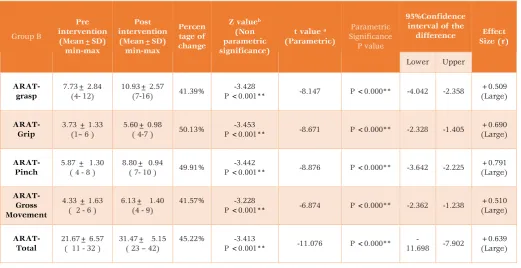
When means were analyzed within the groups (Table-2 & 3) shows that in Group-A and Group-B there is a statistically significant change in means of Action Research Arm Test – grasp, grip, pinch, Gross Movement, total score when means were
analyzed from pre intervention to post
intervention within the groups with p<0.000 with negative percentage of change showing that there is decrease in the post means and positive percentage of change showing there is increase in post means. There is clinical significant improvement with large effect size.
When pre intervention means (Table-4) of Action Research Arm Test – grasp, grip, pinch, Gross Movement, total Score were compared there is no statistically significant difference in grasp and gross movement between the groups and there is a statistically significant difference in grip, pinch and total score between the groups. There is a no clinically significant difference in pre means with small effect size.
When post intervention means (Table-5) of Action Research Arm Test – grasp, grip, pinch, Gross Movement, total Score were compared there is no statistically significant difference in grasp score between the groups and there is a statistically significant difference in grip, pinch, Gross Movement, and total Score between the groups. There is a moderate clinically significant difference in pre means with medium effect size.
Table 1: Basic Characteristics of the subjects studied
Basic Characteristics of the subjects
studied
Group A
(Task specific Motor Imagery with Mental Practice Group)
Group B (Task specific Mirror
Therapy Group)
Between the groups Significance
Total number of
subjects studied (n) 15 15 --
Age in years (Mean± SD)
51.93 ± 5.63 (44-65)
52.67± 7.19
(44-68) p= 0.967 (NS)
Gender
Males 11 10
--
Females 4 5
Duration of stroke(Months)
4.73± 1.09 (3.0-6.0)
5.10± 0.84
(3.5-6.0) p= 0.409 (NS)
Brunstrum stage 4.13± 0.91
(3-5)
4.20± 0.67
(3-5) p= 0.929 (NS)
MAS
1 n=7 46.67% n=6 40%
Table 2: Analysis of Action Research Arm Test within Group A (Pre to post test analysis) Group A Pre intervention (Mean±SD) min-max Post intervention (Mean±SD) min-max Percentage of change
Z valueb
( Non parametric significance)
t valuea
( Parametric) Parametric Significance P value 95%Confidence interval of the
difference
Effect Size
(r)
Lower Upper
ARAT-grasp
7.87± 1.84 (5- 11)
12.60±3.08
(8-17) -60.10%
-3.423
P =0.001** -8.646 P <0.000** -5.907 -3.559
+0.68 ( Large)
ARAT-Grip
2.67± 1.17 (3 – 7 )
7.20± 1.74
( 5 -10) 19.96%
-3.443
P =0.001** -9.906 P <0.000** -3.082 -1.985
+0.83 (Large )
ARAT-Pinch
6.27± 1.66 ( 3 - 9 )
10.20± 2.17
( 8 - 14 ) 62.67%
-3.424
P =0.001** -9.932 P <0.000** -4.783 -3.084
+0.71 (Large)
ARAT-Gross Movement
5.00± 1.60 ( 2 - 7 )
7.33± 1.87 ( 4 - 9)
46.6% -3.462
P =0.001** -11.068 P <0.000** -2.785 -1.881
+0.55 (Large)
ARAT-Total
23.80± 4.93 ( 16 - 31 )
37.33± 8.04 ( 27 - 49)
56.84% -3.410
P =0.001** -11.290 P <0.000** -16.104 -10.962
+0.71 (Large)
** Statistically Significant difference p<0.05; NS- Not significant; a. Pared t test. b. Wilcoxon Signed Ranks Test
Table 3: Analysis of Action Research Arm Test within Group B (Pre to post test analysis)
Group B Pre intervention (Mean±SD) min-max Post intervention (Mean±SD) min-max Percen tage of change
Z valueb
(Non parametric significance)
t value a
(Parametric)
Parametric Significance
P value
95%Confidence interval of the
difference Effect Size (r)
Lower Upper
ARAT-grasp
7.73± 2.84 (4- 12)
10.93± 2.57
(7-16) 41.39%
-3.428
P <0.001** -8.147 P <0.000** -4.042 -2.358
+0.509 (Large)
ARAT-Grip
3.73 ± 1.33 (1– 6 )
5.60± 0.98
( 4-7 ) 50.13%
-3.453
P <0.001** -8.671 P <0.000** -2.328 -1.405
+0.690 (Large)
ARAT-Pinch
5.87 ± 1.30 ( 4 - 8 )
8.80± 0.94
( 7- 10 ) 49.91%
-3.442
P <0.001** -8.876 P <0.000** -3.642 -2.225
+0.791 (Large)
ARAT- Gross Movement
4.33 ± 1.63 ( 2 - 6 )
6.13± 1.40 (4 - 9)
41.57% -3.228
P <0.001** -6.874 P <0.000** -2.362 -1.238
+0.510 (Large)
ARAT-Total
21.67± 6.57 ( 11 - 32 )
31.47± 5.15 ( 23 – 42)
45.22% -3.413
P <0.001** -11.076 P <0.000**
-11.698 -7.902
+0.639 (Large)
Table 4: Comparison of means of Action Research Arm Test between Group A and Group B (PREINTERVENTION COMPARISION) Pre-intervention Group A(Mean±SD) min-max Groups B (Mean±SD) min-max Percentage of difference
Z valueb
( Non parametric)
t value a
( Parametric) Significance P value 95% Confidence interval of the
difference
Effect Size r
Lower Upper
ARAT-grasp
7.87± 1.84 (5- 11)
7.73±2.84
(4- 12) -1.79%
Z= -.358
P=0.720 0.152
P =0.120
(NS) -1.658 1.925
+0.029 (Small)
ARAT-Grip 2.67±1.17 (3 – 7 )
3.73±1.33
(1– 6 ) 33.12%
Z= -1.831
P=0.067 2.033 P =0 .004** -0.007 1.874
+0.390 (Medium)
ARAT-Pinch
6.27±1.66 ( 3 - 9 )
5.87±1.30
( 4 - 8 ) -56.01%
Z=-.616
P=0.538 0.732 P =0.030** -0.719 1.519
+0.133 (Small) ARAT- Gross Movement 5.00±1.60 ( 2 - 7 )
4.33± 1.63
( 2 - 6 ) 21.67%
Z=--1.171
P=0.242 1.128
P =0.058
(NS) -0.544 1.877
+0.203 (Small)
ARAT-Total
23.80±4.93 ( 16 - 31 )
21.67± 6.57
( 11 - 32 ) -9.36%
Z=--.894
P=0.372 1.005 P =0.024** -2.214 6.480
+0.180 (Small)
** Statistically Significant difference p<0.05; NS- Not significant a. Independent t test b. Mann-Whitney Test
Table 5: Comparison of means of Action Research Arm Test between GroupA and Group B (POST INTERVENTION COMPARISION)
Post-intervention Group A(Mean±SD) min-max Groups B (Mean±SD) min-max Percentage of difference
Z valueb
(Non parametric)
t value a
(Parametric)
Significance p value
95% Confidence interval of the
difference Size r Effect
Lower Upper
ARAT-grasp
12.60±3.08 (8-17)
10.93± 2.57
(7-16 -14.19%
Z= -1.507
P=0.132** 1.605
P =0.120
(NS) -0.461 3.794
+0.28 (Small)
ARAT-Grip 7.20± 1.74 ( 5 -10)
5.60± 0.98
( 4-7) -25%
Z= -2.548
P=0.011 3.098 P =0.004** 0.542 2.658
+0.49 (Medium)
ARAT-Pinch
10.20± 2.17 ( 8 - 14 )
8.80± 0.94
( 7- 10 ) -14.73%
Z=-1.736
P=0.083 2.285 P =0.030** 0.145 2.655
+0.386 (Medium)
ARAT- Gross Movement
7.33± 1.87 ( 4 -9)
6.13± 1.40
(4 - 9) -17.83%
Z= -1.912
P=0.056 1.981 P =0.058 * -0.041 2.441
+0.341 (Medium)
ARAT-Total
37.33± 8.04 ( 27 - 49)
31.47± 5.15
( 23 – 42) -17.03%
Z= -1.872
P=0.061 2.379 P =0.024 ** 0.815 10.918
+0.398 (Medium)
Graph - 1: Comparison of post intervention means of Action Research Arm Test between Group A and Group B (Post-intervention comparative analysis)
The above graph shows that that when post intervention means of Action Research Arm Test – grasp, grip, pinch, Gross Movement, total Score were compared there is no statistically significant difference in grasp score between the groups and there is a statistically significant difference in grip, pinch, Gross Movement, and total Score between the groups.
DISCUSSION
The findings from the present study found that there is statistically and clinically significant improvement in hand functions in subjects who received 10 weeks of motor imagery with mental practice and physical practice compared to subjects with task specific mirror therapy.
In the task specific motor imagery with mental practice group, the significant improvement in hand function could be because of the motor imagery with mental practice which is a cognitive process and is thought to be associated with the activation of parieto-occipital network supporting visual-spatial functions involving planning and execution of actions. MP can be considered to be similar to physical practice except that there is no activity in the neuromuscular system. In this study physical practice has been given prior to mental practice with the help of videotape. Traditionally, five sources of response-related input have been identified in regard with motor learning and actual execution of movements such as proprioceptive, tactile, vestibular, visual and auditory information. So mental practice with physical practice where
participants simultaneously observed and
performed congruent movements, motor training
occurs through formation of motor memories. 11, 26,
27 In a recent fMRI study, volunteers were asked to
observe motor acts (e.g. grasping a cup) and showed that the mirror network was active. Because observation of a movement is assumed to facilitate the execution of that movement, both MP and physical practice can be considered to be self-generated, with the aim of improving performance
and promoting motor learning.6, 27 Hence in the
present study, motor imagery with mental practice is found to be an effective additional treatment to improve motor functions of upper extremity through using task oriented model of learning. In the task specific mirror therapy, it is found that, there is a significant improvement in hand
function.21 Mirror therapy (MT) based on the visual
stimulation, is a very simple and promising technique that can be adapted in clinical and home
setting, focusing on missing functions of hand.28 In
stroke, motor neglect or extinction, an
‘‘underutilization of one side, without defects of strength, reflexes, or sensibility” occurs when one hemisphere of the brain is compromised and motor planning systems favor the motor commands from the dominant, unaffected side. Hence, sensory feedback is reduced and motor output is disrupted
reducing movement on the contra lateral side.29
These symptoms of learned nonuse syndrome can be reversed by mirror therapy. It has been proved based on the mirror neuron system that, observation of distal arm movements in the mirror increases corticospinal excitability of the same areas that are excited during normal movements
than when directly viewing the inactive hand.1,28
Thieme H et al., in their Cochrane review summarized the effectiveness of mirror therapy and concluded that it has a significant effect on motor function, activities of daily living, pain with
0
5
10
15
20
25
30
ARAT- Grasp ARAT- Grip ARAT- Pinch ARAT- GM ARAT-Total
12.60
7.20
10.20
7.33
37.33
10.93
5.6 5.87 6.13
31.47
M
e
an
s
of
A
R
A
T
its effects being stable at follow up of six months and that it can be used as an adjunct to normal
rehabilitation for patients after stroke.30
In the present study, when the post interventions means of hand function were compared, task specific MI with MP found greater percentage of improvement than TSMT group. Recent evidences have been in favor of a sensory-motor mapping mechanism such as mirror neurons that are involved in action perception and fire when an individual either performs a given motor act or
observe a similar one.17 It can be considered that in
the present study, both mental practice with motor imagery and mirror therapy are based on mirror neuron system which are multi-input stimulation techniques (combining action observation, mental training, and training in a virtual environment) and can be considered to have a huge impact on
compensation of lost functions after hemiplegia.28
In the TSMT group, it is assumed that mirror provides a form of virtual feedback creating an illusion that the affected side is moving with a normal pattern. Ruud W. Selles et al., in their study confirmed that mirror therapy can facilitate motor learning, but suggested that bimanual movement was less effective than unimanual training. This may be because the movement of the affected limb placed behind the mirror may provide an increased proprioceptive feedback causing an incongruence
between task performance and visual feedback.10
Stroke patients may also experience
somatosensory changes, such as delayed
perception, uncertainty of response, changes in sensory threshold, and changes in sensory adaptation time during mirror therapy resulting in significant differences in degree and time of movement between paretic and non-paretic side. Similarly in the present study, a form of bimanual mirror therapy was administered which could have not contributed to have similar effect as task
specific MI with MP group.31
Several studies have also been done in support of
physical practice combined with mental practice.6
Hence in the present study mental practice was used with physical practice using videotape for observation and performance of the given tasks. So it can be considered to be more effective because the intervention could have reduced the effect by using unaffected arm along with affected arm for unimanual tasks when compared to use of bimanual tasks as in mirror therapy.
Hence from the present study, there is a significant difference between task specific MI with MP when compared to TSMT on improving upper limb
functions for sub-acute hemiplegia. Therefore, null hypothesis is rejected.
Limitations of the study
Subjects with history of ischemic stroke only were studied. In this study, only subjects with 2 to 6 months post stroke were selected. The effect of TSMT and TSMP on spasticity was not evaluated. The number of tasks selected in the study was limited. Only ARAT was used as an outcome measure for evaluation.
Recommendation for future research
Further studies on larger population may be more beneficial. Further studies with use of more tasks for training may be useful. Further studies on effect of these treatments on spasticity and ROM may provide better results for evaluation. Further studies emphasizing on follow up and long term efficacy may be required.
CONCLUSION
The present study concludes that the 10 weeks of task specific motor imagery with mental practice and task specific mirror therapy both shown significant effect on improvement of upper extremity function. However, task specific motor imagery with mental practice found greater percentage of improvement in hand function when compared to task specific mirror therapy. It is clinically important to consider the use of either of the two treatment techniques for recovery of upper extremity for daily activities as an adjunct with a comprehensive rehabilitation program.
Acknowledgement
Authors were expressing their sense of gratitude’s to the people who helped and encouraged them for the guidance and completion of this study.
Conflicts of interest: None
REFERENCES
1. AlessioFaralli, Matteo Bigoni, Alessandro
Mauro, Ferdinando Rossi, Daniela Carulli. Noninvasive strategies to promote functional recovery after stroke. Review Article. Neural Plasticity. 2013;2013:854597.
2. Merritt, Lewit P. Rowland, Randy Rowland.
Merritt's Neurology 10th Edition;2000.
3. Kameshwar Prasad, Deepti Vibha, Meenakshi.
Cerebrovascular disease in South Asia − Part I. A burning problem. J R Soc Med Cardiovasc Dis. 2012;1:20.
4. Jeyaraj Durai Pandian, Paulin Sudhan. Stroke
Epidemiology and Stroke Care Services in India. Journal of Stroke. 2013;15(3):128-134.
5. Yavuzer G, Selles R, Sezer N, Sütbeyaz S,
sub-acute stroke: a randomized controlled trial. Arch Phys Med Rehabil. 2008;89(3):393-8.
6. Th. Mulder. Motor imagery and action
observation: cognitive tools for rehabilitation. J Neural Transm. 2007;114(10): 1265–1278.
7. Andy Wu, Valerie Hermann, Jun Ying, Stephen
J. Page. The Timing of Mentally Versus Physically Practiced Affected Arm Movements in Stroke. Am J Occup Ther. 2010; 64(6): 929– 934
8. Jeanine A Verbunt, Henk AM Seelen, Feljandro
P Ramos, Bernard HM Michielsen, Wim L Wetzelaer, Martine Moennekens. Mental
practice-based rehabilitation training to
improve arm function and daily activity performance in stroke patients: a randomized clinical trial. BMC Neurol. 2008; 8: 7.
9. Andrea Zimmermann-Schlatter, Corina
Schuster, Milo A Puhan, EwaSiekierka, Johann Steurer. Efficacy of motor imagery in post-stroke rehabilitation: a systematic review. J Neuroeng Rehabil. 2008; 5: 8.
10.Ruud W. Selles, Marian E. Michielsen,
Johannes B. J. Bussmann, Henk J. Stam, Henri L. Hurkmans, Iris Heijnen, Daniellede Groot , Gerard M. Ribbers. Effects of a Mirror-Induced Visual Illusion on a Reaching Task in Stroke Patients: Implications for Mirror Therapy Training. Neurorehabil and Neural Repair. 2014;28(7):652-9.
11.Sjoerd de Vries, Theo Mulder. Motor imagery
and stroke rehabilitation: A critical discussion. Review Article. J Rehabil Med. 2007; 39(1): 5– 13.
12.Christian Dohle, Judith Püllen, Antje Nakaten,
JuttaKüst, Christian Rietz, and Hans Karbe. Mirror Therapy Promotes Recovery From Severe Hemiparesis: A Randomized Controlled Trial. Neuro rehabilitation and Neural Repair. 2008.
13.Sneha S. Khandare, R. M. Singaravelan,
Subhash M. Khatri. Comparison of Task Specific Exercises and Mirror Therapy to Improve Upper Limb Function in Subacute Stroke Patients. IOSR-JDMS. 2013; 7(1): 05-14.
14.Daehee Lee, HyolyunRoh,, Jungseo Park,
Sangyoung Lee, Seulki Han. Drinking Behavior Training for Stroke Patients Using Action Observation and Practice of Upper Limb Function. J. Phys. Ther.Sci. 2013; 25: 611–614.
15.Aline Furtado Bastos, Beatriz
CantanhedeCarrapatoso, Marco Orsini, Marco Antonio Araujo Leite, Julio Guilherme da Silva Gabriela Guerra Leal Souza. Functional Recovery of Upper Limb Post-Stroke: Mental Practice with Motor and Non-Motor Imagery. Am. Med. J.2012; 3 (1): 50-55.
16.Cecilia Heyes. Mesmerising mirror neurons.
NeuroImage.2010;51(2):789-91.
17.Giacomo Rizzolatti, Maddalena Fabbri-Destro,
Luigi Cattaneo. Mirror neurons and their
clinical relevance. Nature Clinical Practice
Neurology. 2009; 5: 24-34.
18.Cecilia Heyes, Where do mirror neurons come
from? Neuroscience and Biobehavioral
Reviews. 2009.
19.Justin H. Mathis, Advisor: Jay E. Elliott.
Constraint induced movement therapy and
mirror therapy supporting stroke
rehabilitation: A literature review. 2012.
20.Peter Langhorne, Julie Bernhardt, Gert
Kwakkel. Stroke rehabilitation. Lancet. 2011; 377(9778): 1693–702.
21.Bruce H. Dobkin. Rehabilitation after Stroke. N
Engl J Med. 2005;352:1677-84.
22.Erhan Oztopa, Mitsuo Kawatob, Michael A.
Arbibc. Mirror neurons: Functions,
mechanisms and models. Neurosci Lett. 2013 ;540:43-55.
23.Rothgangel AS, Braun SM. Mirror therapy:
Practical protocol for stroke rehabilitation. 2013.
24.Pamela Duncan, Lorie Richards, Dennis
Wallace, Joni Stoker-Yates, Patricia Pohl, Carl Luchies, Abna Ogle, Stephanie Studenski. A Randomized, Controlled Pilot Study of a Home-Based Exercise Program for Individuals With
Mild and Moderate Stroke. Stroke.
1998;29(10):2055-2060 .
25.Ching-Ljn Hsieh, I-Ping Hsueh, Fu-Mei Chiang,
Po-HsinLjn. Inter-rater reliability and validity of the Action Research arm test in stroke patients. Age and Ageing. 1998; 27(2): 107-1I3.
26.Sjoerd de Vries, Marga Tepper, Bert Otten,
Theo Mulder. Research Article. Recovery of Motor Imagery Ability in Stroke Patients.
Hindawi Publishing Corporation.
Rehabilitation Research and Practice Volume 2011, Article ID 283840.
27.Dickstein R, Deutsch JE. Motor imagery in
physical therapist practice. Phys Ther. 2007;87(7): 942–953.
28.Alina Radajewska, Jo´zef A. Opara,
CezaryKucio, Monika Błaszczyszyn, Krzysztof Mehlich, Jarosław Szczygiel. The effects of mirror therapy on arm and hand function in subacute stroke in patients. Int J Rehabil Res. 2013;36(3):268-74.
29.Candy Mccabe. Mirror visual feedback
therapy.A practical approach. J Hand Ther. 2011;24(2):170–9.
30.Thieme H, Mehrholz J, Pohl M, Behrens J,
function after stroke: Review. The Cochrane Library. 2012.
31.Hyun Jin Kim, Gyu Chang Lee, Chang Ho
Song. Effect of Functional Electrical
Stimulation with Mirror Therapy on Upper Extremity Motor Function in Poststroke Patients. Journal of Stroke and Cerebrovascular Diseases. 2014; 23(4): 655-666.
Citation
Thara. N, Sai Kumar. N, Vinod Babu. K, & Akshata Akalwadi. (2015). COMPARATIVE STUDY BETWEEN TASK SPECIFIC MOTOR IMAGERY WITH MENTAL PRACTICE VERSUS TASK SPECIFIC
MIRROR THERAPY ON UPPER LIMB FUNCTIONS FOR SUB ACUTE HEMIPLEGIA. International