Published online 2019 February 5. Research Article
Plate Augmentation for Nonunion of Femoral Shaft Fractures
Ali Yeganeh
1, 2, Mehdi Abbasi
1, Habib-o-lah Gorgani
1, 2and Mehdi Moghtadaei
1, 2, *1Department of Orthopedic Surgery, Rasool-e-Akram Hospital, Iran University of Medical Sciences, Tehran, Iran
2Bone and Joint Reconstruction Research Center, Shafa Orthopedic Hospital, Iran University of Medical Sciences, Tehran, Iran
*Corresponding author: Department of Orthopedic Surgery, Rasool-e-Akram Hospital, Iran university of Medical Sciences, Tehran, Iran. Email: moghtadaei.m@iums.ac.ir
Received2019 January 04;Revised2019 January 07;Accepted2019 January 25.
Abstract
Background:Femoral nonunion is an important complication, which can occur after intramedullary nailing and it requires sur-gical intervention. Plate augmentation over intramedullary nail is emerging as an acceptable option with satisfactory results for femoral nonunion.
Objectives:The aim of the present study was to determine whether plate augmentation over retained intramedullary nail is an effective treatment for nonunion of femoral shaft fracture.
Methods:Overall, 35 cases of femoral nonunion, initially treated with intramedullary nailing, were managed with plating augmen-tation. Patients with oligotrophic or atrophic nonunion also received iliac cancellous auto graft. The outcome was evaluated by the rate and duration of union and complications were recorded.
Results:All patients achieved bony union during an average time of 21 weeks (±3.94) and no union occurred later than 35 weeks. In plain radiography, evidence of callus formation was seen at mean time of 10 weeks. There was no statistically significant difference in union time among different types of nonunion (P: 0.466) while a significant difference was noticed in the time for callus formation (P < 001). Also, no complications were observed.
Conclusions:Plating augmentation is an effective and safe treatment option for nonunion of femoral shaft fractures.
Keywords:Plate Augmentation, Intramedullary Nail, Femoral Nonunion, Bone Graft
1. Background
Intramedullary nailing with its minimally invasive na-ture is considered the standard treatment for the ma-jority of patients with femoral shaft fractures and it has shown excellent union outcome with reported high union rates (1,2). Femoral nonunion is an important complica-tion, which can occur in up to 57% of patients after in-tramedullary nailing and it requires a surgical interven-tion (3, 4). Several treatment options exist for femoral nonunion after intramedullary nailing, including reamed re-nailing (5,6), dynamization (7), nail removal followed by plating (8), stable fixation with or without bone grafting (9,
10), and external fixation (11).
Currently, exchange nailing with or without bone graft is the standard of care for femoral nonunion. Some studies have reported secondary union rates as low as 53% after ex-change nailing, which questions the efficacy of this treat-ment paradigm (12).
Plate augmentation over intramedullary nail is emerg-ing as an acceptable option with satisfactory results for femoral nonunion. Many studies have indicated high
union rates of this method (13-15).
2. Objectives
This study sought to evaluate the efficacy of plating augmentation over retained intramedullary nail in achiev-ing bony union in patients with nonunion of femoral shaft fracture.
3. Methods
3.1. Study Design and Patients
This was a prospective interventional study conducted between 2015 and 2018. Patients with current unilateral femoral nonunion, initially treated by intramedullary nail-ing, were recruited in the study. Patients were included if they had clinical manifestation of non-union and also depicted no sign of bony union on X-ray for at least six months, postoperatively. Patients with nonunion due to infection were excluded. The outcome parameters were rate and time to union. Patients were also categorized to
three groups, according to their non-union type, including hypertrophic, atrophic, and oligotrophic. The mean weeks to obtain cross-bridge or solid union was reported in the study population and compared between groups.
Femoral shaft was defined as a part of femoral diaph-ysis between 5 cm distal to lesser trochanter and 5 cm prox-imal to adductor tubercle (15). Non-union was defined as the lack of radiographic union at least for six months com-bined with persistent pain at the fracture site (16). Patients were evaluated at the scheduled intervals to investigate for evidence of clinical and radiographic bony union. Union was defined as the absence of clinical symptoms in addi-tion to visualized bridging callus over three-fourths of the diameter of the fracture site of femur on both anteropos-terior and lateral view (15).
3.2. Intervention
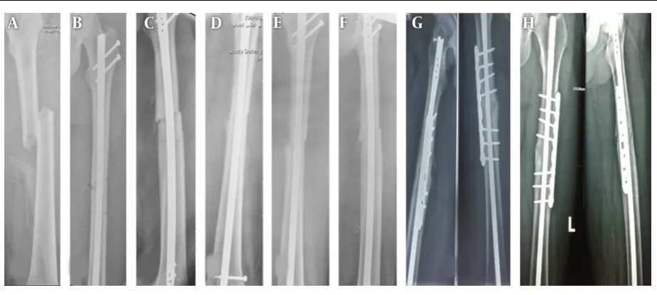
All the procedures were performed by the senior au-thor (AY). Blood tests were obtained from patients to ex-clude infectious causes of non-union, preoperatively. Di-rect lateral approach was used by splitting the tensor fascia latae and vastus lateralis muscles to expose the nonunion site. Following incision on the previous scar, all the fibrotic tissues were resected and the bone edges were completely freshened with curettage, drilling, and rongeur while os-teotome was applied to perform thin layers decortication on the posterior, lateral, and anterior edges of the bone. Samples were taken for laboratory analysis of infection then the researchers applied a 4.5-mm Dynamic Compres-sion Plate (DCP) or anatomical locking plate for the distal part of the shaft. The plates were between 8 to 14 holes and at least three cortical screws were implanted on each side of the fracture based on the type of fracture to gain rigid fixation over the retained intramedullary nail. Fol-lowing plate fixation, 6 to 10 cc of iliac cancellous auto graft was harvested and injected to the nonunion sites in pa-tients with oligotrophic or atrophic nonunion while the non-union gap was closed rigidly (Figure 1). Patients were discharged after 48 hours of empirical antibiotic therapy and allowed protected partial weight bearing gait. Partial weight bearing with the aid of crouches was allowed im-mediately after surgery whereas full weight bearing began after satisfactory bony union and clinical improvement. Also, nutritional supplement of vitamin D and calcium for three months and oral antibiotics for one week were pre-scribed.
3.3. Statistical Analysis
All statistical analysis was performed using SPSS 18.0. Descriptive data were reported as a mean±standard
devi-ation for continuous values and frequency for categorical values. Repeated measure analysis was used to compare the outcome between subgroups. Spearman correlation was also conducted to evaluate the relationship between baseline characteristics of patients and union time. P val-ues below 0.5 were considered statistically significant.
4. Results
The mean±SD age of patients was 34.31±14.59, in-cluding 25 (71.4%) males. Motor vehicle accidents was the major mechanism of fractures and 13 cases initially pre-sented open fractures. Diabetes mellitus occurred in 10 patients as a comorbidity disorder. In 9 patients, frac-tures were located at the distal part of shaft and the rest of patients had fractures located at either proximal or mid-dle third of femoral bone. In terms of non-union typ-ing, 10 fractures revealed hypertrophic non-union, 15 pa-tients depicted oligotrophic and 10 cases had atrophic nonunion. Among these 35 non-union circumstances, 28 patients had ineffective nonunion treatment; 12 cases un-derwent dynamization followed by weight bearing exer-cise, 14 cases sustained re-nailing with replacement of a larger nail and in two patients bone marrow injection was done. The mean time of nonunion between primary surgery of intramedullary nailing and the plate augmenta-tion was 20.03 (±6.37). Mean surgical time was 90 minutes (with a range of 70 to 150 minutes).
All patients achieved solid bony union in a mean time of 21 weeks (±3.94) and no union occurred later than 35 weeks. The mean time of follow-up was 21.09 (±6.22).Table 1summarizes patients’ profile. In the radiographic study, evidence of callus formation was seen at mean time of 10 weeks. In the hypertrophic non-union group, radiographic callus formation was earlier than other nonunion types. There was no statistically significant difference in union time among different types of nonunion (P: 0.466) while a significant difference was noticed in time to visualize cal-lus formation (P < 001) (Table 2).
Statistical evaluation demonstrated that there was no significant relationship between union time and gender (P: 0.986), and primary fracture type (P: 0.193). There was a significant negative relationship between age and union time (P: 0.005, correlation coefficient:- 0.493).
Figure 1.A, Femoral shaft fracture; B, Primary Intramedullary nailing; C and D, 6 months postoperative nonunion of femoral fracture; E and F, 9 months postoperative nonunion of femoral fracture; G, 4 months post-plate augmentation; H, 6 months post-plate augmentation
5. Discussion
Femoral nonunion is a rare yet difficult to treat com-plication of fractures, in which standard treatment, such as re-nailing has failed to produce consistent results in different studies. In the recent years, plate augmen-tation has emerged as a promising treatment option. The current results indicated that the combination of plate augmentation and bone auto graft over retained in-tramedullary nail is an effective and safe treatment op-tion for femoral nonunion with union rate of 100%. In a meta-analysis by Somford et al. applying a plate over existing intramedullary nail had more union rate com-pared with other techniques, including re-nailing (17). Re-ports of results of augmentation plating over retained in-tramedullary nailing in femoral nonunion are limited. The number of patients in these studies are generally less than 50 cases and favorable outcome in all cases were reported (15,18,19). The findings are aligned with the results of pre-vious studies. In the study of Hakeos et al. using a plate over intramedullary nailing associated with bone graft, particularly for complex fractures around the metadiaphy-seal region, resulted in satisfactory outcome and they con-cluded that this procedure appears to be effective in reduc-ing pain and improvreduc-ing function, and predictably leads to radiographic consolidation of the nonunion (20). In a retrospective study by Vaishya et al., this method was performed in 16 cases. All patients had bony union and bone graft was applied in four cases. The average surgical time was acceptable and no major complication occurred
yet they had some cases of limb shortening (21). Ueng et al. as in the current study reported successful bony union after augmentation plating over intramedullary nailing. However, this study additionally applied bone autograft to nonunion sites in atrophic and oligotrophic nonunion pa-tients to stimulate further callus formation (13).
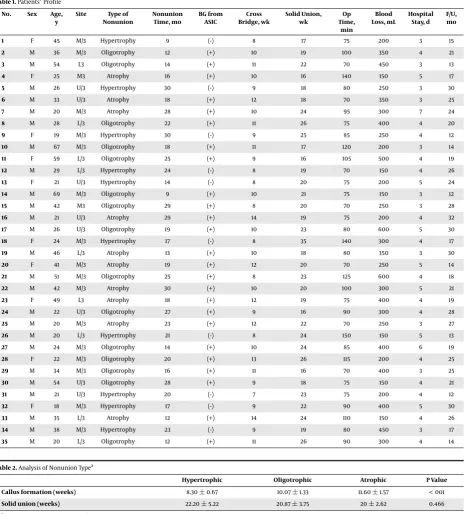
du-Table 1.Patients’ Profile
No. Sex Age, y
Site Type of Nonunion
Nonunion Time, mo
BG from ASIC
Cross Bridge, wk
Solid Union, wk
Op Time,
min
Blood Loss, mL
Hospital Stay, d
F/U, mo
1 F 45 M/3 Hypertrophy 9 (-) 8 17 75 200 3 15
2 M 36 M/3 Oligotrophy 12 (+) 10 19 100 350 4 21
3 M 54 L3 Oligotrophy 14 (+) 11 22 70 450 3 13
4 F 25 M3 Atrophy 16 (+) 10 16 140 150 5 17
5 M 26 U/3 Hypertrophy 30 (-) 9 18 80 250 3 30
6 M 33 U/3 Atrophy 18 (+) 12 18 70 350 3 25
7 M 20 M/3 Atrophy 28 (+) 10 24 95 300 7 24
8 M 28 L/3 Oligotrophy 22 (+) 11 26 75 400 4 20
9 F 19 M/3 Hypertrophy 30 (-) 9 25 85 250 4 12
10 M 67 M/3 Oligotrophy 18 (+) 11 17 120 200 3 14
11 F 59 L/3 Oligotrophy 25 (+) 9 16 105 500 4 19
12 M 29 L/3 Hypertrophy 24 (-) 8 19 70 150 4 26
13 F 21 U/3 Hypertrophy 14 (-) 8 20 75 200 5 24
14 M 69 M/3 Oligotrophy 9 (+) 10 21 75 150 3 12
15 M 42 M3 Oligotrophy 29 (+) 8 20 70 250 3 28
16 M 21 U/3 Atrophy 29 (+) 14 19 75 200 4 32
17 M 26 U/3 Oligotrophy 19 (+) 10 23 80 600 5 30
18 F 24 M/3 Hypertrophy 17 (-) 8 35 140 300 4 17
19 M 46 L/3 Atrophy 13 (+) 10 18 80 350 3 30
20 F 41 M/3 Atrophy 19 (+) 12 20 70 250 5 14
21 M 51 M/3 Oligotrophy 25 (+) 8 23 125 600 4 18
22 M 42 M/3 Atrophy 30 (+) 10 20 100 300 5 21
23 F 49 L3 Atrophy 18 (+) 12 19 75 400 4 19
24 M 22 U/3 Oligotrophy 27 (+) 9 16 90 300 4 28
25 M 20 M/3 Atrophy 23 (+) 12 22 70 250 3 27
26 M 20 L/3 Hypertrophy 21 (-) 8 24 150 150 5 13
27 M 24 M/3 Oligotrophy 14 (+) 10 24 85 400 6 19
28 F 22 M/3 Oligotrophy 20 (+) 13 26 115 200 4 25
29 M 34 M/3 Oligotrophy 16 (+) 11 16 70 400 3 25
30 M 54 U/3 Oligotrophy 28 (+) 9 18 75 150 4 21
31 M 21 U/3 Hypertrophy 20 (-) 7 23 75 200 4 12
32 F 18 M/3 Hypertrophy 17 (-) 9 22 90 400 5 30
33 M 35 L/3 Atrophy 12 (+) 14 24 110 150 4 26
34 M 38 M/3 Hypertrophy 23 (-) 9 19 80 450 3 17
35 M 20 L/3 Oligotrophy 12 (+) 11 26 90 300 4 14
Table 2.Analysis of Nonunion Typea
Hypertrophic Oligotrophic Atrophic P Value
Callus formation (weeks) 8.30±0.67 10.07±1.33 11.60±1.57 < 001
Solid union (weeks) 22.20±5.22 20.87±3.75 20±2.62 0.466
aValues are expressed as mean±SD.
ration of follow-up are required to establish the definitive outcome of augmentation plating for femoral nonunion.
5.1. Conclusions
The current literature and the present study have shown the effectiveness of plate augmentation over
5.2. Compliance with Ethical Standards
Ethical approval was obtained from the ethics commit-tee of Iran University of Medical Sciences. This study was also in accordance with the Helsinki Declaration and its later amendments and prior to entering the study, patients gave their informed consent to participate in this study.
Footnotes
Authors’ Contribution: Study concept and design: Ali Yeganeh and Mehdi Moghtadaei; acquisition of data: Ali Yeganeh, Mehdi Abbasi, Habib-o-lah Gorgani and Mehdi Moghtadaei; analysis and interpretation of data: Ali Yegan, Mehdi Abbasi and Habib-o-lah Gorgani; drafting of the manuscript: Mehdi Abbasi, Habib-o-lah Gorgani and Mehdi Moghtadaei; critical revision of the manuscript for important intellectual content: Ali Yeganeh, Habib-o-lah Gorgani and Mehdi Moghtadaei; statistical analysis: Mehdi Abbasi, Habib-o-lah Gorgani and Mehdi Moghtadaei.
Conflict of Interests: All authors declared that they had no conflict of interests.
Ethical Considerations: Ethical approval was obtained from the Ethics Committee of Iran University of Medi-cal Sciences. This study was also in accordance with the Helsinki Declaration and its later amendments and prior entering the study patients gave their informed consent to participate in this study.
Funding/Support: None.
References
1. Winquist RA, Sigvard J, Clawson DK. Closed intramedullary nailing of femoral fractures. A report of five hundred and twenty cases.Orthop Trauma Direct. 2005;3(4):29–31. doi:10.1055/s-2005-919122.
2. Neumann MV, Sudkamp NP, Strohm PC. Management of femoral shaft fractures. Acta Chir Orthop Traumatol Cech. 2015;82(1):22–32. [PubMed:25748658].
3. Taitsman LA, Lynch JR, Agel J, Barei DP, Nork SE. Risk factors for femoral nonunion after femoral shaft fracture.J Trauma. 2009;67(6):1389–92. doi:10.1097/TA.0b013e318182afd0. [PubMed:19704386].
4. Canadian Orthopaedic Trauma Society. Nonunion following in-tramedullary nailing of the femur with and without reaming. Re-sults of a multicenter randomized clinical trial.J Bone Joint Surg Am. 2003;85-A(11):2093–6. doi: 10.2106/00004623-200311000-00005. [PubMed:14630836].
5. Hak DJ, Lee SS, Goulet JA. Success of exchange reamed intramedullary nailing for femoral shaft nonunion or delayed union.J Orthop Trauma. 2000;14(3):178–82. doi:10.1097/00005131-200003000-00005. 6. Weresh MJ, Hakanson R, Stover MD, Sims SH, Kellam JF, Bosse
MJ. Failure of exchange reamed intramedullary nails for ununited femoral shaft fractures. J Orthop Trauma. 2000;14(5):335–8. doi:
10.1097/00005131-200006000-00005. [PubMed:10926240].
7. Meadows TH, Bronk JT, Chao YS, Kelly PJ. Effect of weight-bearing on healing of cortical defects in the canine tibia.J Bone Joint Surg Am. 1990;72(7):1074–80. doi: 10.2106/00004623-199072070-00018. [PubMed:2384507].
8. Wolinsky PR, McCarty E, Shyr Y, Johnson K. Reamed intramedullary nailing of the femur: 551 cases.J Trauma. 1999;46(3):392–9. doi:
10.1097/00005373-199903000-00007. [PubMed:10088839]. 9. Loomer RL, Meek R, De Sommer F. Plating of femoral shaft
frac-tures: The Vancouver experience.J Trauma. 1980;20(12):1038–42. doi:
10.1097/00005373-198012000-00005. [PubMed:7452747].
10. Smith WR, Morgan SJ. Failure of internal fixation of the femoral shaft. Tech Orthop. 2002;17(4)::448–57. doi: 10.1097/00013611-200212000-00009.
11. Menon DK, Dougall TW, Pool RD, Simonis RB. Augmentative Ilizarov external fixation after failure of diaphyseal union with intramedullary nailing. J Orthop Trauma. 2002;16(7):491–7. doi:
10.1097/00005131-200208000-00007. [PubMed:12172279].
12. Birjandinejad A, Ebrahimzadeh MH, Ahmadzadeh-Chabock H. Aug-mentation plate fixation for the treatment of femoral and tibial nonunion after intramedullary nailing.Orthopedics. 2009;32(6):409. doi:10.3928/01477447-20090511-12. [PubMed:19634823].
13. Ueng SW, Chao EK, Lee SS, Shih CH. Augmentative plate fixation for the management of femoral nonunion after intramedullary nailing. J Trauma. 1997;43(4):640–4. doi:10.1097/00005373-199710000-00013. [PubMed:9356061].
14. Chiang JC, Johnson JE, Tarkin IS, Siska PA, Farrell DJ, Mormino MA. Plate augmentation for femoral nonunion: More than just a salvage tool? Arch Orthop Trauma Surg. 2016;136(2):149–56. doi:
10.1007/s00402-015-2365-9. [PubMed:26646845].
15. Choi YS, Kim KS. Plate augmentation leaving the nail in situ and bone grafting for non-union of femoral shaft fractures. Int Or-thop. 2005;29(5):287–90. doi: 10.1007/s00264-005-0668-0. [PubMed:
16132986]. [PubMed Central:PMC3456636].
16. Lin CJ, Chiang CC, Wu PK, Chen CF, Huang CK, Su AW, et al. Effective-ness of plate augmentation for femoral shaft nonunion after nailing. J Chin Med Assoc. 2012;75(8):396–401. doi:10.1016/j.jcma.2012.06.008. [PubMed:22901724].
17. Somford MP, van den Bekerom MP, Kloen P. Operative treatment for femoral shaft nonunions, a systematic review of the literature. Strate-gies Trauma Limb Reconstr. 2013;8(2):77–88. doi: 10.1007/s11751-013-0168-5. [PubMed:23892497]. [PubMed Central:PMC3732674].
18. Jung HG, Kim DJ, Kim BH, Chung YY. Treatment of the femoral shaft nonunion occurred after intramedullary nailing.J Korean Orthop As-soc. 2007;42(5):653–8.
19. Nadkarni B, Srivastav S, Mittal V, Agarwal S. Use of locking com-pression plates for long bone nonunions without removing ex-isting intramedullary nail: Review of literature and our experi-ence.J Trauma. 2008;65(2):482–6. doi:10.1097/TA.0b013e31817c9905. [PubMed:18695487].
20. Hakeos WM, Richards JE, Obremskey WT. Plate fixation of femoral nonunions over an intramedullary nail with auto-genous bone grafting. J Orthop Trauma. 2011;25(2):84–9. doi:
10.1097/BOT.0b013e3181dfbb33. [PubMed:21245710].