MUSEI NATIONALIS PRAGAE
www.aemnp.eu
ISSN 1804-6487 (online) – 0374-1036 (print)
R E S E A R C H P A P E R
First Anthomyzidae (Diptera) from China:
a new genus, six new species and new records
Jindřich ROHÁČEK
Department of Entomology, Silesian Museum, Nádražní okruh 31, CZ-746 01, Opava, Czech Republic; e-mail: rohacek@szm.cz
Abstract. The family Anthomyzidae (Diptera: Acalyptrata) is recorded from China for the
fi rst time based on 11 species, 6 of them new to science. A distinctive new genus Marshallya
gen. nov. is described, based on single peculiar species, M. platythorax sp. nov. (both sexes)
from Sichuan. Other new species, viz. Amygdalops sevciki sp. nov. (Hainan I.) (both sexes),
Epischnomyia tkoci sp. nov. (Sichuan) (male only), Anthomyza ornata sp. nov. (Sichuan)
(female only), Anthomyza sulphurea sp. nov. (Yunnan) (both sexes) and Arganthomyza
hy-perseta sp. nov. (Shaanxi) (male only) are described and illustrated in detail. Male-female
association of two Amygdalops species is clarifi ed by means of molecular barcoding and the
female of A. bisinus Roháček, 2008 is correctly identifi ed and described. Relationships of all
these taxa are discussed. Five species, viz. Amygdalops bisinus (Hainan I.), Epischnomyia
merzi Roháček, 2009, Anthomyza cuneata Roháček, 1987, Anthomyza trifurca Sueyoshi &
Roháček, 2003 (all from Sichuan) and Arganthomyza versitheca Roháček, 2009 (Shaanxi,
Sichuan) are new additions to the Chinese fauna of Anthomyzidae. DNA sequences of the
barcoding region of COI have been obtained for 3 species, Amygdalops bisinus, Amygdalops
sevciki and Marshallya platythorax. Biology and distribution of all 11 species are discussed. First photographs of living Anthomyzidae from East Asia are presented. Based on knowledge of Anthomyzidae from neighbouring areas the diversity of the Chinese fauna of the family is estimated to include 50–60 species.
Key words. Diptera, Anthomyzidae, Marshallya, biodiversity, biology, distribution, DNA
barcoding, new genus, new records, new species, relationships, taxonomy, China
Zoobank: http://zoobank.org/urn:lsid:zoobank.org:pub:9808C120-13B7-43F8-B735-C13D2B6D43CA
© 2018 The Authors. This work is licensed under the Creative Commons Attribution-NonCommercial-NoDerivs 3.0 Licence. Accepted:
16th April 2018
Published online: 25th April 2018
Introduction
Although dipterology has recently achieved signifi cant
progress in China, fl ies of the family Anthomyzidae have
not yet been studied in this vast territory. Representatives of this small acalyptrate family (currently 134 recent species are described in the world) are delicate slender fl ies related to Opomyzidae. The family Anthomyzidae is currently pla-ced within the superfamily Opomyzoidea and is considered
a sister-group of Opomyzidae (ROHÁČEK 1998, 2006, 2013).
Larvae of most anthomyzids are micro(phyto)sapropha-gous grazers living in graminoids between the leaf base and stem, utilizing (at least partly) rotten tissues usually in plants damaged to some extent. Other species feed in a similar way on decaying soft tissues of dicotyledonous
plants and a few are mycosaprophagous. For more detail on the biology and information on immatures of
Anthomy-zidae see ROHÁČEK (2006, 2009) and ROHÁČEK & BARBER
(2011). The biology of the East Palaearctic and Oriental species (including basic data about habitat and host plant
association) is largely unknown (see ROHÁČEK 2008, 2009;
ROHÁČEK & PRZHIBORO 2016) and, consequently, any
biolo-gical information on them is very valuable. Because China covers a vast territory comprising both Palaearctic and Oriental realms its dipterous fauna is extremely diverse. This is surely also true for Anthomyzidae despite none having been recorded from this country. However, they are known from neighbouring areas including Mongolia in the north (2 species of 2 genera), Far East of Russia
(20 species of 6 genera), Japan (10 species of 4 genera) and the Korean peninsula (10 species of 5 genera) in the
northeast, and Taiwan (4 Amygdalops species), Vietnam
(1 Amygdalops species), Nepal (2 species of 2 genera)
and India (2 Amygdalops species) in the southeast and
south; for published data see SUEYOSHI & ROHÁČEK (2003),
ROHÁČEK (2006, 2008, 2009), and ROHÁČEK & PRZHIBORO
(2016). Therefore we can expect that at least some of these species also occur in China besides hitherto unknown taxa living in this territory.
In this study Anthomyzidae (a total of 11 species) are
recorded from China for the fi rst time, including one
new genus and 6 new species which are described below. However, this treatment is to be considered only a fi rst and relatively small probe into the Chinese anthomyzid fauna where we can expect not only many more species but also further surprising taxonomic discoveries (as is the peculiar new genus treated here) in the future.
Material and methods
Material. The material examined is deposited in collec-tions as follows:
CAU Entomological Museum of China Agricultural University, Bei-jing, China;
CNCI Canadian National Collection of Insects, Arachnids & Nema-todes, Ottawa, Canada;
DEBU University of Guelph Insect Collection, School of Environmental Sciences, University of Guelph, Guelph, Canada;
HNHM Hungarian Natural History Museum, Budapest, Hungary; MBP Miroslav Barták collection, Praha, Czech Republic; NMPC National Museum, Praha, Czech Republic;
SIINH Saigusa Institute of Insect Natural History, Fukuoka, Japan; SMOC Silesian Museum, Opava, Czech Republic;
USNM Department of Entomology, National Museum of Natural His-tory, Smithsonian Institution, Washington D. C., U.S.A.; ZSMC Zoologische Staatssammlung München, München, Germany.
Methods. Specimens have been examined, drawn and
mea-sured using two types of binocular stereoscopic microscopes (Reichert, Olympus SZX10). Male genitalia and female terminalia were examined after their detachment, following treatment in hot 10% KOH, washing in water and dissection of the whole abdomen in a drop of glycerine under a binocu-lar microscope. Detailed examination was performed with a compound microscope (JENAVAL). After examination, all parts were transferred to a small plastic tube in glycerine and pinned below the respective specimens.
Drawing techniques and photography. Legs were
drawn on squared paper using a Reichert binocular microscope with an ocular screen. Details of the male and female terminalia were drawn using Abbe’s drawing apparatus on a compound microscope (JENAVAL) at
hi-gher magnifi cation (130–350×). Wings were photographed
on the same microscope with an attached digital camera
(Nikon COOLPIX 4500). For more details see ROHÁČEK
(2006). Whole adult specimens and their details were
photographed by M. Tkoč using Canon 5DSR digital
camera with a Nikon CFI Plan 10x/0.25NA 10.5mm WD objective attached to an EF 70–200mm f/4L USM lens. The specimens were repositioned upwards between each exposure using a WeMacro Rail (http://www.wemacro.
com/, each step was 5, 10, or 20 μm) and the fi nal photo-graph was compiled from multiple layers (50–150) using
Helicon Focus Pro 5.3. The fi nal images were edited in
Adobe Photoshop CS6.
Measurements. Five main characteristics of adults were
measured: body length (measured from anterior margin of head to end of cercus, thus excluding the antenna), wing length (from wing base to wing tip), wing width (maximum width), C-index (Cs3 : Cs4) (= ratio of length of 3rd costal sector : length of 4th costal sector) and index rm\dm-cu : cu (= ratio of length of section between rm and dm-cu on discal cell : length of dm-dm-cu). All type specimens were measured.
Presentation of faunistic data.Label data of primary
type specimens are presented strictly verbatim including information on colour of all associated labels. Data from other material examined are standardized and presented in full. Phenological and other biological information obtained from the material examined and literature are given in the Biology paragraph; data on occurrence are summarized in the Distribution paragraph.
Barcoding.The specimens used for DNA analysis were
alcohol-preserved (80% ethanol) and/or dry-mounted. The DNA was extracted using NucleoSpin Tissue Kit (Macherey-Nagel, Germany) following manufacturers’ protocols. Scle-rotized remains of all extracted bodies (some representing type specimens) have been preserved in pinned plastic vials in glycerine, labelled with full data and deposited in SMOC. PCRs (polymerase chain reactions) were performed using
specifi c primers for the mitochondrial COI gene barcode
region, LCO1490 (5′
-GGTCAACAAATCATAAAGA-TATTGG-3′) and HCO2198 (5′
-TAAACTTCAGGGTGA-CCAAAAAATCA-3′) (FOLMER et al. 1994). All amplifi ed
products were purifi ed using Gel/PCR DNA Fragments
Extraction Kit (Geneaid, New Taipei City, Taiwan). After
PCR, the amplifi ed DNA samples were sequenced by
Macro-gen Europe (Netherlands). All sequences were assembled, manually inspected, and primers trimmed in SeqTrace (STUCKY 2012). For more details about molecular methods
used in this study see ŠEVČÍK et al. (2016). New sequences
were deposited in the GenBank database and their accession numbers are given in Table 1. The dataset was analyzed using the maximum likelihood (ML) method. The node support values are given in the form of bootstrap value (BV). The analyses were conducted on the CIPRES computer cluster
using RAxML-HPC BlackBox 8.2.10 (STAMATAKIS 2014).
Phylogenetic trees were visualized using Interactive Tree Of Life (iTOL) (LETUNIC & BORK 2016). The species
Fungomy-za albimana (Meigen, 1830) was used as a root following the phylogenetic hypothesis of Holarctic Anthomyzidae by ROHÁČEK & TÓTHOVÁ (2014). The outgroups were
repre-sented by six species: Anagnota bicolor (Meigen, 1838),
Arganthomyza carbo Roháček & Barber, 2013, Typhamyza bifasciata (Wood, 1911), Stiphrosoma fi ssum Roháček,
1996, Anthomyza macra (Czerny, 1928) and F. albimana.
Morphological terminology follows that used in
mo-nographs of Anthomyzidae by ROHÁČEK (2006) and/or
ROHÁČEK & BARBER (2016) including terms of the male
hypopygium and female terminalia. For male genitalia terminology, the ‘hinge’ hypothesis of the origin of the
eremoneuran hypopygium (see ZATWARNICKI 1996), has
been adopted. The following synonymous terms of the male genitalia, emanating from other hypotheses and used
in Diptera manuals (e.g. CUMMING & WOOD 2010), need
to be listed (terms used here fi rst): aedeagus = phallus;
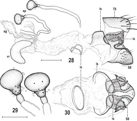
ejacapodeme = ejaculatory apodeme; epandrium = peri-andrium; gonostylus = surstylus; medandrium = bacilli-form sclerite, intraepandrial or intraperiandrial sclerite; phallapodeme = aedeagal apodeme; postgonite = gonite, paramere. Morphological terms of the male postabdomen and genitalia are depicted in Figs 8–22, 45–50, those of the female postabdomen in Figs 23–30. Characters in the generic diagnosis are numbered following the system used by ROHÁČEK (2006, 2009) and ROHÁČEK & BARBER (2016).
Abbreviations of morphological terms used in text and/ or fi gures:
A1 – anal vein ac – acrostichal (seta)
afa – aedeagal part of folding apparatus ag – accessory gland
bm – basal membrane C – costa
ce – cercus
cp – caudal process of transandrium cs – connecting sclerite
Cs3, Cs4 – 3rd, 4th costal sector CuA1 – cubitus
dc – dorsocentral (seta) dm – discal medial cell
dm-cu – discal medial-cubital (= posterior, tp) cross-vein ea – ejacapodeme
ep – epandrium epp – epiphallus f – fi lum of distiphallus f1, f2, f3 – fore, mid, hind femur fc – fulcrum of phallapodeme gs – gonostylus
hl – hypandrial lobe
hu – humeral (= postpronotal) (seta) hy – hypandrium
is – internal sclerite(s) M – media
ma – medandrium npl – notopleural (seta) oc – ocellar (seta) ors – orbital (seta) pa – postalar (seta) pg – postgonite pha – phallapodeme pp – phallophore
ppl – propleural (= proepisternal) (seta) prg – pregonite
prs – presutural (seta) pvt – postvertical (seta) R1 – 1st branch of radius R2+3 – 2nd branch of radius R4+5 – 3rd branch of radius
r-m – radial-medial (= anterior, ta) cross-vein s – saccus of distiphallus
S2–S10 – abdominal sterna sa – supraalar (seta) sc – scutellar (seta) Sc – subcosta sp – spermatheca
stpl – sternopleural (= katepisternal) (seta) T1–T10 – abdominal terga
t1, t2, t3 – fore, mid, hind tibia ta – transandrium
vi – vibrissa vr – ventral receptacle vte – outer vertical (seta) vti – inner vertical (seta)
wpha – wing-shaped lobes of phallapodeme
Results
Marshallya gen. nov.
Type species. Marshallya platythorax sp. nov., here de-signated.
Diagnosis. (1) Head slightly longer than high. (2) Eye large, very convex, elongately ellipsoid, with longest diameter oblique. (3) Frons narrow, fl attened and medially slightly depressed; (4) frontal triangle short and shining. (5) Frontal lunule reduced, indistinct. (6) Occiput strongly concave. (7) Vertex with silvery grey microtomentose stripes lateral to ocellar triangle. (8) Antenna geniculate between pedicel and 1st fl agellomere; pedicel simple, 1st fl agellomere strongly compressed laterally. (9) Arista ciliate (not pectinate) (Fig. 2). (10) Palpus slender, with 1 distinct subapical seta and 2 apical setulae. Cephalic chaetotaxy: (11) pvt small, con-vergent to parallel; (12) vte and/or posterior ors longest of cephalic setae; (13) vti shorter than vte but longer than oc; (14) 2 long ors, widely spaced – posterior in the middle of orbit, anterior close to fore margin of frons; 1 setula and 1 microsetula just in front of anterior ors; (15) postocular se-tulae minute, in single row; (16) 1 long vi and 1 subequally long subvibrissa; (17) peristomal setulae sparse but at least twice as long as postoculars. (18) Posteroventral corner of head (postgena) almost rectangled (Fig. 3). (19) Antenna and face of similar colouring in both sexes (only slightly darker in female).
Table 1. List of species with their GenBank accession numbers and localities.
Taxa COI locality
Amygdalops bisinus Roháček, 2008 (male)
MH012201 China: Hainan I.
Amygdalops bisinus Roháček, 2008 (female)
MH012200 China: Hainan I.
Amygdalops sevciki sp. nov. (male) MH012202 China: Hainan I. Amygdalops sevciki sp. nov. (female) MH012203 China: Hainan I. Amygdalops thomasseti Lamb, 1914 KJ418529 Greece: Crete Anagnota bicolor (Meigen, 1838) KJ418530 Czech Republic
Anthomyza macra Czerny, 1928 KJ418533 Austria
Arganthomyza carbo Roháček & Barber, 2013
KJ418561 Canada: Ontario
Fungomyza albimana (Meigen, 1830)
KJ418547 Czech Republic
Marshallya platythorax sp. nov. (male)
MH012199 China: Sichuan
(20) Thorax narrower than head, subshining. (21) Mesonotum (but not scutellum) dorsally fl attened (Fig. 7). (22) Pleuron with dark longitudinal band at dorsal margin. Thoracic chaetotaxy: (23) 1 hu, 2 npl (anterior longer); (24) 0 prs; (25) 0 sa, 1 short pa; (26) 2 postsutural dc, both in prescutellar portion of scutum, only posterior long, anterior small; (27) ac microsetae in 4 rows in front of suture; (28) 2 sc, apical long, laterobasal short and weak; (29) 0 ppl; (30) 2 stpl, anterior almost as long as posterior, and a few setulae in dorsal half of sternopleuron. Legs: (31) femora and tibiae with dark annulus near knee; (32) f1 of both sexes without ctenidial spine but with a comb of short anteroventral setae in distal two-fi fths; (33) t2 with distinct ventroapical seta; (34) f3 of both sexes with pos-teroventral and anpos-teroventral row of setae some of which in distal half are shortened and thickened. (35) Wing long and narrow; (36) wing membrane darkened, particularly subapically (Fig. 1); (37) C with distinct spinulae among
fi ne setulae on Cs2 (Fig. 4); (38) R2+3 long, slightly
sinu-ous, ending about twice as far from apex of R4+5 than does
M and bent towards C apically; (39) R4+5 slightly bent
apically; (40) R4+5 and M distinctly convergent preapically but parellel apically; (41) M slightly sinuous; (42) dm cell narrow and relatively long; cross-vein r-m situated near its basal two-fi fths to third; (43) CuA1 medium long and
almost reaching wing margin; (44) A1 short but almost
reaching wing margin; (45) anal lobe and alula reduced, both very narrow.
(46) Male abdomen very narrow, elongate. (47) T1
separate from T2; (48) T2–T5 large, long and broad, reaching laterally onto ventral aspect of abdomen, uni-formly dark-pigmented. (49) S1–S5 narrower and very pale-pigmented. Male postabdomen: (50) T6 not deve-loped (membranous); (51) S6–S8 fused dorsolaterally to form asymmetrical synsclerite; (52) S6 and S7 strongly
asymmetrical, fi rmly fused and situated laterally, both
without setae. (53) S8 very long, setose, less asymmetrical and situated dorsally.
Male genitalia (Figs 8, 9, 11–22). (54) Epandrium
rela-tively large, hemispherical, wider than long, densely seto-se, with 2 pairs of markedly longer setae. (55) Anal fi ssure large, high but relatively narrow. (56) Medandrium broad, not high, with submembranous dorsal part; (57) cercus
medium-sized, sclerotized but dorsoventrally fl attened,
fi nely setose. (58) Gonostylus long, slender, with tapered
apex and strongly bent medially, setose mainly on inner side, with micropubescence covering most of outer side.
(59) Hypandrium robust, with fl at lateral lobes well
de-veloped; (60) transandrium (Figs 14, 15, 17) formed by simple medial sclerite, but its lateral robust part projecting in caudal process which is peculiarly connected with both pregonite and postgonite. (61) Pregonite elongate, very low, with 2 groups of setae, fused to hypandrium, only its anterior part projecting medially (see Fig. 15), posterior part very simple but fused with postgonite (and lateral caudal process of transadrium). (62) Postgonite unique because of its lateral position, absence of basal sclerite, short and complex form with several processes and fusion with pregonite and caudal process. (63) Basal membrane
without sclerotized structures and ventrally incised. (64) Aedeagal part of folding apparatus (Fig. 22) relatively short, provided with dark, radially arranged tubercles; (65) connecting sclerite (Fig. 22) extremely robust and heavily sclerotized, distally dilated, darkened and angular. (66) Phallapodeme with basal part expanded into large
fl at wing-like lobes (Fig. 19) embracing base of
disti-phallus. Aedeagus with (67) medium long (posteriorly projecting) phallophore and (68) distiphallus composed of voluminous largely membranous saccus and slender
sclerotized fi lum. (69) Membrane of saccus overgrown
by fi ne spines, mainly on its right side; (70) fi lum formed by 2 long, dark, slender and twisted band-like sclerites, but one of them shortened and ending far from apex. (71) Ejacapodeme small, with slender digitiform projection.
(72) Female abdomen also very elongate but with
somewhat broader terga (T2–T5) and sterna (S2–S5) than in male. (73) Postabdomen (Figs 23–25) relatively long, with terga and sterna (except S6) well sclerotized and dark-pigmented. (74) T6 and S6 relatively large, T6 dark, S6 pale. (75) T7 laterally extended and reaching ventral aspect but not fused with S7 and 7th spiracles free in ple-ural membrane; (76) S7 much smaller than S6, rounded,
characteristically pigmented; (77) T8 simple, fl at, with
rounded corners; (78) S8 protruding posteromedially, with very small posteromedial incision. (79) Internal scleroti-zation of female genital chamber (uterus) well developed (Figs 28, 30), formed by very complex (but symmetrical) posterior sclerites being posteroventrally fused to inner side of S8 and (80) 1 anteroventral, simply transversely ellipsoid annular sclerite. (81) Anterior part of uterus with voluminous vesicular kidney-shaped ventral receptacle (Fig. 28). (82) Accessory glands of usual form, on short simple (non-dilated) ducts. (83) Spermathecae (1+1) shortly pyriform (Figs 28, 29), with well-developed scle-rotized cervix and some grain-like spinulae on smooth body surface; spermathecal duct relatively short. (84) T10 relatively large, rounded, partly dark, with 1 pair of dorsomedial setae; (85) S10 slightly smaller than T10,
simply semicircular, micropubescent besides fi ne setulae.
(86) Cerci moderate but robust, with comparatively short,
fi ne and abundant setae, none of which is enlarged.
Discussion. The combination of diagnostic characters of Marshallya gen. nov. (as listed above) is unique within the Anthomyzidae. These characters include external
features shared with Amygdalops Lamb, 1914 and its
re-latives (while lacking most of their synapomorphies in the male and female terminalia), plesiomorphies widespread
among anthomyzid genera and, most signifi cantly,
apo-morphies unique within the whole family Anthomyzidae.
The type species of Marshallya externally closely
resembles species of the Old World tropical genus
Amyg-dalops and the related Afrotropical genus Margdalops
Roháček & Barraclough, 2003 in having very convex
Figs 1–7. Marshallya platythorax sp. nov. and its habitat (China: Sichuan). 1 – wing (length ca. 3 mm); 2 – antenna, laterally; 3 – head and thorax, late-rodorsally; 4 – detail of C with spine-like setae; 5 – copse of bamboo, habitat and probable host plant in the type locality (with Owen Lonsdale in front of it); 6, 7 – living adults on bamboo stem (body length ca 3.1 mm). Figs 1, 4 based on male paratype, Figs 2, 3 on male holotype. Photos by J. Roháček (1, 4), M. Tkoč (2, 3) and S. A. Marshall (5–7).
frontal lunule, dc macrosetae in prescutellar position
and shortened CuA1 (cf. ROHÁČEK 2004, 2008), and with
Margdalops vertex with silvery microtomentose stripes
and the ciliate (not pectinate) arista (see ROHÁČEK &
BARRACLOUGH 2003). However, more detailed study of
the type species of Marshallya revealed substantive
particularly in having the dorsally fl attened mesonotum, reduced thoracic chaetotaxy (prs, sa, ppl absent, pa short), both male and female with rows of shortened thickened setae on f1 (anteroventrally) and f3 (both anteroventrally and posteroventrally), C with distinct spine-like setae
among fi ne setulae and very elongate slender abdomen.
It is notable that some of these characters can be found as homoplasies in distinctly non-related genera, e. g. the
fl attened mesonotum in the genus Typhamyza Roháček,
1992, the reduced thoracic chaetotaxy in Anagnota Becker,
1902 and Cercagnota Roháček & Freidberg, 1993 (see
ROHÁČEK 2006) and f1 with anteroventral row of thicker
setae in the Afrotropical genus Barbarista Roháček, 1993
(see ROHÁČEK 1993).
Nevertheless, the most important autapomorphies that
characterize Marshallya can be seen in the male genitalia
and female postabdomen as follows: (58) gonostylus elon-gate but strongly bent medially (Fig. 9); (60) transandrium with robust lateral parts, each projecting in caudal process which is uniquely connected with both pregonite and post-gonite; (62) postgonite in unusual lateral position, lacking the basal sclerite, of short and complex form with several processes and fused with (posterior part of) pregonite and (lateral) caudal process (see Figs 16, 17); (63) basal mem-brane without sclerotized structures and ventrally incised; (65) connecting sclerite (Fig. 22) extremely robust and heavily sclerotized, distally dilated, darkened and angular;
(66) phallapodeme basally with large symmetrical fl at
wing-like lobes embracing base of distiphallus (Figs 19, 22); (70) fi lum of distiphallus with (plesiomorphic) 2 long band-like sclerites, but one them shortened and ending far from apex (Fig. 21); (79) internal sclerotization of female genital chamber (Figs 28, 30) well developed, formed by very complex (but symmetrical) posterior sclerites which are posteroventrally fused to inner side of S8 and (80) an anteroventral, simply transversely ellipsoid annular sclerite; (81) ventral receptacle voluminous, vesicular, kidney-shaped (Fig. 28).
The relationships of the genus Marshallya have not
yet been resolved. It is probable that its sister-group will be discovered among the largely unknown Oriental taxa of Anthomyzidae. Morphological and molecular distance
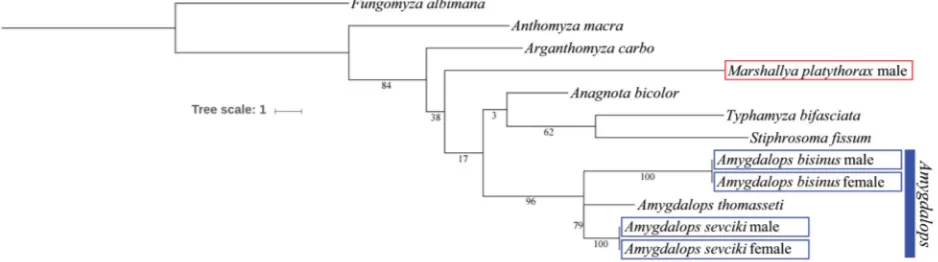
(see Fig. 31) of M. platythorax to the externally similar
Amygdalops species proved to be too large to demonstrate their close alliance. The new genus could possibly be a
more distant relative of the Amygdalops clade (comprising
Amygdalops, Margdalops and an unnamed Neotropical
genus, cf. ROHÁČEK 2008) as is indicated by the shared
external characters discussed above despite large molecular dissimilarity in the barcoding region of COI (Fig. 31).
Species included.Marshallya platythorax sp. nov. (descri-bed below).
Etymology. The new genus Marshallya is dedicated to
my friend, Steve A. Marshall (University of Guelph), the collector of its type species, world renowned Cana-dian dipterist and insect photographer. The name is an
abbreviated conjunction of Marshall + [m]ya, gender
feminine.
Marshallya platythorax sp. nov.
(Figs 1–4, 6–30)
Type material. HOLOTYPE: , labelled: ‘China: Sichuan: Emeishan,
19.v.2016, 800–900m, N 29°34ʹ10.42ʺ E 103°23ʹ27.98ʺ, S. A. Marshall’ and ‘Holotypus , Marshallya platythorax sp. n., J. Roháček det. 2017’ (red label). The specimen is intact, dried from ethanol and mounted on a pinned paper point, with only left mid leg removed and glued to the specimen pin (CAU). PARATYPES: 1 1 , with same label data as the
holotype, only second (type) label is yellow and has ‘Paratypus’ instead of ‘Holotypus’; the male body (without abdomen, one wing and hind leg) was used for DNA extraction and subsequently placed (partly dissected) in glycerine in a pinned plastic microvial; abdomen of both male and female cleared, dissected and preserved in glycerine in a pinned plastic tube but that of male with the removed wing and hind leg in addition (DEBU, 1 ; SMOC, 1 ).
Description. Male. Total body length 3.02–3.18 mm.
Body elongate and very slender (Figs 6, 7), bicolourous, dark brown and whitish yellow. Head slightly longer than high; frons fl attened and dorsomedially depressed. Occiput distinctly concave, dark but not entirely (ochreous to ochre-ous yellow dorsally, particularly behind eyes), subshining and greyish microtomentose (most densely medially above foramen). Vertex with narrow silvery grey microtomen-tose stripes between ocellar triangle and posterior part of orbit. Frontal triangle very short (only in posterior third of frons) and roughly equilateral, shiny because of very sparse microtomentum, including ocellar triangle. Ocelli large and close to each other. Frons largely dark brown, dark grey microtomentose and dull, only its narrow me-dial band pale brown to ochreous yellow (anteriorly) and anterior half of orbit yellow. Orbit dark brown and shining in posterior half but becoming lighter anteriorly to dirty yellow and whitish grey microtomentose in anterior third. Frontal lunule very reduced, not visible in dry specimens. Face dark ochreous laterally, brown and distinctly concave medially; parafacialia and (most of) gena whitish yellow, with silvery white microtomentum but both pale brown margined (on anterior part of gena most broadly); postge-na with posteroventral corner almost rectangled (Fig. 3) but rounded, whitish yellow, dorsally concolourous with adjacent dark brown part of occiput. Mouthparts relatively small, dirty yellow including somewhat darker clypeus. Cephalic chaetotaxy (Fig. 3): pvt small but distinct, con-vergent (usually) to parallel; vti distinctly shorter than vte (about two-thirds of the latter); vte and/or posterior ors longest of cephalic setae; oc strongly proclinate, only about as long as vti, situated outside line connecting ocelli; 2 widely spaced and long ors, posterior as long as or longer than vte, anterior only slightly shorter; 1 setula and 1 much smaller microsetula in front of anterior ors; 2 pairs of microsetulae in interfrontal position in anterior third of frons; 1 relatively weak vi (about as long as oc) and 1 almost equal subvibrissa; peristomal setulae (6–7)
fi ne, becoming longer anteriorly, the foremost about half
very convex, with longest diameter about 1.6 times as long as shortest one. Gena very narrow; its shortest height 0.08 times as long as shortest eye diameter. Antenna (Fig. 2) yellow, strongly geniculate; scape and pedicel of usual form and setosity; 1st fl agellomere strongly laterally fl attened, entirely yellow, at most with slightly darker margin around base of arista, shortly whitish ciliate on apex. Arista also yellowish, including short and thick basal segment, about 2.2 times as long as antenna, with medium long whitish ciliation (Fig. 2).
Thorax narrower than head (with eyes), bicolourous, dark brown dorsally and yellowish white ventrally. Dor-sal part of thorax sparsely microtomentose and relatively shining, pleural part duller. Mesonotum dorsally fl attened (see Figs 6, 7), largely dark brown but with paler ochre-ous brown anterior spots adjacent to humeral callus and also dorsocentral lines somewhat paler (Fig 3). Humeral (postpronotal) callus pale brown and also notopleural area somewhat lighter than surrounding sclerites; pleural part of thorax with rather broad (covering entire propleuron and dorsal half of mesopleuron) blackish brown dorsal band extending from propleuron to haltere and sharply delimited from yellowish white ventral portion of pleuron. Scutellum
slightly convex (not fl at) and blackish brown, as are the
well developed (bulging) postscutellum and metapleuron. Thoracic chaetotaxy (Fig. 3) reduced: ppl, prs and sa ab-sent; 1 relatively weak hu, 2 npl (anterior slightly longer than hu, posterior short), 2 (both postsutural) dc arising close to scutellum; anterior dc short (only twice the length of dc microsetae) and situated immediately in front of the long posterior dc, the latter only slightly shorter than apical sc; ac microsetae small and sparse, in 4 rows on suture, in 2 rows between dc macrosetae and reaching to prescutellar position; 1 short pa (not longer than laterobasal sc); 2 sc, apical long and strong (the longest thoracic seta), latero-basal small, less than one third of length of apical sc and situated rather far from base of scutellum; 2 stpl, posterior only slightly longer than anterior; 5–6 microsetae below stpl and a number (about 10) of fi ne setae on ventral corner of sternopleuron. Scutellum rounded triangular, about 1.5 times as wide as long. Legs bicolourous, largely yellow to yellowish white (coxae lightest) but femora with diffuse brown darkening in distal third to half (except for yellow apical part at knees) and with tibiae similarly (somewhat paler) brown-darkened in proximal fourth to third (cf. Figs
3, 10, 27). Pedal chaetotaxy: f1 with usual posterodorsal
and posteroventral row of longer setae but also with an anteroventral comb of short and thicker setae (3–4 usually more robust than others) in distal two-fi fth (see Fig. 27); f2 without peculiarities, only with some slightly longer and erect ventral setae in proximal third; f3 with posteroventral row of setae along entire length (4–5 of them in distal half shortened and thickened) and with an additional anteroven-tral row of several (yet shorter) thickened setae (see Fig. 10). t1 and t3 uniformly setulose; t2 with usual ventroapical seta, distinctly longer than maximum width of tibia; ba-sitarsi of all legs with 1–3 longer and somewhat thicker ventrobasal setulae, being larger (2–3) on hind basitarsus which also bears a series of thicker ventral setulae among
fi ne setulae. Wing (Fig. 1) with distinctly brown darkened
membrane, darkest along distal half of R2+3 and between
apices of the latter and R4+5; veins dark brown. C (unlike
Amygdalops species) with thicker spine-like, widely spa-ced, setae on dorsal side (cf. Figs 3, 4). Sc well developed,
free proximally, attached to R1 distally and forming with
the latter a very distinct preapical kink at subcostal break. R2+3 very long, running close to C, distally slightly sinuate and apically upcurved to C. R4+5 distally slightly bent and converging preapically to slightly sinuate vein M and ending in parallel. Discal (dm) cell narrow but relatively
long (longer than in most of Amygdalops species); r-m
situated around basal two-fi fths to third of dm cell; apical
section of CuA1 more than twice longer than dm and
al-most ending in wing margin; bm and cup cells small and
narrow; A1 short but almost reaching wing margin due to
reduced anal lobe of wing. Alula small and very narrow
(as in Amygdalops species). Wing measurements: length
2.98–3.02 mm, width 0.83–0.93 mm, Cs3 : Cs4 = 2.08–2.19,
r-m\dm-cu : dm-cu = 3.89–4.06. Haltere with brown stem and large dark brown knob.
Abdomen elongate and very narrow (about 4.6 times as long as wide), blackish brown, greyish microtomentose and subshiny dorsally, pale (whitish) ochreous ventrally. Preabdominal terga large, bent onto lateral to ventrolateral aspects (hence pleural part of segments reduced), uniformly blackish brown and relatively densely but shortly and fi nely setose. T1 distinctly separate and about half length of T2; T2 slightly shorter than T3 and/or T4 (these subequal); T5 shorter than T2 but widest of preabdominal terga. T6 not developed (membranous). Preabdominal sterna very
pale, fi nely sparsely setose, becoming somewhat wider
posteriorly, S5 the widest. S1 shorter than wide, bare; S2 with brownish stripe along anterior margin, slightly shorter than S3 and/or S4 (these subequal) and all distinctly longer than wide; S5 widest but shorter, about as long as wide. S6–S8 asymmetrical, dorsolaterally fused; both S6 and S7 lighter brown, subequal in length, each with anterior darkened marginal ledge-like stripe and situated on left side of postabdomen; S6 ventrally shortened, transversely band-like, laterally dilated, subrhomboidal, bare except for 2 microsetulae; S7 of pentagonal outline, bare, with fusion line with S6 thickened and darkened and its dorsal part embedding large 7th spiracle; 6th spiracle situated in membrane above dorsal corner of S6. S8 situated dorsally, blackish brown, long (almost 3 times as long as S7) and with a number of relatively short setae in posterior half.
Genitaliaof distinctive construction. Epandrium
rela-tively large (compared to postabdominal sclerites), hemi-spherical, not long (Fig. 8) but broad (markedly wider than long, cf. Fig. 9), rather densely setose, with 1 dorsomedial and 1 laterocaudal (this longest and thickest) long setae; anal opening relatively large, high, narrowly semi-ellipsoid (Fig. 9). Cercus characteristically formed, not small but only half length of gonostylus, strongly (dorsoventrally)
fl attened and hidden in anal fi ssure (hence not visible
connecti-Figs 8–13. Marshallya platythorax sp. nov., male paratype (China: Sichuan). 8 – genitalia, laterally; 9 – external genitalia, caudally; 10 – f3 and t3 an-teriorly; 11 – medandrium, anan-teriorly; 12 – apex of gonostylus, anterolaterally (widest extension); 13 – gonostylus, lateroventrally (widest extension). Scales = 0.5 mm (Fig. 10), 0.05 mm (Figs 12, 13), 0.1 mm (others). For abbreviations see p. 37.
on slender and submembranous. Gonostyli symmetrical, strongly bent medially (cf. Fig. 9), with apices crossed or appressed in rest position. Gonostylus (Figs 8, 9, 13) long and relatively slender, roughly sickle-shaped in widest ex-tension view (Fig. 13); its anterior (basal) part with a group of longer fi ne setae ventrally, most of outer side covered
by micropubescence, otherwise fi nely setose on inner side
Figs 23–27. Marshallya platythorax sp. nov., female paratype (China: Sichuan). 23–25 – postabdomen (23 – dorsally; 24 – laterally; 25 – ventrally); 26 – female f3, anteriorly; 27 – female f1, anteriorly. Scales = 0.2 mm. For abbreviations see p. 37.
see Fig. 17) connecting robust lateral parts each projecting into a peculiar stump-like (caudal) process being antero-ventrally connected with both postgonite and posterior part of pregonite (see Figs 14–17, cp). Pregonite (Figs 14, 15), elongate and low, incurved, only its anterior part somewhat projecting medially (Fig. 15, prg) and bearing a group of 5–6 setae; its inconspicuous posterior part with 4–5 setae. Postgonite (Figs 14–18, pg) very unusual, shifted laterally, lacking basal sclerite but anterodorsally fused to
Figs 28–30. Marshallya platythorax sp. nov., female paratype (China: Sichuan). 28 – genital chamber with 8th abdominal segment, laterally; 29 – spermathecae; 30 – internal sclerites of genital chamber and S8 (with setosity omitted), ventrally. Scales = 0.05 mm (Fig. 29) and 0.1 mm (others). For abbreviations see p. 37.
extremely robust, heavily sclerotized, proximally slender but becoming thicker in middle part, distally dilated and somewhat hook-like, with robust blackish angular apex projecting below hypandrial complex (see Fig. 8, cs). Ae-deagal complex (Fig. 22). Phallapodeme relatively robust, having basal part with peculiar (symmetrical) wing-like but ventrally somewhat bent lobes (Fig. 19) embracing basal part of distiphallus (see Fig. 22), apex distinctly widened and lateroventrally projecting; fulcrum also relatively robust. Aedeagus of ancestrally looking type (Fig. 22). Phallophore relatively long, subtriangular in profi le, dar-kest on dorsal corner, with posterior corner more projecting and slightly bent ventrally. Distiphallus composed of short
but relatively voluminous saccus and long slender fi lum.
Saccus largely membranous except some sclerotization in
basal and middle part, and provided with numerous fi ne
spines, mostly on right side of distal membranous part (see Fig. 22). Filum of distiphallus (Fig. 22) very slender, for-med by 2 dark stripe-like twisted sclerites being attenuated
distally but (unlike all other taxa) one of them shortened
(ending far from end of fi lum), and the other terminating
in slightly dilated and fl attened submembranous apex
(see Fig. 21). Ejacapodeme relatively small, with slender
fi nger-like projection (Figs 20, 22).
Female. Similar to male but differing as follows.
Total body length 3.14 mm. Face and clypeus somewhat darker; antenna distinctly darker, ochreous including
1st fl agellomere and arista with somewhat longer cilia.
Very unusually f1 and f3 (see Figs 26, 27) with combs of
shortened thicker setae as in male. t2 with ventroapical
seta somewhat longer and thicker. Wing measurements:
length 3.02 mm, width 0.87 mm, Cs3 : Cs4 = 2.04, r-m\
T3. S1 and S2 as in male but S3–S5 subequal in length and width, all longer than wide.
Postabdomen (Figs 23–25) relatively long. T6 large (but smaller than T5), distinctly wider and much longer than T7 (Fig. 23), slightly tapering posteriorly, with numerous dense, short setae in posterior two-thirds, darker laterally than dorsally and with narrow pale anterior margin. S6 (Fig. 25) distinctly shorter than S5, about as long as wide, slightly widened posteriorly, all pale whitish ochreous, sparsely setose in posterior two-thirds. T7 (Figs 23, 24) narrower and darker than T6, uniformly blackish brown, densely setose as is T6. S7 much smaller than S6, convex, with rounded sides (Figs 24, 25), with characteristic dark brown pattern leaving only anterior marginal stripe and posteromedial wedge-shaped area pale-pigmented; it is sparsely setose, with longest setae in posterior third of
sclerite. T8 relatively fl at, transversely suboblong with
rounded corners, dark brown except for anterior corners
(Fig. 23), with fi ne but longer setae in posterior third,
longest in posterior corners. S8 (Figs 24, 25, 28) smaller than S7, very convex (bulging posteroventrally, cf. Fig.
24), fi nely setulose, having small posteromedial incision
(best visible in Fig. 25) in pale-pigmented posterior part and peculiar in being fused with internal sclerites of female genital chamber situated anterodorsally to S8. Internal sclerotization of genital chamber very conspicuous (see Figs 24, 25, 28, 30, is). Posterior sclerotization complex, formed by 1 pair of lighter bent anterior sclerites which are posteriorly fused to heavily sclerotized and dark-pigmented but symmetrical complex of sclerites projecting posteriorly on each side in 2 processes and being posteroventrally fused with (invaginated parts? of) S8; annular sclerite also unusual, rather simple, forming a transversely ellipsoid ring situated rather far in front of posterior sclerites (Figs 28, 30). Ventral receptacle (Fig. 28) unusually voluminous but weakly sclerotized and pale-pigmented, of slender kid-ney-shaped form with smooth surface. Accessory gland on relatively short and slender (non-dilated) duct. Spermathe-cae (1+1) shortly pyriform (Figs 28, 29), blackish brown, each with a few grain-like spines (mainly on narrowed basal part); duct cervix well developed, comparatively long, pigmented; spermathecal duct relatively short (cf. Fig. 28). T10 (Fig. 23) relatively large and broad (shorter than wide), of suboblong rounded outline, dark brown in
anterior two-thirds, and with a pair of long medial setae beyond middle. S10 (Figs 24, 25) slightly smaller than
T10, semicircular, pale brown, micropubescent, with fi ne
setulae at posterior margin. Cerci (Figs 23–25)
medium-sized but robust, with abundant fi ne setae none of which
is particularly longer or thicker.
Discussion. This new species is peculiar in many
cha-racters (see above under the genus Marshallya) but in
external appearance it is reminiscent of a large and very
slender species of Amygdalops. It particularly resembles
some Amygdalops species having a brown-darkened
an-nulus on the femora and tibiae, such as those known from
the East Palaearctic and Oriental Regions (see ROHÁČEK
2008, 2009). However, it can be easily distinguished from all known species of the latter genus in having the
fi nely ciliate (not pectinate) arista, the dorsally fl attened
mesonotum, no prs and sa, short pa, f1 with a comb of
short thickened anteroventral setae, f3 of both sexes(!)
with 2 rows (anteroventral and posteroventral) of thic-kened and shortened setae and wing with distinct costal
spine-like setae among fi ne setulae. The species of the
Afrotropical genus Margdalops are also externally similar
to M. platythorax, particularly in their ciliate arista and dark wing pattern (cf. ROHÁČEK & BARRACLOUGH 2003) but
they differ (even more distinctly than Amygdalops spp.)
in cephalic, thoracic and pedal chaetotaxies. The striking
genital and postabdominal differences from Amygdalops,
Margdalops and species of other genera of Anthomyzidae
have been discussed above under the genus Marshallya
and need not be repeated here.
Etymology. The name (a Greek noun in apposition) of
the new species refers to the strikingly dorsally fl attened thorax (mesonotum).
Biology. The type specimens were photographed (Figs
6, 7) and subsequently collected by Steve Marshall on stems in a copse of bamboo (Poaceae) (documented by photograph, Fig. 5) on Emei Mountain (at about 800–900 m) on 19 May. Because these peculiar anthomyzids were reasonably abundant in this habitat (but only 3 specimens were collected; S. A. Marshall, personal communication, 2018) it is plausible that this bamboo species is the host plant of M. platythorax.
Distribution. Hitherto known only from the type
speci-mens from China (Sichuan).
Amygdalops Lamb, 1914
The genus Amygdalops Lamb, 1914 is the most diverse
group of Anthomyzidae in the tropical belt of the
Afro-tropical (ROHÁČEK 2004) and Oriental Regions (ROHÁČEK
2008) and a few species of Amygdalops also extend into
the Palaearctic Region (ROHÁČEK 2006, 2009). Therefore,
several species have been expected to occur in China, particularly in its Oriental part. The two species recorded below from Hainan Island. clearly belong to the Oriental
fauna of Amygdalops. Their syntopic occurrence on this
island revealed not only a new species but also a mistake in the previous affi liation of the female sex of Amygdalops bisinus Roháček, 2008. Under the latter name, two species
were mixed by ROHÁČEK (2008: 341–345) due to similarity
of the distinctively sinuate male gonostyli and, moreover,
the female described by him as A. bisinus belongs to
an-other (new) species because the true female of A. bisinus
was unknown at that time. To address the problem of co-rrect male-female affi liation, the specimens from Hainan I. were studied using the barcoding area of COI (see material and methods and Fig. 31). Results of this analysis reve-aled that the female with more complex structures of the
terminalia belongs to A. bisinus (described below) while
the other female is conspecifi c with the male of a new
species, A. sevciki sp. nov. Subsequently, all paratypes of A. bisinus as originally labelled by ROHÁČEK (2008), have
been revised and those belonging to A. sevciki removed
and included in the type series of the latter species.
Amygdalops bisinus Roháček, 2008
(Figs 36–44, 62)
Amygdalops bisinus Roháček, 2008: 341 (partim, male only).
Type material examined. HOLOTYPE: , labelled: ‘THAILAND:
Ban-gkok, Huaykwang, Aug.–Sept. 1962, J. Scanlon –light’ (USNM, genit. prep.) and ‘Holotypus , Amygdalops bisinus sp. n., J. Roháček det. 2007’ (red label). PARATYPE: VIETNAM: Cuc phuong, Ninh binh, 6.–18.v.1966,
1 , Topál leg. (HNHM, genit. prep.). Paratype with same type label as the holotype but it is yellow and has ‘Paratypus’ instead of ‘Holotypus’.
Additional material examined.CHINA: HAINAN I.: Sanya 30 km NE, Sandaozhen env. Xinjiancun, 18°28ʹ44ʺN 109°38ʹ23ʺE, sweeping, 22.v.2016, 1 1 , J. Ševčík leg. (SMOC, both dissected and after being used for DNA extraction remnants of their bodies are preserved in glycerine in plastic pinned microvials, genit. prep.).
Addition to description. Male. Total body length
1.82–1.96 mm. f3 with 5–7 short and thickened setae in
distal third of posteroventral row but these setae distinctly
longer and more widely spaced than in A. sevciki. Wing
measurements: length 1.71–1.91 mm, width 0.50–0.54 mm,
Cs3 : Cs4 = 2.05–2.23, r-m\dm-cu : dm-cu = 3.38–3.56. T5
always with small semicircular, pale ochreous to yellowish, anterolateral spot on each side. Preabdominal sterna pale ochreous, contrasting with dark brown terga. Male genitalia as in Figs 32–37.
Female (fi rst description). Similar to male (for
descrip-tion see ROHÁČEK 2008: 341 and the above addition) but
differing as follows. Total body length 2.32 mm (measured in ethanol). f3 simply setulose, without thickened postero-ventral setae. Wing (Fig. 62) with pattern generally paler
than in most other Amygdalops species, thus with preapical
brownish spot and stripe along R4+5 faded and less distinct,
and area between R4+5 and C only a little lighter. R4+5 and M subparallel or slightly converging preapically and then running parallel apically to margin; r-m situated at middle or slightly in front of midpoint of dm cell. Wing
measure-ments: length 2.14 mm, width 0.64 mm, Cs3 : Cs4 = 2.05,
r-m\dm-cu : dm-cu = 3.23. Preabdominal terga wider and more transverse, dark brown but T4 and T5 with small (and short) yellowish white anterolateral spots, those on T4 being distinctly shorter. Preabdominal sterna paler than in male, yellowish white; S1 short, transverse and bare, S2–S5
fi nely and sparsely setose. S2 and S3 narrow, longer than
broad; S4 and S5 wider than S3, about as long as broad; S5 slightly narrower than S6.
Postabdomen (Figs 38–40) relatively short and broad (at 6th segment). T6 large, broad, relatively shortly but not den-sely setose (with most robust setae at posterior margin), dark brown with small pale-pigmented anterolateral areas being medially connected by transverse stripe (Fig. 38). S6 whitish yellow, relatively large although narrower than T7, with
fi ne and sparse setae (Fig. 40). T7 transversely suboblong, much narrower and darker brown than T6, anteriorly with pale-pigmented emargination, and covered by dense short setae in posterior half (Figs 38, 39). S7 of distinctive shape with rounded sides, brown-pigmented and fi nely setose only in middle third; its anterior third pale and micropubescent, and posterior third also pale-pigmented but bare (Figs 39, 40). T8 transversely suboblong and fl at (Fig. 38), smaller and paler than T7, with sparse fi ne setae at posterior margin.
S8 (Figs 39, 40, 42, 43) peculiarly modifi ed and markedly
different from those of all relatives, relatively short but of complex shape, with dark inclinate digitiform projection on each side, posteromedially narrowly incised, with crescent--shaped dark pigmentation and fi nely setose in middle part, and its pale-pigmented dorsolateral parts with more robust micropubescence. Internal sclerotization of genital chamber very weak, largely unpigmented and poorly visible; annular sclerite many times twisted, thicker and best recognized ventrally (Fig. 43); vaginal part of chamber provided with minute thorn-like spines near genital opening (Figs 42, 43). Ventral receptacle (Fig. 41) submembranous, vesiculate but terminally projecting in a beak-shaped process. Spermathe-cae shortly pear-shaped (Fig. 44), narrowed at duct insertion, with relatively dense and short irregular spines inserted on basal half of spermatheca; duct cervix poorly developed and short. T10 (Fig. 38) small and relatively short, pale, somewhat darkened only laterally, centrally with a spot of
sparse and fi ne micropubescence and with a pair of longer
setae. S10 (Fig. 40) slightly wider and darker than T10, micropubescent, with setulae at posterior margin. Cerci (Figs 38, 39) relatively small and short, micropubescent,
fi nely and shortly setose.
Discussion. As noted above, the original description of Amygdalops bisinus Roháček, 2008 involved a mixture of two species, A. bisinus and A. sevciki sp. nov., and the female described as this species belonged to the latter spe-cies. Therefore, the interpretation of A. bisinus is corrected
above, with the fi rst description of its true female. The
Figs 32–37. Amygdalops bisinus Roháček, 2008, male holotype (Thailand). 32–33 – external genitalia (32 – caudally; 33 – laterally); 34 – gonostylus, lateroventrally (widest extension); 35 – hypandrial complex, laterally; 36 – transandrium, caudally; 37 – aedeagal complex, laterally. Scales = 0.05 mm (Fig. 34), 0.1 mm (others). Adapted from ROHÁČEK (2008: Figs 26–31).
the pyriform spermathecae with thick spines indicate a clo-ser affi nity to some species of the A. nigrinotum subgroup of ROHÁČEK (2008), particularly to A. nigrinotum Sueyoshi
& Roháček, 2003 and A. geniculatus de Meijere, 1916. It also shares larger basal sclerite of postgonite with A.
nig-rinotum (cf. ROHÁČEK 2008: Fig. 104). The relationships of
A. bisinus cannot be solved defi nitively this time inasmuch
as the male of A. geniculatus is unknown and A. bisinus
displays some peculiar modifi cations in the male
caudal process of transandrium in particular, see Figs 35, 36) as well as in the formation of the female S8 (Figs 42,
43) which is unique within the genus Amygdalops.
Differences between A. bisinus and the externally
(including the form of the gonostylus) similar A. sevciki
are stressed below under the latter species. A. bisinus can also be distinguished from the latter species in having differently modifi ed posteroventral setae on the male f3, small pale lateral spots on T5 in the male and on T4 and T5 in the female, a distinctively formed female S7 and pyriform spermathecae. However, the peculiarities in the male hypandrium and female S8 are the best diagnostic characters of A. bisinus.
Biology. Largely unknown because of the small number
of recorded specimens. The holotype was collected at a light and the pair from Hainan I. was swept together with
a pair of A. sevciki from low vegetation under longan
(Dimocarpus longan Lour., Sapindaceae) trees (Fig. 65). The few known specimens were collected in May, August and September.
Distribution. Known only from three neighbouring
coun-tries in the Oriental Region: Thailand, Vietnam (ROHÁČEK
2008) and China (Hainan I.) (fi rst record).
Amygdalops sevciki sp. nov.
(Figs 45–61, 63)
Amygdalops bisinus (misidentifi cation): ROHÁČEK (2008: 341) (partim,
females only).
Type material. HOLOTYPE: , labelled: ‘CHINA: Hainan I.: Sanya 30 km
NE, Sandaozhen env. Xinjiancun, 22.v.2016, 18°28ʹ44ʺN 109°38ʹ23ʺE, sweeping, J. Ševčík leg.’ and ‘Holotypus , Amygdalops sevciki sp. n., J. Roháček det. 2017’ (red label). The specimen is dissected and (after being used for DNA extraction) preserved in glycerine in a pinned plastic microvial; its abdomen is detached and preserved with dissected genitalia in glycerine in a plastic tube pinned on the same pin (SMOC).
PARATYPES: 1 , with same data, dissected, prepared, partly used for
DNA extraction and preserved in glycerine as in the holotype including the abdomen (SMOC). Three further paratypes (originally misidentifi ed and designated as paratypes of Amygdalops bisinus by ROHÁČEK 2008:
341): THAILAND: Mae Fang N. P., No. 14, over & along forest bro-ok, 1.xi.2004, 1 , L. Papp & M. Földvári leg. (HNHM, genit. prep.).
INDO NESIA: Isle Flores: 8,49 S, 121,02 E, eastern periphery of village Mataloko, ca 10 km ESE Badjawa, 200–300 m E mission church and school, X 859, creek valley, open cultivated land (vegetables, manioc, diverse herb. vegetation (2 m height), grazed by buffaloes, 24.ix.1992, 1 1 , M. v. Tschirnhaus leg. (ZSMC, both dried from ethanol, genit. prep.). All paratypes with same type label as the holotype but it is yellow and has ‘Paratypus’ instead of ‘Holotypus’.
Description.Male. Total body length 1.86–1.99 mm. Body bicolourous, brown and yellow. Head slightly longer than high. Occiput concave, entirely blackish brown, subshi-ning, with a pair of large greyish microtomentose spots laterodorsal to foramen. Frontal triangle very long and narrow, almost reaching to anterior margin of frons, with very acute anterior corner, largely bare and shiny, including ocellar triangle. Frons almost entirely dark, paler brown only in anterior fi fth or fourth; stripes delimiting frontal triangle from orbits somewhat depressed, darker, greyish microtomentose and dull. Orbit largely dark brown and shining, with anterior fourth somewhat paler and only its foremost part lateral to base of antenna whitish yellow and dull. Face dark yellow to (ventrally) ochreous; parafacialia
and gena almost white, with silvery white microtomentum but both pale-brown to ochreous margined (very narrowly on gena); postgena with only ventral corner whitish yellow, otherwise concolourous with dark brown occiput. Mouthparts dirty yellow including clypeus and palpus. Cephalic chaetotaxy (partly reconstructed from paratypes): pvt small but distinct and crossed; vti relatively short, only about 1.5 times as long as but thicker than pvt; vte (together with posterior ors) longest of cephalic setae; oc relatively short but somewhat longer than vti; 2 long ors, posterior as long as vte, anterior only slightly shorter; 2 microsetulae in front of anterior ors, anterior microsetula only slightly shorter than posterior; 2 pairs of microsetulae at sides of anterior thirds to fourth of frontal triangle; 1 relatively weak vi and 1 subvibrissa about three-fourths of vi; peristomal setulae sparse, only 4–5, being somewhat longer anteriorly, shorter posteriorly; postocular setulae very minute; palpus with usual subapical seta. Eye large,
covering most of head profi le, very convex, with longest
diameter about 1.5 times as long as shortest one. Gena anteriorly very narrow; its shortest height 0.05 times as long as shortest eye diameter. Antenna strongly geniculate between pedicel and 1st fl agellomere, with yellowish white scape and pedicel but the latter narrowly orange ochreous at anterior margin; 1st fl agellomere dirty yellowish white but distinctly brown darkened in dorsal half, darkest around base of arista, whitish long ciliate on apex. Arista blackish brown, about 1.9 times as long as antenna, long-pectinate. Thorax distinctly narrower than head, bicolourous, dark brown and pale yellow. Mesonotum including scutellum brown to dark brown but somewhat lighter than head. Humeral and notopleural areas ochreous yellow; pleural part of thorax with broad (covering entire propleuron and most of mesopleuron) dark brown dorsal band extending from propleuron to haltere and sharply delimited from whitish yellow ventral portion of pleura. Thoracic chaeto-taxy: 1 relatively short hu, 2 npl (anterior stronger than hu), 1 very small and fi ne prs; only 1 (posterior) long dc about as long as apical sc and situated close to scutellum (anterior dc reduced to microseta); ac microsetae sparse, in 4 rows on suture, in 2 rows between posterior dc; sa shorter than pa, both relatively short; 2 sc, apical long and strong (together with posterior dc longest thoracic setae), laterobasal sc about half length of apical sc; ppl reduced, not visible; 2 stpl, posterior longer and thicker; only 3 additional setae on ventral part of sternopleuron. Scutellum rounded triangular with slightly convex dorsal surface; postscutellum well developed, convex, blackish brown. Legs bicolourous, largely yellow to pale yellow, with femora diffusely brownish darkened in distal fourth to third (except for yellow knees) and with tibiae similarly but less darkened in proximal fourth; these brownish markings less distinct on f1 and t1. Pedal chaetotaxy: f1 with usual posterodorsal and posteroventral row of longer setae, those posteroventral in distal half longest; f2 without peculiarities and t2 with usual ventroapical seta; f3 with posteroventral row of setae along entire length but only 5 of them in distal
Figs 45–50. Amygdalops sevciki sp. nov., male holotype (China: Hainan I.). 45 – hypandrial complex, laterally; 46 – aedeagal complex, laterally; 47 – transandrium, caudally; 48 – gonostylus, lateroventrocaudally (widest extension); 49–50 – external genitalia (49 – laterally; 50 – caudally). Scales = 0.05 mm (Fig. 48), 0.1 mm (others). For abbreviations see p. 37.
preapical spot darker brownish (but paler than in most relatives), rest of wing membrane almost unicolourous, with area between R4+5 and C only a little lighter. R4+5 and M subparallel, with very slight preapical convergence; r-m situated near middle (slightly in front of midpoint of) dm
cell. Anal lobe and alula reduced. Wing measurements:
length 1.78–2.02 mm, width 0.53–0.58 mm, Cs3 : Cs4 =
1.68–1.82, r-m\dm-cu : dm-cu = 3.54–4.00. Haltere with pale brown stem and dark brown knob.
Figs 51–55. Amygdalops sevciki sp. nov., male holotype and female paratype (China: Hainan I.). 51 – male right f3, posteriorly; 52 – female postabdomen, ventrally; 53 – ventral receptacle, laterally; 54 – spermathecae; 55 – female S8, ventrally. Scales = 0.1 mm (Figs 51, 52), 0.05 mm (others).
relatively thick setae, uniformly brown to dark brown, thus without paler laterobasal spots on T4 and/or T5. T6 submembranous, short, bare and very pale. Preabdominal
sterna relatively narrow, and fi nely setose, paler brown
than terga, and becoming somewhat wider posteriorly, hence S5 the largest. S6–S8 dark brown, dorsolaterally fused; S6 ventrally shortened, transversely band-like; S7 almost twice as long as than S6, somewhat rectangular, with ventral side shorter than dorsal; both S6 and S7 with anterior darkened marginal ledge-like stripe (thicker in S6) and each with 2 or 3 small setae; S8 relatively long, with thicker setae (as in T5) in posterior half.
Genitalia. Epandrium hemispherical, medium-long (Figs 49, 50), rather sparsely setose, with 1 dorsomedial and/or 1 dorsolateral pair of longer setae; anal opening relatively small, narrowly subpentagonal (Fig. 50). Cercus small, distinctly shorter than gonostylus, fi nely setose. Medandrium (Figs 49, 50) comparatively high and narrow, ventrally narrower than that of A. bisinus. Gonostylus (Fig.
48–50) relatively small (much shorter than epandrial height),
sinuous in profi le and hence some what resembling that of
A. bisinus but differing from the latter in having distal half more robust in lateral view (Fig. 48) and more tapered in caudal view (Fig. 50); also micropubescent pattern of outer side of gonostylus similar to that of A. bisinus including bare anterior margin and apex; setosity of inner side of gonosty-lus also similar in both species. Hypandrial complex (Fig. 45) markedly dif ferent from that of A. bisinus: hypandrium slender (also anteriorly), with reduced (membranous) inter-nal lobes; transandrium (Fig. 47) simple, relatively slender laterally (not projecting ventrolaterally – cf. Fig. 45), more narrowly concave ventromedially (Fig. 47); caudal process small, narrow, inconspicuous and short, transient to
spinu-lose basal membrane, thus somewhat resembling that of A.
cuspidatus Roháček, 2008 (cf. ROHÁČEK 2008: 347, Fig.
Figs 56–61. Amygdalops sevciki sp. nov., female paratype (Indonesia: Flores I.). 56 – ventral receptacle, laterally; 57 – postabdomen, dorsally; 58 – the same, ventrally; 59 – internal sclerites of genital chamber, ventrally; 60 – the same + S8, laterally; 61 – spermathecae. Scales = 0.1 mm (Figs 57, 58), 0.05 mm (others). Adapted from ROHÁČEK (2008: Figs 33–37, as A. bisinus sp. nov.).
along posterior half of its ventral side. Postgonite (Fig. 45) very slender, knife-shaped, apically slightly bent and acu-tely pointed, having a few sensilla on outer side and 1 seta near middle of anterior margin; basal sclerite of postgonite
markedly different from that of A. bisinus (cf. Fig. 35),
slender and about as long as postgonite. Aedeagal part of folding apparatus (Fig. 46) not darkened dorsally, relatively
short and its external side with rather small fl at tubercles
(resembling that of A. cuspidatus); connecting sclerite
A. cuspidatus type. Phallapodeme relatively slender, but with somewhat dilated and bifurcate base and its apex with well-developed lateral projections; also fulcrum relatively slender. Aedeagus with short frame-like phallophore closely
resembling that of A. cuspidatus (cf. ROHÁČEK 2008: Fig.
42) and large distiphallus. Saccus of distiphallus relatively voluminous, membranous except for very short basal part
and provided with two groups of fi ne spines, one in basal
half, the other on right side of apex of the membranous part (see Fig. 46). Filum of distiphallus very slender, formed by 2 dark stripe-like twisted sclerites which are closely attached except for distal third and terminate in a rounded membranous apex. Ejacapodeme small, short, with slender
fi nger-like projection.
Female. Similar to male unless mentioned otherwise.
Total body length 2.22–2.50 mm. Face somewhat darker, ochreous to pale brown; also mouthparts darker, with clypeus brown and palpus ochreous-brown; 1st antennal
fl agellomere more extensively brownish. t2 with ventroapi-cal seta longer and thicker; f3 posteroventrally simply fi nely setulose, lacking a group of shortened ventral setae. Wing measurements: length 2.22–2.42 mm, width 0.67–0.73 mm,
Cs3 : Cs4 = 1.75–2.15, r-m\dm-cu : dm-cu = 3.57–3.92.
Abdomen with preabdominal terga shorter, more transverse and all uniformly dark brown. T3–T6 becoming narrower posteriorly. Preabdominal sterna smaller, narrower and paler than in male, pale ochreous-yellow, only lateral margins of S3–S5 may be narrowly darkened; S3 and S4 subequal (or S3 slightly narrower) and distinctly narrower than S5; the latter as broad as S6 but longer.
Postabdomen (Figs 52, 57–58) moderately long. T6 large, markedly wider and slightly longer than T7, tapering posteriorly, with numerous dense, short and thick setae (Fig. 57), dark brown with pale anterior margin. S6 (Figs 52, 58) narrower than T7, pale ochreous both anteriorly and poste-riorly and characteristically brown-darkened in the middle part, fi nely setose. T7 (Fig. 57) blackish brown, anteriorly shallowly emarginate and with anterolateral corners exten-ded onto ventral aspect and embedding 7th spiracle (see Figs 52, 58), densely setose like T6 but mainly in posterior half. S7 relatively small and narrow, abruptly tapered an-teriorly (Figs 52, 58) and sometimes gradually narrowed posteriorly (Fig. 58), with characteristic pattern composed of light narrow medial area bordered by larger brown lateral parts being anteriorly blackish brown patterned; fi ne setae concentrated in front of posterior pale-pigmented part of S7. T8 dark brown and unusually narrow (Fig. 57), tape-ring posteriorly due to ventrally bent sides, with a few (1 longer) fi ne setae in posterior third. S8 (Figs 52, 55, 58, 60) relatively long and narrow, brown in posterior two-thirds to four-fi fths and setose in posterior third to half, and with prominent posteromedial bulge (Fig. 55) having the usual mediodorsal incision very narrow. Internal sclerotization of
genital chamber formed by 2 pairs of fused fl at bent pale
brown sclerites being widened anteriorly (Fig. 59); annular sclerite very thin and twisted several times; vaginal area
fi nely spinulose (see Figs 59, 60). Ventral receptacle vesi-culate, submembranous, roughly bell-shaped, with smooth surface and digitiform, somewhat variably bent (Figs 53,
56) terminal projection. Spermathecae spherical (Figs 54, 61), relatively large (one distinctly larger than other) each with a few grain-like spines in basal part; duct cervix short to medium long. T10 (Fig. 57) small and narrow, about as long as wide, brown, with scattered microtomentum and 1 pair of longer posteromedial setae. S10 (Figs 52, 58) also small, slightly larger than T10, brown, micropubescent, posteromedially projecting, with fi ne setulae at posterior margin. Cerci (Figs 52, 57) medium-sized, brown, with moderately long fi ne setae.
Discussion. Although Amygdalops sevciki sp. nov. strikingly resembles A. bisinus Roháček, 2008 in the form of the go-nostylus and some external characters (which led to previous
confusion of these two species by ROHÁČEK 2008) these
two species proved not to be closely related (see also their dissimilarity in COI, Fig. 31). This new species belongs to the A. cuspidatus subgroup of ROHÁČEK (2008). This can be
demonstrated by a number of similarities in the construction of the male internal genitalia (cf. pregonite, basal sclerite of postgonite, caudal process of transandrium, armature of basal membrane and aedeagal part of folding apparatus, connecting sclerite) as well as in the female terminalia (cf. structure of S7, spherical spermathecae with short blunt
spines). Amygdalops sevciki sp. nov. seems to be
particu-larly related to A. cuspidatus Roháček, 2008 resembling the latter in most of the above male characters and to A. sp. cf. cuspidatus of ROHÁČEK (2008: Figs 52–57) from Taiwan
in the shape of the female S7 and spermathecae. Actually, the latter (hitherto unnamed) species is possibly the closest relative of A. sevciki but this can only be demonstrated as and when its male is found and described. Female A. sp. cf. cuspidatus differs distinctly from that of A. sevciki sp. nov. in having yellow legs, broad and maculate S6 and wider,
more transverse T6, T7, T8, T10, S8 and S10 (cf. ROHÁČEK
2008: Figs 53, 54). Amygdalops sevciki can also be easily
distinguished from A. cuspidatus by the sinuate lateral out-line of the gonostylus, the more slender hypandrium and some detail in armature of the basal membrane and saccus of the distiphallus (for these structures in A. cuspidatus see ROHÁČEK 2008: Figs 38–43), and (in female) also by the
shape and pigmentation of T7, T8, S6, S7, S8, S10 and the
more densely spinulose spermathecae (cf. ROHÁČEK 2008:
Figs 45, 46, 48).
Externally, A. sevciki is diagnosed by its very long and narrow frontal triangle, only 1 (posterior) dc seta, femora and tibiae with brownish annulus (in contrast to uniformly yellow legs in other species of the A. cuspidatus subgroup),
male f3 with a comb of only 5 short thickened
posterovent-ral setae (Fig. 51), simplifi ed wing pattern (Fig. 63) and both male and female preabdominal terga unicolourous brown, without paler lateral spots. Nevertheless, for its safe identifi cation the examination of the male and female terminalia is recommended because the existence of further unnamed and similarly coloured species in the Oriental Region cannot be excluded.
Figs 62–67. Amygdalops and Epischnomyia species and their habitats. 62 – A. bisinus Roháček, 2008, female (China: Hainan I.), wing (length 2.1 mm); 63 – A. sevciki sp. nov., female paratype (China: Hainan I.), wing (length 2.3 mm); 64 – same specimen, detail of costal setosity; 65 – habitat of Amygda-lops species in the type locality of A. sevciki; 66 – mating pair of E. merzi Roháček, 2009 (body length of male 3.1 mm, of female 3.8 mm); 67 – habitat of E. merzi in Sichuan: Laohegou. Photos by J. Roháček (62–64), J. Ševčík (65) and S. A. Marshall (66, 67).
Biology. The male holotype and female paratype of A.
sevciki were netted by J. Ševčík on Hainan I. together
with a pair of A. bisinus from lush vegetation near growth
of longan (Dimocarpus longan) trees (Fig. 65). Other
paratypes were found on herbaceous (sometimes partly
grazed) vegetation beside streams in forested valleys.
Adult occurrence was confi rmed in May, September and
November.