Bartimaeus et al. World Journal of Pharmaceutical and Life Sciences
THE ANTIOXIDANT EFFECT OF VITAMIN E PRETREATMENT AGAINST
ACETAMINOPHEN-INDUCED TOXICITY IN ALBINO RATS
*1Bartimaeus E. S., 2Waribo H. A., 1Nduka N. and 3Nwanjo H. U.
1
Department of Medical Laboratory Science, Rivers State University, P.M.B. 5080, Nkpolu-Oroworukwu. Port Harcourt, Nigeria.
2School of Medical Laboratory Science, Rivers State College of Health Science and Technology, Oro-Owo Rumueme.
Port Harcourt, Nigeria.
3
Department of Medical Laboratory Sciences, Imo State University, Owerri, Nigeria.
Article Received on 24/10/2017 Article Revised on 14/11/2017 Article Accepted on 05/12/2017
INTRODUCTTION
Acetaminophen is one of the most widely used drugs that are hepatotoxic. At therapeutic doses, it is very safe but could cause liver failure in over dose.[1] It is usually recommended for the treatment of fever, headache and other pains[2] and is readily available without prescription. Its link to hepatotoxicity arises from the drug’s ability to generate free radicals capable of causing variety of liver diseases and disorders.[3] The metabolism of drugs by the liver using the cytochrome P450 pathway often result in the generation of N-acetyl-p-benzoquinoneimine (NAPQI), which is a highly toxic reactive intermediate normally readily detoxified by
conjugation with glutathione (GSH)[4,5] but if detoxification could not occur, series of activties are triggered resulting to liver damage.[6,3] The resultant effect of sustained over use of acetaminophen is the build up of N-acetyl-p-benzoquinoneimine (NAPQI) which covalently bind to cellular macro molecules causing acute hepatic necrosis.[7-9]
Oxidative stress is usually occasioned by increased level of highly reactive species, NAPQI via lipid peroxidation causing hepatotoxicity.[10] Oxidative stress is caused by accumulation of reactive oxygen species (ROS)[11] produced as normal by-products of cellular *Corresponding Author: Dr. Bartimaeus E. S.,
Department of Medical Laboratory Science, Rivers State University, P.M.B. 5080, Nkpolu-Oroworukwu, Port Harcourt, Nigeria.
ABSTRACT
Acetaminophen usually recommended for the treatment of fever, headache and other pains and possess the ability to generate free radicals capable of causing variety of liver diseases and disorders. The study was designed to determine the anti-oxidant effect of vitamin E pretreatment against acetaminophen-induced hepatotoxicity in albino rats. A total of 40 male and female albino rats weighing between 80-120g were used.They were divided into 4 groups. Group 1 serving as the control received distilled water only, group 2 as the toxicity control received distilled water and intoxicated with 800mg acetaminophen intraperitoneally, group 3 was pretreated with low dose 25mg/kg of vitamin E and 800mg acetaminophen whie group 4 was pretreated with high dose 50mg/kg vitamin E and 800mg acetaminophen by oral gavage. The testing involved pretreatment with Vitamin E for 7 and 28 days, then intraperitoneal administration of the acetaminophen on the 8th and 29th day. Five out of the 10 animals in each group were sacrificed on the 8th day while others were further subjected to the pretreatment for (21 days) before they were intoxicated with acetaminophen, and sacrificed under chloroform anesthesia. The antioxidants malondialdehyde (MDA), catalase (CAT), superoxide dismutase (SOD), and glutathione peroxidase (GPX) were dtermined using standard procedures. Comparison of the levels of the antioxidants shows that the administration of acetaminophen induced a significant decrease (p<0.05) in MDA and significant increase in CAT, SOD and GPX levels in the albino rats treated for 7 and 28 days in all the groups. In the groups pre-treated with vitamin E, significant decrease (p<0.05) in MDA and increase in CAT, SOD and GPX was observed. The liver of pre-treated rats showed recovering cellular matrix and emerging canaliculi, filled portal tract with cells such as erythroid, myeloid, immune cells and kupffer cells which are evidence of the protective antioxidant and hepatoprotective potentials of vitamin E. This finding suggest that vitamin E can be used as a safe, cheap, and effective chemopreventive and protective agent in the management of liver diseases especially in acetaminophen over dosage.
KEYWORDS: Acetaminophen, oxidative stress, hepatic, antioxidants, toxicity, enzymes.
World Journal of Pharmaceutical and Life Sciences
WJPLS
metabolism.[12] Antioxidants have been reported to act against disease processes by causing elevations in the level of endogenously produced antioxidant enzymes, such as superoxide dismutase (SOD), catalase (CAT), reduced glutathione (GSH) and glutathione peroxidase (GPX), thus decreasing lipid peroxidation.[13-15]
Vitamin E has been shown to be hepato-protective as a result of its anti-oxidant properties.16,17 (Iwu, 1982; Traber and Stevens, 2011). However, there is pausity of literatures on the protective effect of vitamin E pretreatment against acetaminophen induced hepatotoxicity. Therefore, the present study was designed to determine the anti-oxidant effect of vitamin E pretreatment against acetaminophen-induced hepatotoxicity in albino rats.
MATERIALS AND METHODS
Experimental Animals
A total number of 40 albino male and female albino rats weighing between 80-120g were procured from the animal house of the Department of Pharmacology, Faculty of Basic Medical Science, University of Port Harcourt. The animals were kept in a well ventilated cage with 12 hours natural light/dark cycle. They were divided into 4 groups (1, 2, 3 and 4) of 10 rats each and allowed to acclimatize for 2 weeks to enable them get used to the handling process during the research process. They were fed with commercially prepared rat feed (finisher) which was purchased from the Top feed Company, Eastern Premier Feed Mill Ltd, Aba, Abia State and had access to water (ad libitum) throughout the period. The conditions of the animals were in conformity with standards as outlined by the National Academy of Science.[18-20]
Acetaminophen and vitamin E
Commercially available acetaminophen and alpha tocopherol acetate (vitamin E) were purchased from Carbosynth Company, Unit 8 and 9, Old Station Business PK, Compton, RG20 SNE United Kingdom. Other reagents and chemicals used in this research work were of analytical grade and purest quality.
Experimental Design
The testing involved pretreatment with Vitamin E by gavage administration for seven days, then intraperitoneal administration of the acetaminophen on the eight day. The first one week (7days), five out of the animals in each group were sacrificed while others were further subjected to the pretreatment for the next three weeks (21days) before they were intoxicated with acetaminophen, then sacrificed under chloroform anesthesia.[21]
Blood was collected for biochemical analysis by cardiac puncture into a plain tube, allowed to clot and serum obtained by centrifuging at 3000 rpm for 10mins in a Wisperfuge centrifuge (Model 1384, Tamsa Holland).
A portion of the liver was fixed in 10% formal saline for histopathological studies and stained with haematoxylin and eosin (H&E) stains [22] and morphological changes were observed.
Pretreatment Plan
Group 1 (Control Group): Made up of 10 rats with average weight of 120g and receiving normal feed and distilled water. Isotonic 0.9% NaCl was given on the eighth day.
Group 2 (Acetaminophen – induced toxicity): This is the nephrotoxicity control group, receiving distilled water for seven days and intoxicated with 800mg acetaminophen intraperitoneally on the eight day.
Group 3 (Low dose vitamin E + acetaminophen): This group receives only 25mg/kg of vitamin E pretreatment for seven days and then intoxicated with 800mg acetaminophen intraperitoneally on the eight day.
Group 4 (high vitamin E + acetaminophen): This
group receives only 50mg/kg of vitamin E pretreatment through oral gavage for seven days and then intoxicated with 800mg acetaminophen intraperitoneally on the eight day.
Animals were fasted overnight after the acetaminophen was given intraperitoneally and then sacrificed under chloroform anesthesia.
After the first week, the remaining animals under took same pattern of treatment for the next three weeks before acute toxicity was induced and subsequent sacrifice.
Collection /Preparation of Serum
Blood samples were collected by cardiac puncture into plane bottles and then allowed to stand for proper clotting and retraction. Serum was gotten from centrifuging at 3000 rpm for 15mins in a bench centrifuge. The clear supernatant was used for the biochemical analysis.
Estimation of Malondialdehyde (MDA)
Malondialdehyde (MDA), was estimated using spectrophotometric method of Ohkawa et al.[23] Malondialdehyde and other thiobarbituric reactive substances (TBARS) condense with thiobarbituric acid to give a fluorescent rod derivative and this was assayed assayed spectrophotometrically.
Estimation of catalase activity
The spectrophotometric method of Aebi [24] was used for the determination of catalase activity based on the principle that hydrogen peroxide in the presence of catalase, an enzyme is decomposed to water and oxygen.
2H2O2 2H2O + O2
Estimation of Superoxide Dismutase (SOD)
Superoxide dismutase activity was determined using the auto-oxidation method.[25] Superoxide activity was determined by measuring the rate of oxidation of epinephrine. The rate of auto-oxidation is proportional to the superoxide dismutase activity.
Estimation of Glutathione Peroxidase (GPX)
Glutathione peroxidase enzyme was determined according to Rotruck et al.[26] A known amount of enzyme preparation was allowed to react with hydrogen peroxide in the presence of GSH for a specific time period. Then the remaining GPX was measured by the method of Ellman.[27]
Statistical analysis
Analysis of means, standard devaiation, analysis of variance, and student t-test on the data obtained from the study was done using the Graph Pad Instant Version 3.10.12 bit for Windows. The Tukey Multiple Comparison Test was used to compare variation of means of test groups against the control group. Results were were considered as significant at p<0.05.
RESULTS
The mean±SD of malondialdehyde (MDA) in the control group ie rats that were given distilled water for 7 days and received 0.9% NaCl isotonic solution on the 8th day before being sacrificed was 2.35±0.02 µmol/mL. Upon intoxication with 800mg acetaminophen on the 8th day (group 2) before the rats were sacrificed, the mean±SD of MDA increased to 6.68 ± 0.36 µmol/mL. The mean±SD of MDA in the rats in group 3 that received a low dose of vitamin E (25 mg/kg) body for 7 days and 800mg of acetaminophen intraperitoneally on the 8th day was 2.82 ±0.18 µmol/mL while in the group 4, where the rats were given a high dose of vitamin E (50 mg/kg) body weight, the mean±SD of MDA was 2.87± 0.01 µmol/mL. Comparison of the means of MDA using analysis of variance (ANOVA) showed significant difference (p<0.05, F=550.6) between the means of the various groups. The Tukey Multiple Comparison Test was used to identify variations in the mean of the groups that were signicantly different from the mean of the control group and between other groups. A significant (p<0.05) increase in the value of MDA was observed between the control group and the acetaminophen induced toxicity group. Results also show appreciably decreased significant difference (p<0.05) between the mean of MDA in the vitamin E pretreated groups (3 and 4) and that of the acetaminophen induced toxicity group 2 (table 1). After 28 days of pretreatment with Vitamin E, the mean±SD of MDA in groups 1, 2, 3 and 4 were 3.03±0.05 µmol/mL, 5.89±0.04 µmol/mL, 3.68±0.08 µmol/mL and 3.89± 0.10 µmol/mL respectively. The means of MDA in the rats in all the groups after the 28 days of pretreatment with vitamin E also showed significant difference (p<0.05, F=1480) and Tukey Multiple Comparison Test similarly showed significant difference (p<0.05) between the means of all the groups
when compared with that of the control and between the other groups (table 2).
Similarly, the mean ±SD of catalase in the rats in group 1, group 2 and groups 3 and 4 after 7 days of treatment with vitamin E were 0.47±0.02, 0.15 ±0.01, 0.37 ±0.02 and 0.44 ±0.02 U/mg respectively. Analysis of variance of the means of the rats in the four groups showed significant (p<0.05, F=305.2) variation. Tukey Multiple Comparison Test of the means of groups 2, 3, and 4 and group 1 showed significant difference (p<0.05) between group 1 and groups 2, 3, and 4. The result of the effect of pretreatment with vitamin E on the rats in groups 3 and 4, the control group and the acetaminophen-induced toxicity group after 28 days showed similarly significant difference (p<0.05, F=160.5). Significant increase (p<0.05) in the catalase level was observed between all the groups after 28 days of pretreatment (table 2).
The effect of vitamin E on superoxide dismutase (SOD) in the animals during the 7 days of treatment for groups 1, 2, 3 and 4 is also shown in table 1. The table shows that the mean±SD of SOD in the various groups were 7.54 ±0.06, 4.89 ±0.06, 7.53 ±0.02and 7.85 ±0.15 µg/ml respectively. The value of SOD decreased significantly (p<0.05) in the acetaminophen -induced toxicity group (group 2) when compared with control (group 1). The comparison of the means of SOD between the groups showed a significant difference (p<0.05, F=853.5). The results of Dunnett Multiple Comparison Test which compared the means of SOD in groups 2, 3 and 4 against group 1 showed significant variation (p<0.05) (table 1). Similarly, the results of prolonged treatment for 28 days is shown in table 2. The results showed that significant (p<0.05) variation in mean was observed between group 1 and 2 and the comparison of the means of SOD in all the test groups using ANOVA showed significant difference (p<0.05, F=853.5). Dunnett Multiple Comparison Test of the means of the test groups against the control was also significant (p<0.05) (table 2).
Table 1: Mean±SD of oxidative parameters in albino rats after 7 days pretreatment.
Groups
Oxidative Parameters MDA
(µmol/ml ± SD)
Catalase (U/mg ± SD)
SOD (µg/ml ± SD)
Glutathione peroxidase (µg/ml ± SD) 1(Control) 2.35 ±0.02a 0.47 ±0.02a 7.54 ±0.06a 29.67 ±0.41ab
2 6.84 ±0.36 a 0.15 ±0.01 a 4.89 ±0.06 a 20.57 ±0.02 a 3 2.82 ±0.18 a 0.37 ±0.02 a 7.53 ±0.02 a 29.58 ±0.03ab 4 2.87 ±0.01 a 0.44 ±0.02 a 7.85 ±0.15 a 30.72 ±0.21ab
F value 550.6 305.2 665.2 2097
p value p<0.0001 p<0.0001 p<0.0001 p<0.0001
Values are presented in mean ± SD. n= 5. p < 0.05 . MDA- Malondialdehye, SOD-Superoxide dismutase. a- significantly different from each other. b- not significantly from the each other.
Table 2: Mean±SD of oxidative parameters in albino rats after 28 days pretreatment.
Groups
Oxidative Parameters MDA
(µmol/ml ± SD
Catalase (U/mg ± SD)
SOD (µg/ml ± SD)
Glutathone Peroxidase (µg/ml ± SD) 1 (control) 3.04 ±0.05a 0.22 ±0.02a 7.14 ±0.02a 24.6 ±1.67ab
2 5.89 ±0.04 a 0.11 ±0.03 a 4.74 ±0.11 a 15.6 ±1.52 a 3 3.68 ±0.08 a 0.32 ±0.01 a 8.02 ±0.13 a 26.0 ±1.23ab 4 3.89 ±0.10 a 0.36 ±0.02 a 7.71 ±0.15 a 27.4 ±1.67 ab
F value 1480 160.5 853.5 60.31
p value p<0.0001 p<0.0001 p<0.0001 p<0.0001
Values are presented in mean ± SD. n= 5. p < 0.05. MDA- Malondialdehye, SOD-Superoxide Dismutase. a- significantly different from each other. b- Not significantly different from each other.
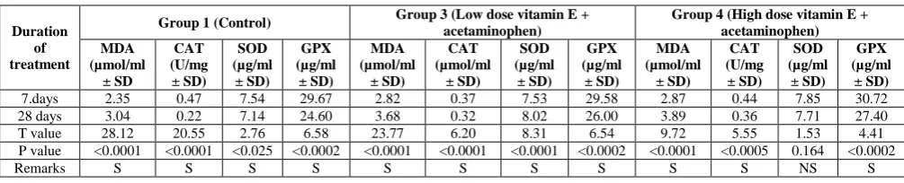
Table 3: Effect of duration of vitamin E pretreatment on the level of oxidative stress markers.
Duration of treatment
Group 1 (Control) Group 3 (Low dose vitamin E + acetaminophen) Group 4 (High dose vitamin E + acetaminophen)
MDA (µmol/ml
± SD
CAT (U/mg ± SD)
SOD (µg/ml
± SD)
GPX (µg/ml
± SD)
MDA (µmol/ml
± SD)
CAT (µmol/ml
± SD)
SOD (µg/ml
± SD)
GPX (µg/ml
± SD)
MDA (µmol/ml
± SD)
CAT (U/mg ± SD)
SOD (µg/ml
± SD)
GPX (µg/ml
± SD)
7.days 2.35 0.47 7.54 29.67 2.82 0.37 7.53 29.58 2.87 0.44 7.85 30.72
28 days 3.04 0.22 7.14 24.60 3.68 0.32 8.02 26.00 3.89 0.36 7.71 27.40
T value 28.12 20.55 2.76 6.58 23.77 6.20 8.31 6.54 9.72 5.55 1.53 4.41
P value <0.0001 <0.0001 <0.025 <0.0002 <0.0001 <0.0001 <0.0001 <0.0002 <0.0001 <0.0005 0.164 <0.0002
Remarks S S S S S S S S S S NS S
Table 3 shows the effect of duration of vitamin E (antioxidant) pretreatment on the levels of oxidative sress markers in the albino rats. The level of MDA in the albino rats was not favourably affected by increase in the number of days of pretreatment with vitamin E. The MDA level was lowest in the rats pretreated for 7 days in the rats that received both low and high dose of the vitamin when compared with results of the control rats. The SOD level, however, was lowest in the rats pretreated for 7 days with low dose vitamin E than that pretreated for 28 days with high dose vitamin E. However, the catalase and glutathione preoxidase levels were lowest in the rats pretreated for 28 days with high dose vitamin E.
Histological examination of liver tissues
The histological examination of the liver tissues of the control animals shows healthy hepatic matrix with normal central vein and normal hepatic lobule (plate 1). The rats in the acetaminophen-induced toxicity group
Plate 1: Photo micrographic slide of liver organ of group 1 control 1 (distilled water + isotonic 0.9% NaCl) H & E X100. CV-Normal central vein and normal hepatic lobule.
Plate 2a: Photo micrographic slide of liver organ of Group 2 week 1 (Acetaminophen-induced toxicity only) H & E X100.DCV –markedly dilated central vein filled with red blood cells, surrounded by swollen hepatocytes. Field showing a disrupted hepatic matrix.
Plate 3a: Photo micrographic slide of liver organ of Group 7 week 1 (low dose vitamin E + acetaminophen) H & E X100. Disrupted cellular matrix and architectural integrity.CV partially filled with cellular debris. Presence of vacuolation.
Plate 3b: Photo micrographic slide of liver organ of Group 3 week 4 (low dose vitamin E + acetaminophen) H & E X100. Recovering cellular matrix and emerging nuclear canaliculi. Emergence of inflammatory cells as a response of pretreatment.
Plate 4b: Photo micrographic slide of liver organ of Group 8 week 4 (high dose vitamin E + acetaminophen) H & E X100. Slow recovery of matrix cellular integrity with vacuolations in the matrix.
DISCUSSION
This study seeks to evaluate the antioxidant effect of vitamin E pretreatment against acetaminophen induced toxicity in albino rats. A lot of drugs or chemical agents such as acetaminophen, carbon tetrachloride and dimethylnitrosamine at excessive dose exposure have been reported to induce acute liver injury which is often characterized by abnormality of hepatic function and degeneration, necrosis or apoptosis of hepatocyte.[28] Vitamin E is a lipid-soluble antioxidant with the potential to effectively break chains within the cell membrane where it also performs the function of protecting membrane fatty acids from lipid peroxidation.[29,30]
Whenever there is oxidative stress, both enzymatic and non enzymatic antioxidants are mobilized by the cells inorder to respond to the oxidative stress under physiological conditions.[31,32] In this study, the antioxidant enzymes such as CAT, SOD, and GPX were used as indexes to evaluate the level of oxidative stress arising from acetaminophen induced hepatotoxicity in albino rats pretreated with vitamin E. The need to determine these enzymatic antioxidants was informed by the fact that they all function in concert. SOD acts as a pro-oxidant producing H2O2 requiring the activities of
CAT and GPX to ensure that an imbalance in their ratio which is dangerous to the body physiological system is not produced.[33]
In humans, acetaminophen (APAP) hepatotoxicity has been reported to be a leading cause of acute liver failure in many Western countries.[34,35] This observation was seen in this as 800mg/kg of acetaminophen appropriately induced hepatotoxic effect on the liver cells of the albino rats studied as evidenced by the significant increase
(p<0.0001) in the level of malondialdehyde (MDA) and the disrupted hepatic matrix with spots of clusters of inflamed cells in the sinusoids devoid of nuclear precursors and encroached swollen filled sinusoidal space of Disse with cellular debris (plates 2a and 2b) in the rats in the groups that stayed for either 7 or 28 days. Drugs such as zoledronic acid has been reported to cause significant elevation in MDA and nitric oxide levels, and reduction in GSH levels, which indicates that zoledronic acid could induce oxidative stress and decrease antioxidant level in liver.[36] The levels of MDA were significantly elevated by morphine while the activities of SOD and CAT were appreciably reduced.[37] The activities of SOD, CAT and GPX were significantly decreased (p<0.0001) in the albino rats following acute adminstration of acetaminophen indicating that the findings in this study is in conformity with the findings of other observers. These toxic events have been proposed to occur as a result of the metabolic activation of APAP by the cytochrome P450 enzyme system, resulting in the generation of the reactive metabolite N-acetyl-p-benzoquinone imine (NAPQI) which is responsible for the hepatotoxic manifestations seen in both rodents and humans. When the formation of NAPQI after APAP overdose is excessive, it results in the depletion of cellular glutathione (GSH), adduction of proteins including mitochondrial proteins, and induction of mitochondrial oxidant stress and dysfunction.[38]
freely catalyze this reaction. This agrees with the observed significantly decreased GPX activity in hepatotoxic rats compared with the control rats in the present study. The significant decrease of hepatic CAT and SOD activities observed in the acetaminophen-induced hepatotoxic rats in this study may be due to increased free radical production caused by administration of acetamoniphen.[41]This is because studies have shown that antioxidant enzymes such as SOD and CAT are easily inactivated by lipid peroxides or reactive oxygen species leading to decreased activities of these enzymes in acetaminophen -induced liver toxicity in this study.
Cellular glutathione (GSH) depletion which occurs in acute hepatotoxicity usually affects liver functions and causes severe necrosis of the hepatocyte, liver failure or death. Since oxidative stress and GSH depletion are reported to play major contributing roles in acetaminophen induced liver injury; substances and agent(s) with antioxidant properties and/or possessing the potential of enhancing the reserving ability of GSH may provide preventive effect against the progression of hepatocellular injury.[42] In recent times, acetaminophen-induced hepatotoxicity has been employed in most clinical experiments for testing phytotherapeutics and other hepato-protective interventions. In this study, hepato-protective intervention was done by pretreatment of the rats with vitamin E for either 7 or 28 days before acetaminophen-induced hepatoxicity was performed. The result show consistent and significant decrease in malondialdehyde concentration in rats pretreated with either low or high dose vitamin E for either 7 or 28 days. The hepatic architecture also exhibited recovery from the toxic effects of acetaminophen induction arising from from either low or high dose vitamin E pretreatment (plates 3 and 4). Also, hepatic levels of SOD, GPX, and CAT were remarkably increased in the vitamin E pretreated rats. This hepatoprotective effect might be associated with the improvement of antioxidant enzymes capacity, primarily via enhancement of the tissue redox system and protection of the antioxidant system in the liver as reported by Yi et al.[43] Reduced glutathione which is also a substrate for glutathione related enzymes, and a regenerator for alpha tocopherol; might have also played an important role in the antioxidant defense system of vitamin E and therefore agrees with the findings of Meister.[44] The study also shows that pretreatment with vitamin E for 7 days produced better hepatoprotective effect in the antioxidant defense system than that observed administration for 28 days (table 3). This is evident from significant decrease (p<0.05) in the level of MDA and increase in the activities of SOD, CAT and GPX.
CONCLUSION
Conclusively, vitamin E in this study has demonstrated a potent hepatoprotective action upon acetaminophen-induced oxidative stress in rat. The hepatoprotective effect of vitamin E could be correlated directly with its
ability to increase activity of antioxidant enzymes and enhance antioxidant defense status. The findings of this study suggest that vitamin E can be used as a safe, cheap, and effective chemopreventive and protective agent in the management of liver diseases especially in acetaminophen over dosage
REFERENCES
1. Lewerenz V, Hanett S, Nasterska C, El-Balay C, Rohrdanz E, Kahl R. Antioxidant protect primary rate hepatocytes cutures against acetaminophen-induced DNA strand breaks but not against acetaminophen-induced cytotoxicity. Toxicol, 2003; 191: 179–187.
2. Mazer M, Perrone J. (2008). Acetaminophen-induced nephrotoxicity: pathophysiology, clinical manifestations, and management. J. Med Toxicol, 2008; 4: 2-6.
3. Nelson SD. (1990). Molecular mechanisms of the hepatotoxicity caused by acetaminophen. Sem. liver Dis, 1990; 10: 267-278.
4. Mittal DK, Joshi D, Shukia S. Protective effects of Polygonum bistort (Linn.) and its active principle against
acetaminophen-induced toxicity in rats. Asian J. Exper. Biol. Sc, 2010; 1: 951-958.
5. Dahlin DC, Miwa GT, Lu AY, Nelson SD. N-acetyl-P-benzoquinone imine: a cytochrome P-450-mediated oxidation product of acetaminophen. Proc. Natl Acad. Sci, 1984; 81: 1327-1331.
6. Mitchell JR., Jollen DJ, Potter WZ, Giletten JR, Brodie BB. Acetaminophen-inducal hepatic necrosis. IV Protective role of glutathione. J Pharm. Exper. Therap, 1973; 187(1): 211 – 217.
7. Vermeulen NPE, Bessems JGM, Vande Streat R. Molecular aspects of paracetamol induced hepatotoxicity and its mechanism based prevention. Drug Metab. Revolut, 1992; 24: 367-457.
8. Park B, Pirmohamed M, Kittereingham N. Idiosyncratic drug reactions: A mechanistic evaluation of risk factors. Brit J Clin Pharm, 1992; 34: 377-395.
9. Somchit MN, Zuraini A, Bustaman AA, Somchit N, Sulaiman MR., Noratunlina, R. Activity of Tumeric (Curcuma longa) in Paraetamol induced Hepatotoxicity in Rats. Inter. J Pharm, 2005; 1(3): 252-256.
10. Hinston JA, Reid, AB, McCullough SS, James LP. Acetaminophen-induced hepatotoxicity: role of metabolic activation, reactive oxygen/ nitrogen species, and mitochondrial permeability transition. Drug Metab. Revolut, 2004; 36: 805-822.
11. Shyur LF, Tsung JH, Chen CY, Lo CP. Antioxidant properties of extracts from medical plants popularly used in Taiwan. Inter J App. Sc. Eng, 2005; 3: 195-202.
12. Breusegem FV, Mittler R. Reactive oxygen species in plant science topics, 2008;
13. Aruoma OI. Nutrition and health aspects of free radicals and antioxidants. Food Chem. Toxicol, 1994; 32: 671-683.
14. Wu J, Danielsson A, Zern M.A. Toxicity of hepatotoxins: new insights into mechanisms and therapy. Expert Opin. Investig. Drugs, 1999; 8: 585-607.
15. Bansal AK, Bansal M, Soni G, Bhatnagar, D. N-nitrosodiethylamine induced oxidative stress in rat liver. Chem. Biol. Interact, 2005; 156: 101-111. 16. Iwu, M. M. Traditional Igbo Medicine. Institute of
African studies, University of Nigeria, Nsukka, 1982.
17. Traber, MG, Stevens JF. “Free Radical Biology and Medicine - Vitamins C and E: Beneficial effects from a mechanistic perspective”. Free Radical Bio Med, 2011; 51(5): 1000-1013.
18. Public Health service Policy on Humane care and use of laboratory animals. Publication of the Department of Health and Human Services. National Institute of Health. Office of Laboratory Animal Welfare, 2015.
19. Guideline for the care and use of laboratory animal.8th edition. National Academic Press. Washington D.C. 2011.
20. Institution Animal Care and Use Committee Guidebook. 2nd Edition NIH publication. Besthesda, 2002; 1-230.
21. Guidelines for Euthanasia of Animals. 2013 Edition, American Vertinary Medical Association, 2013; 1-102.
22. Barker FJ, Silverton RE, Pallister CJ. Introduction to histology. In: Baker & Silverton’s Introduction to Medical Laboratory Technology, Seventh edition Bounty Press Limited, London, 2009; 175-242. 23. Ohkawa H, Ohishi N, Yagi K. (1979).Assay for lipid
peroxides in animal tissues by thiobarbituric acid reaction. Analy Biochem, 1979; 95(2): 351-358. 24. Aebi H. Catalase in-vitro. Methods in Enzymology,
1984; 105: 121-126.
25. Misra HP, Fridovich I. The role of superoxide anion in the antioxidation of epinephrine and a simple assay of Superoxide Dismutase. J Biol Chem, 1972; 247: 3170-3175.
26. Rotruck JT, Pope AL, Ganther HE, Swanson AB, Hafeman DG, Hoekstra WG. Selenium: biochemical role as a component of glutathione peroxidase. Science, 1973; 179(4073): 588-590.
27. Ellman GC. Tissue sulfhydryl groups. Ach Biochem Biophy, 1959; 82: 70-77.
28. Higuchi H, Gores GJ. Mechanisms of liver injury: an overview. Curr. Mol. Med, 2003; 3: 483–490. 29. Halliwell B. Free radicals, antioxidants and human
disease: curiosity, cause or consequences? Lancet, 1994; 344: 721-724.
30. Jacob RA. The integrated antioxidant system. Nutr Res, 1995; 15(5): 755-66.
31. Medina J, Moreno-Otero R. Pathophysiological basis for antioxidant therapy in chronic liver disease. Drugs, 2005; 65: 2445–2461.
32. Dey A, Lakshmanan J. The role of antioxidants and other agents in alleviating hyperglycemia mediated oxidative stress and injury in liver. Food Funct, 2013; 4: 1148–1184.
33. Marrocco I, Altieri F, Peluso I. (2017). Measurement and clinical significance of biomarkers of oxidative stress in humans. Oxid Med Cell Long, 2017; 2017: Article ID 6501046, 32 pages https://doi.org/10.1155/2017/6501046. 34. Budnitz DS, Lovegrove MC, Crosby AE.
Emergency department visits for overdoses of acetaminophen-containing products, Am. J. Prev. Med, 2011; 40: 585–592.
35. Manthripragada AD, Zhou DS, Budnitz EH, Lovegrove MC, Willy ME. (2011). Characterization of acetaminophen overdose-related emer gency department visits and hospitalizations in the United States, Pharmacoepidemiol. Drug Saf, 2011; 20: 819–826.
36. Karabulut AB, Gui M, Karabulut E, Kiran TR, Ocak SG, Otlu O. Oxidant and antioxidant activity in rabbit livers treated with zoledronic acid. Transplant. Proc, 2010; 42: 3820–3822.
37. Samarghandian, S.; Afshari, R.; Farkhondeh, T. Effect of long-term treatment of morphine on enzymes, oxidative stress indices and antioxidant status in male rat liver. Int. J. Clin. Exp. Med, 2014; 7: 1449–1453.
38. Jaeschke H, McGill MR, Ramachandran A. (2012). Oxidant stress, mitochondria, and cell death mechanisms in drug-induced liver injury: lessons learned from acetaminophen hepatotoxicity, Drug Metab. Rev, 2012; 44: 88–106.
39. Linares V, Alonso V, Albina ML, Belles M, Sirvent JJ, Domingo JL, Sanchez DJ. Lipid peroxidation and antioxidant status in kidney and liver of rats treated with sulfasalazine. Toxicol, 2009; 256: 152–156. 40. Oz M, El Nebrisi EG, Yang K-H S, Howarth F C, Al
Kury LT. (2004). Cellular and Molecular Targets of Menthol Actions, Front. Pharmacol, 2014;| https://doi.org/10.3389/fphar.2017.00472.
41. Bessems JG, Vermeulen NP (2001). Paracetamol (acetaminophen)-induced toxicity: molecular and biochemical mechanisms, analogues and protective approaches. Critical Rev Toxicol, 2001; 1(31): 55-138.
42. Baudrimont I, Ahouandjivo R, Creppy EE (1997). Prevention of lipid peroxidation induced by ochratoxin-A in Vitro cells in culture by several agents. Chem-Biol Interact, 1997; 104: 29-40. 43. Yi J, Xia W, Wu J, Yuan L, Wu J, Tu D, Fang J,
Tan Z. Betulinic acid prevents alcohol-induced liver damage by improving the antioxidant system in mice. J. Vet. Sci, 2014; 15: 141–148.