The Thirty-Third AAAI Conference on Artificial Intelligence (AAAI-19)
Non-Local Context Encoder: Robust
Biomedical Image Segmentation against Adversarial Attacks
Xiang He,
1Sibei Yang,
2Guanbin Li,
1∗Haofeng Li,
2Huiyou Chang,
1Yizhou Yu
31School of Data and Computer Science, Sun Yat-sen University, China 2The University of Hong Kong, Hong Kong 3Deepwise AI Lab, China
[email protected], [email protected], [email protected] [email protected], [email protected], [email protected]
Abstract
Recent progress in biomedical image segmentation based on deep convolutional neural networks (CNNs) has drawn much attention. However, its vulnerability towards adversarial sam-ples cannot be overlooked. This paper is the first one that discovers that all the CNN-based state-of-the-art biomedical image segmentation models are sensitive to adversarial per-turbations. This limits the deployment of these methods in safety-critical biomedical fields. In this paper, we discover that global spatial dependencies and global contextual in-formation in a biomedical image can be exploited to de-fend against adversarial attacks. To this end, non-local con-text encoder (NLCE) is proposed to model short- and long-range spatial dependencies and encode global contexts for strengthening feature activations by channel-wise attention. The NLCE modules enhance the robustness and accuracy of the non-local context encoding network (NLCEN), which learns robust enhanced pyramid feature representations with NLCE modules, and then integrates the information across different levels. Experiments on both lung and skin lesion segmentation datasets have demonstrated that NLCEN out-performs any other state-of-the-art biomedical image seg-mentation methods against adversarial attacks. In addition, NLCE modules can be applied to improve the robustness of other CNN-based biomedical image segmentation methods.
Introduction
Biomedical image analysis catches people’s eyes due to its popular application in computer-aided diagnosis and medi-cal plan recommendation. Biomedimedi-cal image segmentation is fundamental in biomedical image analysis, which per-forms pixel-level annotation for regions of interest (e.g. organs, substructures, and lesions) on biomedical images (e.g. X-ray, Magnetic Resonance Imaging, Computerized Tomography). However, it is challenging to obtain accu-rate segmentation because of the large shape and size varia-tions of regions of interest, and the diversity of images pro-duced by different biomedical imaging equipments (Hwang
∗
Xiang He and Sibei Yang contributed equally to this work. Corresponding author is Guanbin Li (Email: [email protected]). This work was partially supported by the National Natural Science Foundation of China under Grant No.61702565 and the Fundamental Research Funds for the Cen-tral Universities under Grant No.18lgpy63.
Copyright c2019, Association for the Advancement of Artificial Intelligence (www.aaai.org). All rights reserved.
SLSDeep NWCN CDNN Ours
Input GT
Perturbation
Adversarial Image
Adversarial Segmentation
Segmentation
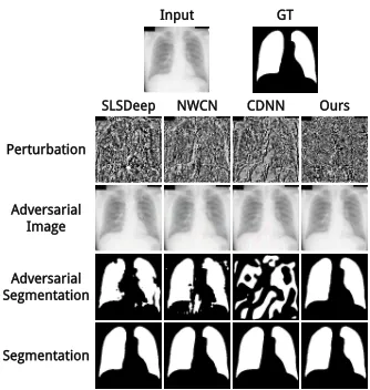
Figure 1: Sample adversarial attacks on SLSDeep (Sarker et al. 2018), NWCN (Hwang and Park 2017), CDNN (Yuan 2017) and our NLCEN. The input chest radiograph and its ground-truth segmentation are shown in the first row. Ad-versarial perturbations and images generated for models by the Iterative FGSM attack method (Kurakin, Goodfellow, and Bengio 2016) with adversarial intensity set to 16 are shown in the second and third rows respectively. Segmen-tation results on the adversarial images and the input image are shown in the fourth and fifth rows respectively.
and Park 2017; Sarker et al. 2018). State-of-the-art biomed-ical image segmentation methods are based on fully con-volutional networks (FCN) (Long, Shelhamer, and Darrell 2015), which is a type of deep convolutional neural net-works (CNNs) designed for semantic segmentation in com-puter vision. The accuracy of CNN-based biomedical image segmentation has been beyond that of traditional ones (Lit-jens et al. 2017; Sarker et al. 2018; Hwang and Park 2017; Ronneberger, Fischer, and Brox 2015; Novikov et al. 2018; Yuan 2017). In addition to the accuracy of biomedical image segmentation, its stability and robustness are also essential for the fault-free clinical practice.
2018), the vulnerability of CNNs to adversarial attacks can-not be overlooked (Szegedy et al. 2013). Adversarial sam-ples are legitimate samsam-ples with human-imperceptible per-turbations, which attempt to fool a trained model to make incorrect predictions with high confidence (Szegedy et al. 2013). Such human-imperceptible perturbations, that CNNs are very sensitive to, are called adversarial noise. By exploit-ing the gradient-based error back-propagation mechanism for CNN training, adversarial attacks generate adversarial noise in an input image by back-propagating the error gra-dient induced by an intended incorrect prediction through a trained CNN model.
Recent work shows that complex semantic segmenta-tion models, which are trained with an independent cross-entropy loss at each pixel on an image, are threatened by adversarial attacks (Xie et al. 2017; Arnab, Miksik, and Torr 2018). Although biomedical image segmentation mod-els share a similar deep learning framework with semantic segmentation models, adversarial attacks targeted at them have not been well explored. Since biomedical image seg-mentation does not have sufficient high-quality training sam-ples, trained models can easily experience overfitting and exhibit a weak generalization capability, which make them more sensitive to noise. This property makes the models more vulnerable when facing adversarial attacks, and chal-lenges their use in safety-critical biomedical fields. An ex-ample of adversarial attacks on lung segmentation is shown in Figure 1.
The common defense strategy against adversarial attacks is adversarial training, which injects adversarial samples into training data to improve the robustness of trained models. Tram`er et al. 2018 show that if the adversarial samples are taken as augmented data, the consequence of adversarial at-tacks can be alleviated. However, this defense strategy is limited because the adversarial samples are obtained from specific models and their corresponding adversarial attack methods. Therefore, instead of a limited training strategy, we wish to design a generic module, which can be easily integrated into CNN-based biomedical image segmentation networks to improve their robustness.
The robustness of biomedical image segmentation can be improved effectively by global spatial dependencies and global contextual information. Therefore, we propose to model them with a module called non-local context encoder (NLCE). In order to better introduce the effectiveness of global spatial dependencies and global contexts, we use a single pixel as an example, and the situation of a single pixel can be easily extended to the entire image because seg-mentation models are trained with independent loss at ev-ery pixel. First, global spatial dependencies are vev-ery impor-tant in defending against adversarial attacks. Given a pixel, capturing its global spatial dependencies means finding all highly related pixels within the entire image, and the pre-diction at this pixel is affected by all those pixels. There are two perspectives to understand the effectiveness of global dependencies. One is that if an incorrect label was given to a pixel, the incorrect loss at the pixel would be passed to all other related pixels by back-propagation, which in-creases the intensity of perturbation, and makes the
adver-sarial sample significantly different from the original im-age. The other is that the noise at a pixel can be gradually weakened by the fusion with its highly related pixels in the process of forward-propagation. Second, global contextual information has a positive effect in defending against adver-sarial attacks because the configuration of the human body is relatively stable. For example, in lung image segmentation, the left and right lungs provide geometric contextual infor-mation by learning their geometric relationship with respect to each other. Because of the association between the left and right lungs, the right lung needs to receive the same perturbation-based attacks when the left lung is attacked. Therefore, the intensity of the required perturbation is in-creased. Unfortunately, on one hand, CNNs have difficulty in capturing global dependencies because convolution op-erations only capture short-range dependencies by process-ing one local neighborhood at a time. Although stacked con-volution operations are capable of capturing long-range de-pendencies by enlarging receptive fields (Fukushima 1980; Lecun et al. 1989), they increase the difficulty of optimiza-tion and may face the problem of gradient vanishing. On the other hand, current biomedical image segmentation methods do not make full use of global contextual information.
Inspired by the above analysis, in this paper, we propose a robust non-local context encoder module for biomedical image segmentation. The NLCE module captures the global spatial dependencies within a feature map by obtaining the response at a position of the feature map as a weighted sum of the features at all positions, and strengthens the fea-tures with channel-wise attention computed from the en-coded global contextual information. In principle, the pro-posed robust NLCE module can also be applied to all CNN-based biomedical image segmentation methods and is able to improve the robustness of these models against adversar-ial attacks.
Moreover, we design and implement a medical image seg-mentation framework, named non-local context encoding network (NLCEN), which consists of two phases, the global phase and the refinement phase. Our global network is based on the feature pyramid network (FPN) (Lin et al. 2017) and our NLCE modules. It learns global feature representations at different levels. The refinement network fuses features at different levels to obtain sharp boundaries. We conduct experiments on two common benchmark biomedical image segmentation datasets, the JSRT dataset for lung segmenta-tion (Shiraishi et al. 2000) and the ISBI 2016 dataset (Gut-man et al. 2016) for skin lesion segmentation. Experimental results show that our NLCEN with NLCE modules has both high segmentation accuracy and robustness against adversar-ial attacks, and the NLCE modules practically help improve the segmentation accuracy of other biomedical image seg-mentation methods when they face adversarial attacks.
In summary, this paper has the following contributions: • This is the first paper, to the best of our knowledge,
• It proposes non-local context encoder (NLCE), which is a robust biomedical image segmentation module against adversarial attacks. The NLCE module is able to cap-ture distance-independent dependencies and global con-textual information. And it can be easily applied to other CNN-based image segmentation methods.
• It introduces non-local context encoding network (NL-CEN), which achieves high segmentation accuracy and is robust on adversarial samples with different levels of adversarial perturbations.
Related Work
Biomedical Image Segmentation
The state-of-the-art biomedical image segmentation meth-ods have similar frameworks to CNNs-based semantic seg-mentation models, but with fewer convolutional blocks and fewer network parameters to avoid overfitting. The U-net network is the most well-known segmentation method for biomedical image segmentation, and it is based on FCNs, but its upsampling phase and the downsampling phase use the same number of convolution operations in each level and the skip connection is used to connect the downsam-pling layer to the upsamdownsam-pling layer (Ronneberger, Fischer, and Brox 2015). InvertedNet is an improved version of U-net that has fewer parameters to reduce overfitting, and for more accurate localization, it adopts delayed subsampling and learns higher resolution features (Novikov et al. 2018). In or-der to use contextual information while maintaining resolu-tion, NWCN adopts an atrous convolution-based model and utilizes a multi-stage training strategy to refine the prelimi-nary segmentation results (Hwang and Park 2017). CDNN is also based on FCNs and it designs a loss function based on Jaccard distance (Yuan 2017). SLSDeep, consisting of skip-connections, dilated residual and pyramid pooling, is an effi-cient skin lesion segmentation model from dermoscopic im-ages. Its loss function, including negative log likelihood and end point error loss, is designed to obtain sharp boundary (Sarker et al. 2018).
Adversarial Attacks
Since the adversarial attacks to deep neural networks have been proposed by Szegedy et al., they have received exten-sive attention. They are designed to generate the adversar-ial samples to fool a trained model to make incorrect pre-dictions with high confidence. The adversarial perturbation is estimated by solving penalized optimization problem by using L-BFGS optimization method (Szegedy et al. 2013). Goodfellow, Shlens, and Szegedy believe that the main rea-son why neural networks are vulnerable to adversarial at-tack is their linear behavior in high-dimensional space and propose the single-step fast gradient sign method (FGSM) to generate adversarial samples directly and efficiently. The single-step targeted attack is a modified version of FGSM, which aims at reducing the loss function of target category instead of the increasing the loss function of the original cat-egory (Kurakin, Goodfellow, and Bengio 2016). In addition, the proposed basic iterative method can increase the success
rate of attacks. The adversarial samples generated by iter-ative methods are less transferable than those generated by single-step attacks (Kurakin, Goodfellow, and Bengio 2016; Arnab, Miksik, and Torr 2018). Xie et al. are the first to ex-plore adversarial attacks on image segmentation and detec-tion on large datasets and propose the density adversary gen-eration to generate effective adversarial samples by consid-ering all the targets simultaneously. Arnab, Miksik, and Torr present the first rigorous evaluation on the robustness of the state-of-the-art semantic segmentation models to single-step adversarial attacks and iterative adversarial attacks.
Global Modeling
The global information modeling of images is an important part of the visual recognition field, and global information is utilized in many visual recognition tasks, e.g. scene segmen-tation (Li et al. 2016), saliency detection (Li and Yu 2016; Li et al. 2017) and semantic segmentation (Zhang et al. 2018). Getting global image information for CNN-based models is challenging, and it needs to consider both local dependencies and long-range dependencies. Stacked convo-lutional blocks can only capture local information due to re-stricted receptive fields. LSTM-CF treats spatial feature map obtained by CNNs as horizontal and vertical sequences re-spectively. It adopts multiple bi-directional long short term memory networks (LSTMs) in vertical direction to capture vertical short and long-range context, then the context is fused to get global spatial information by applying another bi-directional LSTMs in horizontal (Li et al. 2016). How-ever, recurrent operations, like LSTMs, are still progress a local neighbor at a time, and the connection between two distant points must pass through the intermediate points. To capture the long-distance dependency, (Wang et al. 2018) proposed a fast and direct method, which considers the fea-tures at all the positions to capture the dependencies at a position in a low-level feature map. Zhang et al. takes the entire dataset into account and learns a set of global inher-ent represinher-entative of features to capture the global context for images. The global information for a feature map is ob-tained by encoding the relationships between its all features and the representative features.
Methodology
e
Codebook
...
K1x
1
Co
nv
θ:
1
x1
φ
: 1
x1
g:
1
x1
(H, W, C)
(HW, C')
(C', HW)
(HW, C') (HW x HW)
softmax
(HW, C')
(H, W, C') (H, W, C'')
FC
1x
1
Co
nv
(C'',)
(H, W, C) (C,) (H, W, C)
Non-local Context Encoder
Encoding
(K, C'')
Figure 2: The architecture of our proposed non-local context encoder (NLCE). Our NLCE module first enhances and denoises the feature map by modeling global spatial dependencies and then applies channel-wise feature map attention by using encoded global context computed from a learned codebook.
H
ea
d
Supervise Input
Output
La
ye
r 2
La
ye
r 3
La
ye
r 4
La
ye
r 5
N
LC
E
N
LC
E
N
LC
E
N
LC
E
Bo
tt
le
Bo
tt
le
Bo
tt
le
Bo
tt
le
Co
nc
at
Bo
tt
le
E5 E4 E3 E2
P5 P4 P3 P2
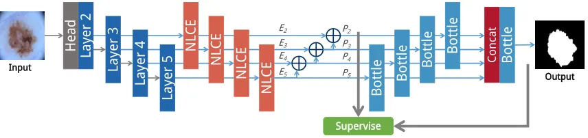
Figure 3: The overall architecture of our proposed non-local context encoding network (NLCEN). The left part is based on a ResNet backbone and a feature pyramid. An NLCE module is added to bottom-up feature activations before lateral connections at different levels, and independent supervision is applied to predictions at all levels. The multi-scale information fused from all the pyramid features are used to refine the prediction and produce segmentation.
Non-Local Context Encoder
Our non-local context encoder takes anH ×W ×C fea-ture map as input. It capfea-tures spatial short- and long-range dependencies in the feature map by following the design by Wang et al. 2018. It considers the feature map as a set ofC -dimensional featuresX ={x1, ...xN}, whereN =H×W is the total number of features. We define the pairwise func-tionfthat learns a relationship between any two featuresxi andxjas
f(xi,xj) = exp θ(xi)Tφ(xj)
, (1)
whereθ(xi) = Wθxiandφ(xj) = Wφxj are feature em-beddings, whereWθandWφare learned weight matrices.
The non-local responseyifor featurexiis defined as
yi=
1
C(x)
N X
j=1
f(xi,xj)g(xj), (2)
where the unary function g is a mapping with a learned weight matrixWg to compute the representationg(xj) = Wgxj of xj.C(x)is the normalization factor, defined as C(x) = PN
i=1f(xi,xj). The non-local responseyi cap-tures short- and long-range dependencies via considering all features in the above non-local operation.
Next, the enhanced featureszi =Wzyi+xi (Wzmaps
yi to the C-dimensional space), which combine the non-local response yi with the original featurexi, are fed into
the context encoder discussed below. The feature map with the size ofH×W ×Cconstructed from the enhanced fea-tures is denoted as Fz. Inspired by Zhang et al. 2018, we learn a global codebookD ={d1, ...dK}, which contains K C00-dimensional codewords. The codebook represents global statistical information about the non-local enhanced features, and each codeword represents a visual center. We transform the enhanced features to the same dimensionality as the codewords via a1×1convolution, and the resulting
C00-dimensional features are denoted asZ0 = {z01, ...z0N}. The normalized residual eik between an enhanced feature
z0iand a codeworddkis defined as
eik=
exp −skkrikk2
R(ei)
rik, (3)
whererik=z0i−dk is the residual between featurez0iand codeword dk, sk is a learned smoothing factor for code-word dk, and R(ei) = P
K
l=1exp(−slkrilk2)is the nor-malization factor for feature xi. Thus, the residual infor-mation for all features captured by the codeworddk is de-fined asek = P
N
i=1eik, and the global context is defined ase=PK
k=1σ(ek), whereσdenotes Batch Normalization with ReLU.
wise feature map attention by predicting a channel-wise scaling factor γ = sigmoid(Wγe), where Wγ is a learned weight matrix. The output from the NLCE module,
Fz⊗γ, is a channel-wise multiplication between the non-local enhanced feature mapFzand the channel-wise scaling factorγ.
The architecture of our NLCE module is shown in Figure 2. The NLCE module first captures short- and long-range spatial dependencies to denoise and strengthen the feature map, and then scales the feature map channels by scaling factors predicted using the encoded global context. Global dependencies and global contexts reduce the negative im-pact of adversarial noise, and give rise to the robustness of the NLCE module against adversarial attacks. Fusing infor-mation from the highly related pixels or the global context in forward propagation gradually weakens adversarial noise to a pixel or a semantic proposal.
Non-Local Context Encoding Network (NLCEN)
Our proposed coarse-to-fine non-local context encoding net-work (NLCEN) takes one biomedical image as input and produces a segmentation of organs or lesions at the pixel level. NLCEN has two phases, and its overall architecture is shown in Figure 3.
The architecture of the global phase is based on the ResNet backbone (He et al. 2016) and feature pyramid network. The fused information of low-level and high-level features by upsampling high-high-level features can cap-ture rich contextual information with high resolution. An NLCE module is attached to the last residual block of conv2 through conv5 respectively to obtain multi-level robust non-local feature maps, denoted asE2, ..., E5. Following FPN, the fused feature mapPi(i= 2,3,4) is obtained by element-wise addition betweenEi and the1×1convolved and up-sampledPi+1, andP5is obtained by attaching a1×1 con-volutional layer toE5. Feature mapsP2, ..., P5are used to independently produce segmentation results by feeding each of them through a distinct3×3convolution filter and a bi-linear interpolation layer. Supervision is directly applied to each of these segmentation results.
Multi-level feature maps are fused together via upsam-pling and concatenation after going through bottleneck operations (He et al. 2016), and the refined segmenta-tion predicsegmenta-tion is produced directly from the fused feature map with multi-scale information. The number of bottle-neck operations is respectively 0,1,2,3 for feature maps
P2, P3, P4, P5. During testing, the final output is produced from the refined segmentation prediction.
The loss function for a single map prediction is defined as the sum of cross-entropy losses at individual pixels between the ground truth and the predicted segmentation map:
Ls=
|I| X
i
logpi,gi, (4)
where |I| denotes the total number of pixels, gi is the ground-truth label at pixeli,pi,giis the probability that pixel
iis classified to categorygi.
We denote the loss for the segmentation predictions ob-tained fromP2, ..., P5asL2g, ..., L5g, and the loss for the re-fined segmentation asLr. The total loss is defined as:
L= 1
4 5 X
i=2
Lig+λLr, (5)
whereλ= 0.25is a weight balancing multiple coarse pre-dictions from the global phase and the refined prediction from the refinement phase.
Experimental Results
Datasets
We have conducted evaluations on two commonly used benchmark biomedical image datasets, the Japanese Soci-ety of Radiological Technology (JSRT) dataset for lung seg-mentation (Shiraishi et al. 2000) and the International Sym-posium on Biomedical Imaging (ISBI 2016) dataset for skin lesion segmentation (Gutman et al. 2016).
The JSRT dataset was first introduced to help diagnostic training and testing for tuberculosis. It contains 154 nodule and 93 non-nodule post-anterior (PA) chest radiographs with a2048×2048high resolution and wide density range. We split chest radiographs into a training set of 124 images and a test set of 123 images by following previous practices in the literature (Hwang and Park 2017). The ground truth for the JSTR dataset is provided in (Van Ginneken, Stegmann, and Loog 2006).
The ISBI 2016 dataset provides 900 training images and 379 testing images with binary masks of skin lesion. The size of the images ranges from524×718to2848×4288.
Adversarial Attacks
We adopt the target Iterative FGSM attack method (Kurakin, Goodfellow, and Bengio 2016) to generate adversarial sam-ples for a concrete model because the iterative white-box attacking methods have a high success rate. An attack sets the target as the inverse of ground-truth masks, denoted as
St, and the adversarial sample of a single example in each iteration is defined as:
xadvt+1=clip(xadvt −α·sign(∇xadv t Lr(f(x
adv
t ;θf)), ), (6)
wherexadv0 is initialized tox, the intensity of the adversarial perturbation is, the step size of iterations is denoted asα, andθfrepresents network parameters.
Following Kurakin, Goodfellow, and Bengio, we setα= 1, the number of iterations tomin (+ 4,d1.25e), and the
L∞ norm of adversarial perturbation to intensity. We gen-erate adversarial samples by setting adversarial intensity to every value from{0.5,1,2,4,6,8,10,12,14,16,18,20,22,
24,26,28,30,32}.
Evaluation Metrics
(DIC) and Jaccard similarity coefficient (J SC) are com-monly used accuracy metrics in biomedical image segmen-tation.DICandJ SCare computed as follows:
DIC= 2·T P
2·T P +F N+F P, (7)
J SC = T P
T P+F N+F P, (8)
whereT P,T N,F P,F Nare the number of pixel-level true positives, true negatives, false positives, and false negatives, respectively.
Implementation
Our proposed NLCEN with NLCE modules has been im-plemented on the open source deep learning framework, Py-Torch(Paszke et al. 2017). We follow the same experimental setups as in Hwang and Park and Sarker et al.. Horizontal flips, vertical flips and random rotations with±10degrees are used as data augmentation operations on the ISBI 2016 dataset while no data augmentation is applied to the JSRT dataset during training. We set the mini-batch size to 8, and all input images are resized to 256×256. The Adam op-timizer is adopted to update network parameters with the learning rate set to0.001initially and reduced by10% when-ever the training loss stops decreasing until0.0001. We use a weight decay of0.0001and an exponential decay rate for the first moment estimates and the second moment estimates of 0.9and0.999respectively. It takes2hours to train a model on the JSRT dataset in a single NVIDIA TITAN GPU and2 more hours to generate adversarial samples for testing when an intensity of adversarial perturbation is given. The train-ing and testtrain-ing times on the ISBI 2016 dataset are4 hours respectively.
Comparison with the State of the Art
We compare the robustness of our proposed NLCEN with that of five state-of-the-art methods for lung segmenta-tion and skin lesion segmentasegmenta-tion, including dilated resid-ual and pyramid pooling networks (SLSDeep) (Sarker et al. 2018), network-wise training of convolutional works (NWCN) (Hwang and Park 2017), convolutional net-works for biomedical image segmentation (UNet) (Ron-neberger, Fischer, and Brox 2015), fully convolutional archi-tectures for multi-class segmentation (InvertNet) (Novikov et al. 2018) and segmentation with fully convolutional-deconvolutional networks (CDNN) (Yuan 2017). All the evaluations of the above networks are conducted on both the JSRT and ISBI 2016 datasets. According to the scale of the datasets, we adopt a ResNet-18 backbone for the JSRT dataset and a ResNet-50 backbone for the ISBI 2016 dataset. On each dataset, we first train a benchmark segmentation model on the training set and compute segmentation accu-racy metrics (DIC andJ SC) on the testing set; and then, under each given intensity of perturbation, we generate ad-versarial samples of the testing set on the basis of the bench-mark model and test its segmentation accuracy on the gen-erated adversarial samples.
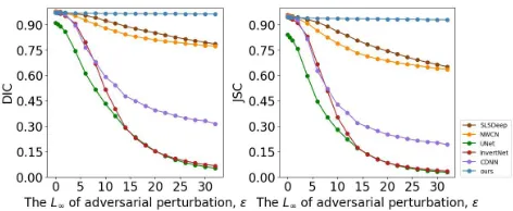
Quantitative EvaluationFigures 4 and 5 show
evalua-tion results in terms ofDICandJ SCon the JSRT and ISBI
Figure 4: Comparison of quantitative results in terms of
DICandJ SCon the JSRT lung segmentation dataset.
Figure 5: Comparison of quantitative results in terms of
DIC and J SC on ISBI 2016 skin lesion segmentation dataset.
2016 datasets respectively. In these figures, we can find that NLCEN achieves the highest accuracy on clean skin lesion images, and achieves almost the top performance on clean lung images. Even when the strongest adversarial perturba-tion ( = 32) is exerted, it still maintains the highest ac-curacy. Its accuracy drops by only0.01(0.971to0.963) in
DICand0.02(0.945to0.929) inJ SCon the JSRT dataset, and drops by0.11(0.907to0.801) inDICand0.14(0.844 to0.704) inJ SCon the ISBI 2016 dataset. The results show that adversarial attacks have almost no effects on our lung segmentation model. The drop in accuracy on the ISBI 2016 skin dataset is larger than that on the JSRT dataset because there is very little contextual information in skin lesion im-ages. Even though, our NLCEN is still the most robust one of all the models. Moreover, this experiment also indicates that the outstanding robustness of our model against adver-sarial samples with different levels of perturbation intensity.
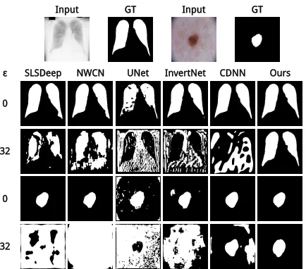
Qualitative EvaluationFigure 6 visually compares
seg-mentation results from our model and five existing methods when they are under the attack of targeted Iterative FGSM with= 32. Refer to the supplementary materials for more results.
0
32
SLSDeep NWCN UNet InvertNet CDNN Ours
ε
0
32
Input GT Input GT
Figure 6: Comparison of segmentation results obtained from SLSDeep, NWCN, UNet, InvertNet, CDNN and our NL-CEN when they are attacked by targeted Iterative FGSM with= 32.
and CDNN are appalling, and all the other methods fail on skin lesion segmentation.
Ablation Studies
As discussed in the Methodology section, the robustness of our NLCE modules against adversarial attacks comes from global spatial dependencies and global contextual informa-tion. To verify their validity and necessity, we compare NL-CEN with its three variants (i.e. NLNL-CEN without NLCE modules (w/o NLCE), NLCEN without modeling global de-pendencies (w/o NL) and NLCEN without capturing global contexts (w/o CE)), which are trained and tested on the JSTR dataset. For the fairness of the comparison, we train the w/o NLCE model first. Then, we fine-tune the w/o NL, w/o CE and NLCEN models separately by freezing the layers of the w/o NLCE model. Finally, we fine-tune NLCEN without freezing any layer to obtain the fine-tuned model.
The robustness of these models are evaluated and the re-sults are shown in Figure 7. The non-local dependencies part or the global context part alone can already improve robust-ness, and the complete NLCE module with both parts can
Figure 7: Ablation study on our non-local context encoding network.
enhance the robustness further. That demonstrates the ne-cessity of global dependencies and global contexts as well as the possibility of cooperation between them. In addition, the accuracy and robustness can be further enhanced by fine-tuning the NLCEN without freezing any layer.
Figure 8: Comparison of robustness with and without non-local context encoder in other biomedical image segmenta-tion methods.
Generalization
To verify that our non-local context encoder can be easily integrated into other networks, we instantiate an NLCE ver-sion for SLSDeep, NWCN, UNet, InvertNet and CDNN net-works, respectively. Except for NWCN, we add one NLCE module between the last downsampling layer and the first upsampling layer in each network. For NWCN, we add two NLCE modules because it has two subnetworks. Finally, we train these updated networks from scratch and test those net-works with NLCE modules on the JSRT dataset.
Figure 8 shows a comparison between methods with NLCE modules and those without on the JSRT dataset. Methods with NLCE modules achieve significantly higher
DICandJ SCthan those without. This reveals NLCE mod-ules are compatible with other biomedical image segmenta-tion methods to strengthen their defense against adversarial attacks.
Conclusions
References
Arnab, A.; Miksik, O.; and Torr, P. H. 2018. On the robust-ness of semantic segmentation models to adversarial attacks. InProceedings of CVPR.
Fukushima, K. 1980. Neocognitron: A self-organizing neural network model for a mechanism of pattern recogni-tion unaffected by shift in posirecogni-tion. Biological Cybernetics
36(4):193–202.
Goodfellow, I.; Shlens, J.; and Szegedy, C. 2015. Explain-ing and harnessExplain-ing adversarial examples. InProceedings of ICLR.
Gutman, D.; Codella, N. C. F.; Celebi, M. E.; Helba, B.; Marchetti, M. A.; Mishra, N. K.; and Halpern, A. 2016. Skin lesion analysis toward melanoma detection: A challenge at the international symposium on biomedical imaging (isbi) 2016, hosted by the international skin imaging collaboration (isic). CoRRabs/1605.01397.
He, K.; Zhang, X.; Ren, S.; and Sun, J. 2016. Deep residual learning for image recognition. InProceedings of CVPR, 770–778.
Hwang, S., and Park, S. 2017. Accurate lung segmenta-tion via network-wise training of convolusegmenta-tional networks. In
Deep Learning in Medical Image Analysis and Multimodal Learning for Clinical Decision Support. Springer. 92–99. Kurakin, A.; Goodfellow, I.; and Bengio, S. 2016. Adver-sarial machine learning at scale. InProceedings of ICLR. LeCun, Y.; Bengio, Y.; and Hinton, G. 2015. Deep learning.
nature521(7553):436.
Lecun, Y.; Boser, B. E.; Denker, J. S.; Henderson, D.; Howard, R. E.; Hubbard, W.; and Jackel, L. D. 1989. Back-propagation applied to handwritten zip code recognition.
Neural Computation1(4):541–551.
Li, G., and Yu, Y. 2016. Visual saliency detection based on multiscale deep cnn features. IEEE Transactions on Image Processing25(11):5012–5024.
Li, G., and Yu, Y. 2018. Contrast-oriented deep neural net-works for salient object detection. IEEE Transactions on Neural Networks and Learning Systems.
Li, Z.; Gan, Y.; Liang, X.; Yu, Y.; Cheng, H.; and Lin, L. 2016. Lstm-cf: Unifying context modeling and fusion with lstms for rgb-d scene labeling. In Proceedings of ECCV, 541–557. Springer.
Li, G.; Xie, Y.; Lin, L.; and Yu, Y. 2017. Instance-level salient object segmentation. InProceedings of CVPR, 247– 256.
Li, G.; Gan, Y.; Wu, H.; Xiao, N.; and Lin, L. 2018. Cross-modal attentional context learning for rgb-d object detection.
IEEE Transactions on Image Processing.
Lin, T.-Y.; Doll´ar, P.; Girshick, R. B.; He, K.; Hariharan, B.; and Belongie, S. J. 2017. Feature pyramid networks for object detection. InProceedings of CVPR, volume 1, 4.
Litjens, G.; Kooi, T.; Bejnordi, B. E.; Setio, A. A. A.; Ciompi, F.; Ghafoorian, M.; van der Laak, J. A.; Van Gin-neken, B.; and S´anchez, C. I. 2017. A survey on deep
learning in medical image analysis.Medical image analysis
42:60–88.
Long, J.; Shelhamer, E.; and Darrell, T. 2015. Fully convolu-tional networks for semantic segmentation. InProceedings of CVPR, 3431–3440.
Novikov, A. A.; Lenis, D.; Major, D.; Hladuvka, J.; Wim-mer, M.; and B¨uhler, K. 2018. Fully convolutional archi-tectures for multi-class segmentation in chest radiographs.
IEEE Transactions on Medical Imaging.
Paszke, A.; Gross, S.; Chintala, S.; Chanan, G.; Yang, E.; DeVito, Z.; Lin, Z.; Desmaison, A.; Antiga, L.; and Lerer, A. 2017. Automatic differentiation in pytorch. InNIPS-W. Ren, S.; He, K.; Girshick, R.; and Sun, J. 2015. Faster r-cnn: Towards real-time object detection with region proposal networks. InProceedings of NIPS, 91–99.
Ronneberger, O.; Fischer, P.; and Brox, T. 2015. U-net: Convolutional networks for biomedical image segmenta-tion. InProceedings of International Conference on Med-ical image computing and computer-assisted intervention, 234–241. Springer.
Sarker, M.; Kamal, M.; Rashwan, H. A.; Banu, S. F.; Saleh, A.; Singh, V. K.; Chowdhury, F. U.; Abdulwahab, S.; Ro-mani, S.; Radeva, P.; et al. 2018. SLSDeep: Skin lesion segmentation based on dilated residual and pyramid pool-ing networks. InProceedings of International Conference on Medical Image Computing and Computer Assisted Inter-vention.
Shiraishi, J.; Katsuragawa, S.; Ikezoe, J.; Matsumoto, T.; Kobayashi, T.; Komatsu, K.-i.; Matsui, M.; Fujita, H.; Kodera, Y.; and Doi, K. 2000. Development of a digital im-age database for chest radiographs with and without a lung nodule: receiver operating characteristic analysis of radiolo-gists’ detection of pulmonary nodules. American Journal of Roentgenology174(1):71–74.
Szegedy, C.; Zaremba, W.; Sutskever, I.; Bruna, J.; Erhan, D.; Goodfellow, I.; and Fergus, R. 2013. Intriguing proper-ties of neural networks. arXiv preprint arXiv:1312.6199. Tram`er, F.; Kurakin, A.; Papernot, N.; Goodfellow, I.; Boneh, D.; and McDaniel, P. 2018. Ensemble adversarial training: Attacks and defenses. InProceedings of ICLR. Van Ginneken, B.; Stegmann, M. B.; and Loog, M. 2006. Segmentation of anatomical structures in chest radiographs using supervised methods: a comparative study on a public database. Medical image analysis10(1):19–40.
Wang, X.; Girshick, R.; Gupta, A.; and He, K. 2018. Non-local neural networks. InProceedings of CVPR.
Xie, C.; Wang, J.; Zhang, Z.; Zhou, Y.; Xie, L.; and Yuille, A. 2017. Adversarial examples for semantic segmentation and object detection. InProceedings of ICCV.
Yuan, Y. 2017. Automatic skin lesion segmentation with fully convolutional-deconvolutional networks. arXiv preprint arXiv:1703.05165.