R E S E A R C H A R T I C L E
Open Access
Gene alterations in monocytes are
pathogenic factors for immunoglobulin a
nephropathy by bioinformatics analysis of
microarray data
Yingbo Guo
1†, Wenfeng Gao
2†, Danyang Wang
3, Weijing Liu
3and Zhongjie Liu
3*Abstract
Background:Immunoglobulin A nephropathy (IgAN) is the most frequent primary glomerulopathy worldwide. The study aimed to provide potential molecular biomarkers for IgAN management.
Methods:The public gene expression profiling GSE58539 was utilized, which contained 17 monocytes samples (8 monocytes samples isolated from IgAN patients and 9 monocytes samples isolated from healthy blood donors). Firstly, differentially expressed genes (DEGs) between the two kinds of samples were identified by limma package. Afterwards, pathway enrichment analysis was implemented. Thereafter, protein-protein interaction (PPI) network was constructed and key nodes in PPI network were predicted using four network centrality analyses. Ultimately, gene functional interaction (FI) was constructed according to expressions in each sample, and then module network was extracted from FI network.
Results:A total of 678 DEGs were screened out, of these, 72 DEGs were identified as crucial nodes in PPI network that could well distinguish IgAN and healthy samples. In particular,IL6,TNF,IL1B,PRKACAandCCL20were closely related to pathways such as hematopoietic cell lineage, apoptosis and Toll-like receptor (TLR) signaling pathway. Moreover, 12 genes in the FI network belonged to the 72 identified key nodes, such asCCL20,HDAC10,FPR2andPRKACA, which were also key genes in 4 module networks.
Conclusions:Several crucial genes were identified in monocytes of IgAN patients, such asIL6,TNF,IL1B,CCL20,PRKACA, FPR2andHDAC10. These genes might co-involve in pathways such as TLR and apoptosis signaling during IgAN progression.
Keywords:Immunoglobulin a nephropathy, Network centrality analysis, Functional interaction, Toll-like receptor signaling, Apoptosis
Background
Worldwide, immunoglobulin A nephropathy (IgAN) is the most frequent primary glomerulopathy. Reportedly, 20–50% of adults who suffered with IgAN would progress to end-stage renal diseases [1]. Therefore, it is pivotal for IgAN patients to identify predictors of prognosis. Numer-ous risk factors associated with IgAN progression have
been reported. A study in Chinese population identifies three risk factors, including renal impairment, hyperten-sion as well as advanced histological involvement [2]. Besides, another study reveals that expressions of renal leukocyte infiltrations and cytokines, such as leukocyte common antigen (LCA), CD3, CD68 and interleukin-1Beta (IL1B), are highly correlated with IgAN [3]. Currently, bio-chemical and genetic data indicate that aberrantly glycosyl-ated IgA1 play significant roles in pathogenesis of IgAN [4–6]. Moreover, alteration on the glycan structure of IgA1 causes the deposition of nephritogenic immune complexes, which induce resident mesangial cells proliferation and * Correspondence:[email protected]
†Yingbo Guo and Wenfeng Gao contributed equally to this work. 3Department of Nephropathy and Endocrinology, Dongzhimen Hospital
Affiliated to Beijing University of Chinese Medicine, No. 5 Haiyuncang, Dongcheng District, Beijng City 100700, China
Full list of author information is available at the end of the article
extracellular matrix proteins expression, and subsequently lead to the loss of glomerular function [7]. Based on the pathogenesis, several biomarkers have been identified, such as levels of urinary secretory (sIgA) [8], serum galactose-deficient immunoglobulin A1 (Gd-IgA1) [9] and the tandem repeats polymorphism of MUC20 gene [10]. However, cellular events involved in the IgAN pathogenesis are unclear.
Recently, it is found that abnormality of IgAN disease is related to IgA immune system and peripheral blood leucocytes, especially the peripheral blood mononuclear cells [11, 12]. Monocytes, a kind of the phagocytes that formed in bone marrow, can differentiate into macro-phages and dendritic cells (DCs) in peripheral tissues. Monocytes have a crucial part in immune response and may contribute to the pathogenesis of IgAN [13]. Thus, a guideline for target therapy of IgAN will be obtained through identifying gene alterations in monocytes of IgAN patients. Moreover, Cox et al. uncover that the al-tered genes in IgAN monocytes are mainly associated with apoptotic pathway and mitochondrial dysfunction [13]. In particular, the expression of NADH: ubiquinone oxidoreductase core subunit S3 (NDUFS3) and TNF receptor superfamily member 1A (TNFRSF1A) proteins are upregulated, thus verifying the altered mitochondrial respiratory system and death receptor homeostasis. Add-itionally, the TNF expression in monocytes of IgAN pa-tients are reduced compared with those in healthy blood donors (HBDs) [13]. However, other critical genes and their interaction have not been investigated.
In the present study, we re-analyzed GSE58539 profil-ing usprofil-ing a more comprehensive bioinformatics. After identifying the differentially expressed genes (DEGs) in monocytes between IgAN patients and HBDs, functional enrichment and protein-protein interactions (PPIs) net-work analyses were carried out, followed by key nodes prediction of the network through four network central-ity analyses. Notably, in order to reveal potential interac-tions of DEGs that involved in similar funcinterac-tions and pathways, gene functional interaction (FI) network and the module network analyses were performed based on gene expressions of each sample. The study aimed to further uncover the pathogenesis and progression of IgAN, and thus provide potential molecular biomarkers for the diagnosis and targeting therapy of IgAN.
Methods
Data resource
The microarray data GSE58539 [13] was downloaded from Gene Expression Omnibus (GEO, http://www.ncbi.nlm.-nih.gov/geo) database. This dataset contained 17 mono-cytes samples, including 8 monomono-cytes samples isolated from IgAN patients (IgAN group) and 9 monocytes sam-ples isolated from HBDs (healthy group). The platform of
the dataset was Illumina HumanHT-12 V4.0 expression beadchip (Illumina, San Diego, California, USA).
Data preprocessing
We used the robust multi-array average (RMA) method in Linear Models for Microarray Analysis (limma,http://
www.bioconductor.org/packages/release/bioc/html/lim-ma.html) package of R [14] to preprocess the
non-normalized raw data by performing background correction, quantile normalization and microarray data condensation. Afterwards, the probe identification num-bers (IDs) were transformed into gene symbols utilizing illuminaHumanv4.db [15] and annotate [16] software in R package and the probes were eliminated which did not correspond to gene symbols. Finally, the average value of different probes would serve as the final expression of the gene if different probes were mapped to the same gene.
DEGs identification
Non-paired t-test method in limma package was utilized to calculate significance p-value of the DEGs between IgAN and healthy samples. The thresholds for DEG se-lection werep-value < 0.05 and log2|fold change|≥0.58. Subsequently, coupled two-way clustering analysis (CTWC) was conducted using gplots tools [17] in R package.
Enrichment analysis of the DEGs
The Database for Annotation, Visualization and Integra-tion Discovery (DAVID, http://david.abcc.Ncifcrf.gov/) [18] tool was used to conduct Gene ontology (GO) and Kyoto Encyclopedia of Genes and Genomes (KEGG, http://www.genome.jp/kegg/pathway.html) [19] pathway enrichment analyses for DEGs. The number of enrich-ment genes (count number)≥2 and p-value < 0.05 were chosen as cut-off criteria.
Construction of the PPI network
The Search Tool for the Retrieval of Interacting Genes (STRING, http://string-db.org/) [20] database was used to predict potential interactions among proteins encoded by the DEGs. Relevant parameters were as follows: spe-cies was “Homo”, the input genes were DEGs and the PPI score (referred to medium confidence) was set as 0.4. A protein in the PPI network serves as a node. The network was visualized by the Cytoscape (http://cytosca-pe.org/) software [21].
Prediction of key nodes in the PPI network
centrality, subgraph centrality and closeness centrality of key genes. Generally, degree was used for describing the importance of protein nodes in the network, and be-tweenness centrality is a kind of indicator that describes the global topological properties of the network. Besides, subgraph centrality was used to measure the importance of nodes in the network based on the combination of network topology and protein complex information. Closely centricity represented the closely connection de-gree of a certain node and all other nodes [22–25].
A cytoscape plug-in, CytoNCA [26], was used to per-form the above analyses. Nodes with high values in the above four network centrality analyses were screened out to predict key genes, and the genes influence on sample clustering were observed using gplots packages. Detailed steps for the selection of key genes were: (1) the top ten genes with high values calculated by each network centrality analysis were selected and then were integrated; (2) if these integrated genes could not well distinguish the IgAN and healthy samples, more nodes
were gradationally selected based on their ranked values to conduct the clustering analysis till they could distin-guish completely the two kinds of samples. These key genes were then defined as feature genes of IgAN and healthy samples.
FI network analysis
Based on gene expression value of each sample, the gene FI network was established using Cytoscape app-ReactomeFI [27]. The input dataset was the expression matrix of all DEGs. The FI network was analyzed utilizing ReactomeFI and the gene functional interaction in the PATHWAY of the Reactome database, thereafter modules from the FI net-work were obtained through Monte Carlo Localization clustering algorithm [28]. In addition, co-expression rela-tionships of genes in each module were determined accord-ing to their expression value. The selection parameters in ReactomeFI network were module size≥7 and average cor-relation ≥0.25. Subsequently, pathway enrichment analysis was carried out for each functional module to identify
potential biological pathways associated with genes in each module, and the threshold for significant pathway selection was false discovery rate (FDR) < 0.05.
Results
DEGs identification
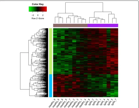
Here, we obtained a total of 453 up-regulated and 225 down-regulated DEGs. As indicated in the clustering heat map (Fig.1), these DEGs could well distinguish the IgAN and healthy samples completely.
Pathway enrichment analysis of the DEGs
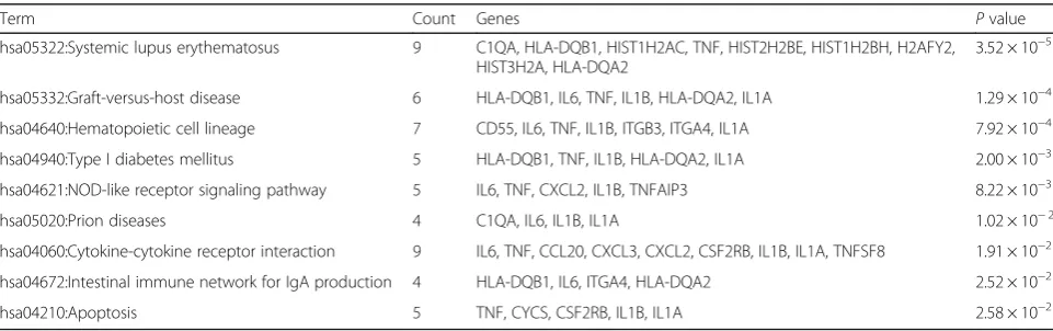
Unfortunately, the up-regulated DEGs were not enriched in any pathways. However, the down-regulated DEGs were significantly enriched in nine pathways. The enriched pathways were listed in Table 1, including hematopoietic cell lineage (pathway, p-value = 7.92 × 10− 4; which involved interleukin-6 (IL6), tumor necrosis
fac-tor (TNF) and interleukin 1 beta (IL1B)), NOD-like recep-tor signaling pathway (pathway, p-value = 8.22 × 10⁻³; which involvedIL6,TNFandIL1B), cytokine-cytokine re-ceptor interaction (pathway, p-value = 1.91 × 10⁻²; which involvedIL6,TNF,IL1Band C-C motif chemokine ligand 20 (CCL20)), intestinal immune network for IgA produc-tion (pathway, p-value = 2.52 × 10⁻²; which involvedIL6) and apoptosis (pathway, p-value = 2.58 × 10⁻²; which in-volvedTNFandIL1B).
PPI network of the DEGs
As presented in Fig.2, the PPI network with 379 nodes and 692 interactions was constructed. The hub nodes (whose degree > 10) mainly included TNF (degree = 31), PRKACA (degree = 26), IL6 (degree = 23), YWHAZ (de-gree = 19), MYB (de(de-gree = 15), TYK2 (de(de-gree = 14), FPR2 (degree = 13), IL1B (degree = 13), CCL20 (degree = 12), GNA11 (degree = 11).
Key nodes in the PPI network
Combined with the integrating results by four network centrality analyses, nodes with higher degree were used to cluster the two different kinds of samples. As a result, a total of 72 genes were identified that could well distin-guish IgAN and healthy samples (Fig. 3). Among them, genes such asIL6,TNF,IL1B,PRKACA,TYK2andCCL20 were closely related to five the pathways, including NOD-like receptor signaling pathway, cytokine-cytokine receptor interaction, hematopoietic cell lineage, apoptosis, and Toll-like receptor signaling pathway (Table2).
FI network analysis
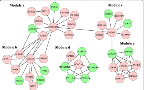
The FI network of the DEGs was constructed utilizing ReactomeFI, which included 42 genes and 71 interaction edges (Fig.4). Moreover, five modules (module a-e) were extracted from the FI network, and the absolute average correlation of genes in module a-e was 0.58, 0.4222, 0.5069, 0.4709 and 0.4275, respectively. Genes such as PRKACAin module a was enriched in integration of en-ergy metabolism, morphine addiction and glutamatergic synapse pathways; in module b, genes such as CCL20, FPR2and GNA11were related to GPCR ligand binding, GPCR downstream signaling and Gastrin-CREB signal-ing pathway via PKC and MAPK pathways; in module c, the gene NFYB was highly associated with RNA binding-related pathways; while in module d, the gene HDAC10 was significantly enriched in two pathways, al-coholism and chromatin modifying enzymes (Table 3) . Genes in module e were not enriched in any pathways.
Notably, we found that 12 genes in the FI network also belonged to the hub genes, such as CCL20, FPR2, and PRKACA.
Discussion
In the present study, a total of 72 crucial nodes in the PPI network were identified via re-analyzing the dataset GSE58539, which could well distinguish the IgAN and healthy samples. Among which, genes such as IL6,TNF,
Table 1Pathway enrichment analysis of the down-regulated differentially expressed genes in monocytes of IgAN patients
Term Count Genes Pvalue
hsa05322:Systemic lupus erythematosus 9 C1QA, HLA-DQB1, HIST1H2AC, TNF, HIST2H2BE, HIST1H2BH, H2AFY2, HIST3H2A, HLA-DQA2
3.52 × 10⁻⁵
hsa05332:Graft-versus-host disease 6 HLA-DQB1, IL6, TNF, IL1B, HLA-DQA2, IL1A 1.29 × 10⁻⁴
hsa04640:Hematopoietic cell lineage 7 CD55, IL6, TNF, IL1B, ITGB3, ITGA4, IL1A 7.92 × 10⁻⁴
hsa04940:Type I diabetes mellitus 5 HLA-DQB1, TNF, IL1B, HLA-DQA2, IL1A 2.00 × 10⁻³
hsa04621:NOD-like receptor signaling pathway 5 IL6, TNF, CXCL2, IL1B, TNFAIP3 8.22 × 10⁻³
hsa05020:Prion diseases 4 C1QA, IL6, IL1B, IL1A 1.02 × 10⁻²
hsa04060:Cytokine-cytokine receptor interaction 9 IL6, TNF, CCL20, CXCL3, CXCL2, CSF2RB, IL1B, IL1A, TNFSF8 1.91 × 10⁻²
hsa04672:Intestinal immune network for IgA production 4 HLA-DQB1, IL6, ITGA4, HLA-DQA2 2.52 × 10⁻²
IL1B,PRKACA, andCCL20were closely related to the fol-lowing pathways: NOD-like receptor signaling pathway, cytokine-cytokine receptor interaction, hematopoietic cell lineage, apoptosis and Toll-like receptor signal-ing pathway. Moreover, 12 genes in the FI network belonged to the 72 identified key nodes, such as
CCL20, HDAC10, FPR2 and PRKACA. Besides, the
12 genes were also the key genes in 4 module net-works correlating with pathways of integration of en-ergy metabolism (module a), GPCR-related pathways (module b), RNA binding-related pathways (module c), alcoholism and chromatin modifying enzymes (module d).
The cytokine encoded byIL6has great roles in inflam-mation and regulation of immune response [29]. Toll-like receptors (TLRs) are major factors that initiate the im-mune reaction. Most TLRs promote imim-mune response (including innate and adaptive) via inducing expression of
proinflammatory cytokines [30]. Increased TLRs, such as TLR-4, has been detected in circulating monocytes of patients with IgAN [31]. Expression of IL6 protein is also increased in mouse proximal tubular epithelial cells, ac-companying by the upregulation of TLR4 mRNA [32]. IL1B, encoded byIL1Bgene, is a member of interleukin 1 cytokine family and crucial for the regulation of inflamma-tory response [33]. In response to the external infections, gene expressions of the proinflammatory cytokines (e. g. IL1A, IL1B and IL6) are always upregulated simul-taneously [34, 35]. In particular, IL1B is implicated in the TLR-4 induced immune response in chronic pain [36]. In our study, IL6 and IL1B were both downregu-lated and enriched in TLR signaling pathway. These results suggested that IL6 and IL1B might be co-regulated in TLR signaling pathway and contribute to the abnormality of the immune response in mono-cytes of IgAN patients.
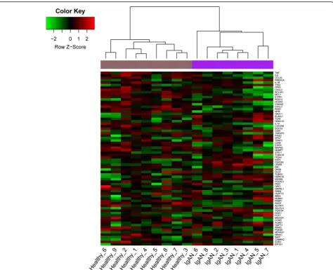
Fig. 3Heat map of clustering analysis of key genes predicted by four network centrality analyses in different samples. X-axis represents samples, and Y-axis represents gene expressions
Table 2Enrichment analysis of hub genes in the protein-protein interaction network
Term Genes Pvalue
hsa05020:Prion diseases IL6, IL1B, PRKACA, HSPA1B, IL1A 1.30 × 10⁻⁴
hsa04630:Jak-STAT signaling pathway TYK2, STAT4, IL6, SOCS2, PIAS3, CSF2RB, CISH 1.03 × 10⁻³
hsa04621:NOD-like receptor signaling pathway IL6, TNF, CXCL2, IL1B, TNFAIP3 1.20 × 10⁻³
hsa05332:Graft-versus-host disease IL6, TNF, IL1B, IL1A 3.15 × 10⁻³
hsa04060:Cytokine-cytokine receptor interaction IL6, TNF, CCL20, CXCL3, CXCL2, CSF2RB, IL1B, IL1A 3.31 × 10⁻³
hsa04640:Hematopoietic cell lineage IL6, TNF, IL1B, ITGA4, IL1A 4.00 × 10⁻³
hsa04210:Apoptosis TNF, CSF2RB, IL1B, PRKACA, IL1A 4.17 × 10⁻³
hsa04620:Toll-like receptor signaling pathway IL6, TNF, IL1B, TLR6, TRAF3 7.08 × 10⁻³
TNF is a multifunctional proinflammatory cytokine. Reportedly, TNF expression is dramatically increased in Mycoplasma penetrans-infected IgAN mice model, and the protein is proposed to involve in the induction of renal damage in IgAN [37]. Moreover, levels of serum TNF receptors are also elevated in IgAN patients
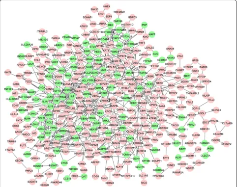
compared with healthy control [38]. However, Cox et al. uncover that TNF expression is obviously reduced in monocytes of IgAN patients, compared with those of HBDs [13]. The finding indicates the downregulated TNF may lead to the monocytes apoptosis. Moreover, the inhibition of TNF-αis proposed as a causative factor Fig. 4Gene functional interaction network of the differentially expressed genes. Circle in red denotes upregulated genes, and in green denotes downregulated genes
Table 3Enrichment analysis of genes in each functional interaction network module
Module GeneSet FDR Genes
a Integration of energy metabolism(R) 8.62 × 10⁻⁵ STK11, GNG10, GNB4, PRKACA
a Morphine addiction(K) 8.62 × 10⁻⁵ GNG10, GNB4, PDE4D, PRKACA
a Glutamatergic synapse(K) 1.39 × 10⁻⁴ GNG10, GNB4, PRKACA, GRIN3A
b GPCR ligand binding(R) 7.31 × 10⁻⁷ PTGIR, CCL20, LPAR6, FFAR2, FPR2, NTSR1, NPFF
b GPCR downstream signaling(R) 4.07 × 10⁻⁶ PTGIR, CCL20, LPAR6, FFAR2, GNA11, FPR2, NTSR1, NPFF
b Gastrin-CREB signaling pathway via PKC and MAPK(R) 6.99 × 10⁻⁶ LPAR6, FFAR2, GNA11, FPR2, NTSR1, NPFF
c overview of telomerase rna component gene hterc transcriptional regulation(B)
1.29 × 10⁻² NFYB
c DNA Double Strand Break Response(R) 1.62 × 10⁻² KDM4B
c Regulation of cholesterol biosynthesis by SREBP (SREBF)(R) 1.62 × 10⁻² NFYB
d Alcoholism(K) 1.74 × 10⁻¹⁰ HIST1H2AC, HIST2H2BE, H2AFY2, HIST1H2BH, HDAC10, HIST3H2A
d Systemic lupus erythematosus(K) 6.92 × 10⁻⁹ HIST1H2AC, HIST2H2BE, H2AFY2, HIST1H2BH, HIST3H2A
d Chromatin modifying enzymes(R) 1.55 × 10⁻⁴ HIST2H2BE, HIST1H2BH, HDAC10
of IgAN [39]. Therefore, it might be speculated that the apoptosis of monocytes induced by downregulation of TNF contribute to IgAN progression.
CCL20 is a small cytokine that also involves in immune regulation and inflammation [40]. Combination of CCL20 with CCR6 (the CCL20 receptor) cause the recruitment of leukocyte subsets, which finally promote immune-mediated kidney damage [41]. Additionally, CCL20 is one of the chemokines that take part in the host response to pathogens invasions by activating inflammatory cells, and it has the similar effects on monocytes [42]. Therefore, the downregulated CCL20in monocytes of IgAN patients might cause alteration in immune response, and thereby influence the IgAN development.
Three novel genes, protein kinase, CAMP-dependent, catalytic, alpha (PRKACA), formyl peptide receptor 2 (FPR2) and histone deacetylase 10 (HDAC10) were firstly predicted in monocytes of IgAN. PRKACA protein encoded byPRKACAgene is one subunit of protein kin-ase A that participates in apoptosis. At present, PRKACAamplification is served as a method for identi-fying genetic defect correlated with Cushing’s syndrome [43]. Somatic mutations ofPRKACAhave been detected in adenomas of the adrenal cortex [44]. Moreover, PRKACA mediates apoptosis-related signaling pathways in many cancer diseases, such as breast cancer and fol-licular thyroid cancer cells [45,46]. In the present study, PRKACAwas up-regulated and significantly enriched in apoptosis pathway, suggesting it might exert its function in monocytes via regulating apoptosis during IgAN pro-gression. FPR2 is known to activate the G-protein coupled receptor and N-formyl peptide receptor. FPR2 is found in adipose tissues as a receptor for the pro-resolving mediators [47], which contribute to the restoration of in adipose inflammation and treatment of obesity-related glomerulopathy [48]. HDAC10, contain-ing two catalytic sites, is highly expressed in numerous human tissues such as kidney [49]. In lung cancer, de-creased HDAC10 is associated with the advanced stage and adverse outcome [50]. However, there are rare re-ports on the relationship of IgAN and HDAC10. In the
current study, FPR2 and HDAC10 were hub
up-regulated genes in both PPI network and FI network, implying they might co-function in monocytes of IgAN patients. One limitation of this study is the lack of ex-pression validation. However, we will do more experi-ments to verify our conclusions once we collect the samples in the future.
Conclusions
In conclusion, several crucial genes were identified in monocytes of IgAN patients, such as IL6, TNF, IL1B, CCL20, PRKACA,FPR2 and HDAC10. They might have
co-functions and their dysregulations might alter activ-ities of pathways such as TLR and apoptosis signaling, which might finally promote IgAN progression. The study is of great value for the prediction of key regula-tors in monocytes of IgAN and the identification of tar-geting therapeutic management for IgAN.
Abbreviations
DCs:Dendritic cells; DEGs: Differentially expressed genes; FDR: False discovery rate; FI: Functional interaction;FPR2: Formyl peptide receptor 2; Gd-IgA1: Galactose-deficient immunoglobulin A1; GEO: Gene Expression Omnibus; HBDs: Healthy blood donors;HDAC10: Histone deacetylase 10; IDs: Identification numbers; IgAN: Immunoglobulin A nephropathy; IL1B: interleukin-1Beta;IL6: Interleukin-6; KEGG: Kyoto encyclopedia of genes and genomes; LCA: Leukocyte common antigen; PPI: Protein-protein interaction; RMA: Robust multi-array average; STRING: Search tool for the retrieval of interacting genes; TLRs: Toll-like receptors; TNF: Protein Kinase, CAMP-Dependent, Catalytic, Alpha; TNF: Tumor necrosis factor
Authors’contributions
YG and WG designed the research and drafted the manuscript. ZL draft the manuscript and revised manuscript for important intellectual content. DW and WL acquired data, analyze data and Statistical analysis. All authors read and approved the final manuscript.
Ethics approval and consent to participate Not applicable.
Consent for publication Not applicable.
Competing interests
The authors declare that they have no competing interests.
Publisher’s Note
Springer Nature remains neutral with regard to jurisdictional claims in published maps and institutional affiliations.
Author details
1Department of Nephropathy, Dongfang Hospital Affiliated to Beijing
University of Chinese Medicine, Beijng 100078, China.2Department of Urology, Dongzhimen Hospital Affiliated to Beijing University of Chinese Medicine, Beijng 100700, China.3Department of Nephropathy and Endocrinology, Dongzhimen Hospital Affiliated to Beijing University of Chinese Medicine, No. 5 Haiyuncang, Dongcheng District, Beijng City 100700, China.
Received: 8 December 2017 Accepted: 7 June 2018
References
1. Nakanishi K, Yoshikawa N. Immunoglobulin a nephropathy. Springer Berlin Heidelberg. 2009;7(4):275–398.
2. Lv J, Zhang H, Zhou Y, Li G, Zou W, Wang H. Natural history of
immunoglobulin a nephropathy and predictive factors of prognosis: a long-term follow up of 204 cases in China. Nephrology. 2008;13(3):242–6. 3. Myllymäki J, Honkanen T, Syrjänen J, Helin H, Rantala I, Pasternack A,
Mustonen J. Severity of tubulointerstitial inflammation and prognosis in immunoglobulin a nephropathy. Kidney Int. 2007;71(4):343–8. 4. Kiryluk K, Novak J, Gharavi AG. Pathogenesis of immunoglobulin a
nephropathy: recent insight from genetic studies. Annu Rev Med. 2013; 64:339.
5. Novak J, Julian BA, Tomana M, Mestecky J. IgA glycosylation and IgA immune complexes in the pathogenesis of IgA nephropathy. Seminars in nephrology. 2008;28(1):78–87.
6. Wyatt RJ, Julian BA. IgA nephropathy. N Engl J Med. 2013;368(25):2402–14. 7. Mestecky J, Raska M, Julian BA, Gharavi AG, Renfrow MB, Moldoveanu Z,
8. Tan Y, Zhang JJ, Liu G, Zhang H, Zhao MH. The level of urinary secretory immunoglobulin A (sIgA) of patients with IgA nephropathy is elevated and associated with pathological phenotypes. Clin Exp Immunol. 2009;156(1): 111–6.
9. Kiryluk K, Moldoveanu Z, Sanders JT, Eison TM, Suzuki H, Julian BA, Novak J, Gharavi AG, Wyatt RJ. Aberrant glycosylation of IgA1 is inherited in both pediatric IgA nephropathy and Henoch–Schönlein purpura nephritis. Kidney Int. 2011;80(1):79–87.
10. Li G, Zhang H, Lv J, Hou P, Wang H. Tandem repeats polymorphism of MUC20 is an independent factor for the progression of immunoglobulin a nephropathy. Am J Nephrol. 2006;26(1):43–9.
11. Canaud G, Audard V, Kofman T, Lang P, Legendre C, Grimbert P. Recurrence from primary and secondary glomerulopathy after renal transplant. Transpl Int. 2012;25(8):812–24.
12. Coppo R, Camilla R, Alfarano A, Balegno S, Mancuso D, Peruzzi L, Amore A, Dal Canton A, Sepe V, Tovo P. Upregulation of the immunoproteasome in peripheral blood mononuclear cells of patients with IgA nephropathy. Kidney Int. 2009;75(5):536–41.
13. Cox SN, Serino G, Sallustio F, Blasi A, Rossini M, Pesce F, Schena FP. Altered monocyte expression and expansion of non-classical monocyte subset in IgA nephropathy patients. Nephrol Dial Transplant. 2015;30(7):1122–232. 14. Smyth GK. Limma: linear models for microarray data. Bioinformatics and computational biology solutions using R and Bioconductor. 2005. p. 397– 420.
15. Dunning M, Lynch A, Eldridge M: illuminaHumanv4. db: Illumina HumanHT12v4 annotation data (chip illuminaHumanv4). R package version. 2013, 2(0).
16. Gentleman R: annotate: Annotation for microarrays. R package version. 2003, 1(0):19.
17. Warnes GR, Bolker B, Bonebakker L, Gentleman R, Huber W, Liaw A, Lumley T, Maechler M, Magnusson A, Moeller S: gplots: Various R programming tools for plotting data. R package version. 2009, 2(4).
18. Huang DW, Sherman BT, Lempicki RA. Systematic and integrative analysis of large gene lists using DAVID bioinformatics resources. Nat Protoc. 2009;4(1):44.
19. Kanehisa M, Goto S. KEGG: kyoto encyclopedia of genes and genomes. Nucleic Acids Res. 2000;28(1):27–30.
20. Szklarczyk D, Franceschini A, Kuhn M, Simonovic M, Roth A, Minguez P, Doerks T, Stark M, Muller J, Bork P. The STRING database in 2011: functional interaction networks of proteins, globally integrated and scored. Nucleic Acids Research. 2011;39(suppl_1):561–8.
21. Smoot ME, Ono K, Ruscheinski J, Wang P-L, Ideker T. Cytoscape 2.8: new features for data integration and network visualization. Bioinformatics. 2011; 27(3):431–2.
22. Latora V, Marchiori M. A measure of centrality based on network efficiency. New J Phys. 2007;9(6):188.
23. Estrada E. Virtual identification of essential proteins within the protein interaction network of yeast. Proteomics. 2006;6(1):35–40.
24. Estrada E, Rodriguez-Velazquez JA. Subgraph centrality in complex networks. Phys Rev E. 2005;71(5):056103.
25. Amitai G, Shemesh A, Sitbon E, Shklar M, Netanely D, Venger I, Pietrokovski S. Network analysis of protein structures identifies functional residues. J Mol Biol. 2004;344(4):1135–46.
26. Tang Y, Li M, Wang J, Pan Y, Wu F-X. CytoNCA: a cytoscape plugin for centrality analysis and evaluation of protein interaction networks. Biosystems. 2015;127:67–72.
27. Wu G, Dawson E, Duong A, Haw R, Stein L. ReactomeFIViz: a Cytoscape app for pathway and network-based data analysis. F1000Research. 2014;3:146. 28. Röfer T, Jüngel M. Vision-based fast and reactive Monte-Carlo localization. In: IEEE international conference on robotics and automation: 2003: IEEE; 1999; 2003. p. 856–61.
29. Rossignol J, Boyer C, Thinard R, Remy S, Dugast AS, Dubayle D, Dey ND, Boeffard F, Delecrin J, Heymann D. Mesenchymal stem cells induce a weak immune response in the rat striatum after allo or xenotransplantation. J Cell Mol Med. 2009;13(8b):2547–58.
30. Akira S, Uematsu S, Takeuchi O. Pathogen recognition and innate immunity. Cell. 2006;124(4):783–801.
31. Coppo R, Camilla R, Amore A, Peruzzi L, Daprà V, Loiacono E, Vatrano S, Rollino C, Sepe V, Rampino T. Toll-like receptor 4 expression is increased in circulating mononuclear cells of patients with immunoglobulin a nephropathy. Clin Exp Immunol. 2010;159(1):73–81.
32. Zhu X-L, Wang Y-J, Yang Y-Ζ, Yang R-C, Zhu B, Zhang YΗ, Lin Y, Lu Y, Li X-F, O'Byrne KT. Suppression of lipopolysaccharide-induced upregulation of toll-like receptor 4 by emodin in mouse proximal tubular epithelial cells. Mol Med Rep. 2012;6(3):493–500.
33. Dai D, Wang L, Xu L, Tang L, Xu X, Ye H, Zhou X, Chen C, Pan G, Ru P. A comprehensive meta-analysis of the association between three 1B polymorphisms and rheumatoid arthritis. Adv Biosci Biotechnol. 2014;5(2): 108–16.
34. Günther J, Esch K, Poschadel N, Petzl W, Zerbe H, Mitterhuemer S, Blum H, Seyfert H-M. Comparative kinetics of Escherichia coli-and Staphylococcus aureus-specific activation of key immune pathways in mammary epithelial cells demonstrates that S. aureus elicits a delayed response dominated by interleukin-6 (IL-6) but not by IL-1A or tumor necrosis factor alpha. Infect Immun. 2011;79(2):695–707.
35. Chen C-J, Ou Y-C, Lin S-Y, Raung S-L, Liao S-L, Lai C-Y, Chen S-Y, Chen J-H. Glial activation involvement in neuronal death by Japanese encephalitis virus infection. J Gen Virol. 2010;91(4):1028–37.
36. Guo L-H, Schluesener H. The innate immunity of the central nervous system in chronic pain: the role of toll-like receptors. Cell Mol Life Sci. 2007;64(9): 1128–36.
37. Jiang X, Lv Y-Q, Zhang J-N, Shi Y-L, Xu F-F. Mycoplasma penetrans infection is a potential cause of immunoglobulin a nephropathy: a new animal model. J Nephrol. 2013;26:470–5.
38. Sonoda Y, Gohda T, Suzuki Y, Omote K, Ishizaka M, Matsuoka J, Tomino Y. Circulating TNF receptors 1 and 2 are associated with the severity of renal interstitial fibrosis in IgA nephropathy. PLoS One. 2015;10(4):e0122212. 39. Wei S, Sinniah R. Adalimumab (TNFαinhibitor) therapy exacerbates IgA
glomerulonephritis acute renal injury and induces lupus autoantibodies in a psoriasis patient. Case Reports Nephrol. 2013;2013(4):1–4.
40. Starner TD, Barker CK, Jia HP, Kang Y, Jr MCP. CCL20 is an inducible product of human airway epithelia with innate immune properties. Am J Respir Cell Mol Biol. 2003;29(5):627.
41. Meng T, Li X, Ao X, Zhong Y, Tang R, Peng W, Yang J, Zou M, Zhou Q. Hemolytic Streptococcus may exacerbate kidney damage in IgA nephropathy through CCL20 response to the effect of Th17 cells. PLoS One. 2014;9(9):e108723. 42. Buonaguro L, Monaco A, Aricò E, Wang E, Tornesello ML, Lewis GK,
Marincola FM, Buonaguro FM. Gene expression profile of peripheral blood mononuclear cells in response to HIV-VLPs stimulation. BMC Bioinformatics. 2008;9(2):1.
43. Lodish MB, Yuan B, Levy I, Braunstein GD, Lyssikatos C, Salpea P, Szarek E, Karageorgiadis AS, Belyavskaya E, Raygada M. Germline PRKACA
amplification causes variable phenotypes that may depend on the extent of the genomic defect: molecular mechanisms and clinical presentations. Eur J Endocrinol. 2015;172(6):803–11.
44. Beuschlein F, Fassnacht M, Assié G, Calebiro D, Stratakis CA, Osswald A, Ronchi CL, Wieland T, Sbiera S, Faucz FR. Constitutive activation of PKA catalytic subunit in adrenal Cushing's syndrome. N Engl J Med. 2014;370(11): 1019–28.
45. Moody SE, Schinzel AC, Singh S, Izzo F, Strickland MR, Luo L, Thomas SR, Boehm JS, Kim SY, Wang ZC. PRKACA mediates resistance to HER2-targeted therapy in breast cancer cells and restores anti-apoptotic signaling. Oncogene. 2015;34(16):2061–71.
46. Grosse J, Warnke E, Wehland M, Pietsch J, Pohl F, Wise P, Magnusson NE, Eilles C, Grimm D. Mechanisms of apoptosis in irradiated and sunitinib-treated follicular thyroid cancer cells. Apoptosis. 2014;19(3):480–90. 47. Clària J, Dalli J, Yacoubian S, Gao F, Serhan CN. Resolvin D1 and
resolvin D2 govern local inflammatory tone in obese fat. J Immunol. 2012;189(5):2597–605.
48. Nolan E, O'Meara YM, Godson C. Lipid mediators of inflammation in obesity-related glomerulopathy. Nephrol Dial Transplant. 2013;28(suppl 4):iv22–9.
49. Tong JJ, Liu J, Bertos NR, Yang X-J. Identification of HDAC10, a novel class II human histone deacetylase containing a leucine-rich domain. Nucleic Acids Res. 2002;30(5):1114–23.