R E S E A R C H A R T I C L E
Open Access
Long-term survival without
graft-versus-host-disease following infusion of
allogeneic myeloma-specific V
β
T cell
families
S. Yado
1, G. Luboshits
1,2, O. Hazan
3, R. Or
3and M. A. Firer
1,2,4*Abstract
Background:Despite chemo-induction therapy and autologous stem cell transplantation (ASCT), the vast majority of patients with Multiple Myeloma (MM) relapse within 7 years and the disease remains incurable. Adoptive Allogeneic T-cell therapy (ATCT) might be curative for MM, however current ATCT protocols often lead to graft versus host disease (GvHD). Transplanting only tumor reactive donor T cells that mediate a graft-versus-myeloma (GvM) but not GvHD may overcome this problem.
Methods:We used an MHC-matched/miHA-disparate B10.D2→Balb/c bone marrow transplantation (BMT) murine model and MOPC315.BM MM cells to develop an ATCT protocol consisting of total body irradiation, autologous-BMT and infusion of selective, myeloma-reactive lymphocytes of T cell receptor (TCR) Vβ2, 3 and 8.3 families (MM-auto BMT ATCT).
Results:Pre-stimulation ex vivo of allogeneic T cells by exposure to MOPC315.BM MM cells in the presence of IL-2, anti-CD3 and anti-CD28 resulted in expansion of the myeloma-reactive T cell TCRVβ2, 3 and 8.3 subfamilies. Their isolation and infusion into MM-bearing mice resulted in a vigorous GvM response without induction GvHD and long-term survival. Repeated infusion of naïve myeloma-reactive T cell TCRVβ2, 3 and 8.3 subfamilies was also effective.
Conclusions:These data demonstrate that a transplantation protocol involving only selective tumor-reactive donor T cell families is an effective immunotherapy and results in long-term survival in a mouse model of human MM. The results highlight the need to develop similar ATCT strategies for MM patients that result in enhanced survival without symptoms of GvHD.
Keywords:Bone marrow transplantation, Graft-versus-host disease, Graft-versus-myeloma, Adoptive allogeneic T-cell therapy; T T-cell–receptor Vβfamilies;
Background
Survival of patients with multiple myeloma (MM) beyond 7 years remains rare even after autologous stem cell trans-plantation (ASCT) and treatment with novel agents [1]. Consequently, immunotherapies aimed at augmenting the anti-MM immune response, such as Adoptive Allogeneic
T-cell Therapy (ATCT) have become attractive alternatives [2–4]. Much of the curative potential of allografts is attrib-uted to the graft-versus-tumor (GvT) response that aims to destroy residual tumor cells that persist after induction therapy and ASCT [5]. Nonetheless, ATCT remains con-troversial [6] because the bulk donor T cells that mediate the GvT effect [7] can also induce graft versus-host disease (GvHD), a major cause of morbidity and mortality in ATCT recipients [8]. Various approaches to diminish the GvH response have had limited success [9–13].
© The Author(s). 2019Open AccessThis article is distributed under the terms of the Creative Commons Attribution 4.0 International License (http://creativecommons.org/licenses/by/4.0/), which permits unrestricted use, distribution, and reproduction in any medium, provided you give appropriate credit to the original author(s) and the source, provide a link to the Creative Commons license, and indicate if changes were made. The Creative Commons Public Domain Dedication waiver (http://creativecommons.org/publicdomain/zero/1.0/) applies to the data made available in this article, unless otherwise stated.
* Correspondence:firer@ariel.ac.il
1Chemical Engineering and Biotechnology, and Adelson School of Medicine, Ariel University, 40700 Ariel, Israel
Since GvT responses involve T-cell recognition of tumor-specific peptides presented by MHC molecules [14], it may be possible to identify and select donor T cells that provide beneficial GvT responses but minimal GvHD risk. In this regard, immune-transcriptome analyses of T cell receptor (TCR) VβCDR3-size and sequence is being used to characterize alloreactive versus tumor-specific T-cell responses. Korngold and colleagues identified donor alloreactive CD8+ and CD4+ Vβ families responsible for GvHD in several animal models of bone marrow trans-plantation (BMT) [15–18]. Binsfeld et al. studied the Vβ families involved in the GvM and the GvH response in an MM-BMT model, finding the Vβ2, 3 and 8.3 families of T cells as those specifically involved in the GvM response [19]. The implication of these results would be that myeloma-specific T cell subfamilies might be positively se-lected from the donor inoculum and infused to myeloma patients post ASCT, to afford separation of allo- from tumor-reactive T cells without the prior need to define specific target antigens.
To test this rationale, we used the allogeneic B10.D2→ Balb/c BMT model with MOPC315.BM myeloma cells. We first demonstrated that myeloma bearing-Balb/c mice ini-tially respond clinically to irradiation and auto-BMT but eventually relapse, similar to MM patients undergoing in-duction therapy and ASCT. By then infusing the animals with B10.D2 T cells from only the TCR Vβ2, 3 and 8.3 families appropriately pre-activated in vitro, we saw a vigor-ous GvM response without any clinical or histological signs of GvHD or disease relapse, which translated into long-term, disease-free survival. These data highlight the possi-bility that tumor-specific ATCT may lead to long-term disease-free survival without GvHD in patients with MM.
Methods
Ethical statement
All experimental procedures were performed in accord-ance with protocols approved by the Ariel University In-stitutional Animal Care and Use Committee. Animal welfare was assessed at least daily. After completion of experiments mice were euthanized in a CO2chamber.
Animals
Balb/c (H-2d) mice were obtained from Envigo La-boratories (Jerusalem, Israel). B10.D2 (H-2d) mice were purchased from Jackson Laboratories (Bar Har-bor, ME, USA) and bred in the Ariel University Ani-mal Facility. For all experiments, Ani-male mice between the ages of 10 and 14 weeks were used as donors and recipients. Treated mice were kept in a pathogen-free environment in autoclaved microisolator cages and were provided with acidified (pH 2.5) water and auto-claved food ad libitum.
Myeloma cell line and model
MOPC315.BM cells [20] was kindly provided by Prof. Bjarne Bogen (University of Oslo, Norway). They were cultured at 37 °C in 5% CO2 in RPMI 1640
(Sigma-Al-drich, Rehovot, Israel) supplemented with 10% FBS, 1% MEM NEAA 100x (Gibco), 0.005% 1 M I-thioglycerol, 0.03% Gensumycin 40 mg/ml (Sigma-Aldrich) and 2 mM L-glutamine (Biological Industries, Beit Haemek, Israel). I.v. injection of MOPC315.BM cells results in tumor development in the bone marrow (BM) and spleen and is associated with osteolytic lesions, validat-ing the model as resemblvalidat-ing human MM disease [21]. In advanced disease stages (within 3–4 weeks), the mice de-velop paraplegia through spinal cord compression. They were sacrificed when presenting signs of paraplegia, de-terioration of general condition or apathy.
Experimental transplantation design (Fig.1a)
Balb/c mice were injected i.v. into the tail vein with 1 × 10⁶MOPC315.BM cells in 100μl RPMI 1640. Preliminary experiments showed that paraplegia developed 38 days post injection (Additional file 1: Figure S1). At day 35, mice were irradiated with 6.5 Gy (Total Body Irradiation) using an X ray source (Kimtron Polaris 320) and injected 6 h later with an infusion of syngeneic 10 × 106BM and 70 × 106spleen cells from healthy Balb/c donors (Day 0). BM cells were collected by flushing the femurs and tibias into sterile PBS. Spleens were crushed through a 70-μm cell strainer into sterile PBS (Biological Industries) and Red blood cells lysed (RBC lysis buffer, eBioscience, San Diego, USA). Animals that received this transplant proto-col are referred to as“MM-Auto-BMT” mice. For ATCT experiments, on day 10 and in some experiments also on day 17 post MM Auto-BMT, mice received an infusion of 1 × 10⁶or 2.5 × 10⁶B10.D2 or Balb/c Vβ2, 3 and 8.3 posi-tive T cells (MM-Auto-BMT-ATCT group) or unselected spleenocytes. These Myeloma-reactive T cells (MT-cells) were isolated with antibody-coated magnetic beads from donor spleenocytes, either pre-activated by MOPC315.BM cells or not (naïve cells) (see below).
Recipient mice were checked daily for morbidity and mortality and sacrificed after appearance of symptoms of myeloma (See Additional file1: Video S1) and/or GvHD. Three mice from each experimental condition were eutha-nized on days −2 (before), + 7 and + 14 post auto-BMT and at the end point. BM and spleens were harvested and analyzed by flow cytometry for the presence of MOPC315.BM cells and to monitor repopulation of T cell subsets. Before sacrifice, a blood sample was obtained for measurement of M315 myeloma paraprotein.
In vitro T-cell activation and cytotoxicity
growth. Following washing, they were then co-cultured in complete medium (RPMI 1640, 10% FBS, 1% Penicil-lin/Streptomycin, 2 mM L-glutamine and 50μg/mL 2-mercaptoethanol) supplemented with recombinant IL--2 (20 U/mL, Biolegend) for 4 days at a ratio of 20:1 with 5 × 10⁶ spleenocytes isolated from healthy B10.D2 or Balb/c mice. In later experiments, cells were co-cultured for 2 days in medium containing 50 U/mL rIL-2,
anti-CD3 (5μg/ml) and anti-CD28 (2μg/ml) (eBioscience) antibodies. Vβ 2, 3 and 8.3 T cells activated with this second protocol are referred to as “IL-2/Ab” activated allo- (B10.D2) or auto- (Balb/c) MT-cells. Following co-culture, spleenocytes were analyzed by flow cytometry and used for cytotoxicity assays. MT cells were isolated by incubation with 0.5 mg/ml of PE-conjugated mono-clonal antibodies: anti-Vβ 2 (clone B20.6), anti-Vβ 3
(clone KJ25), and anti-Vβ 8.3 (clone 1B3.3) (BD Phar-mingen, San Jose, CA) followed by anti-PE mAb– conju-gated magnetic beads and separation using the SuperMacs system (Miltenyi Biotec, Auburn, CA). The positive fraction was typically > 90% PE positive as deter-mined by flow cytometry.
To test the cytotoxicity of donor B10.D2 or Balb/c MT cells, 107 fresh MOPC315.BM target cells/mL were la-beled with 1μM carboxyfluorescein succinimidyl ester (CFSE) (eBioscience) for 10 min at RT. The reaction was stopped by addition of 4–5 volumes of cold complete media and 5-min incubation on ice. After washing with complete medium, the target cells were resuspended in complete medium at 1 × 10⁶cells/mL dispensed into 96-well microtiter plates (100μL/well). MT cell populations were added in 20:1 10:1 and 5:1 effector-to-target ratios in a total volume of 250μL complete medium and plates were incubated at 37 °C in 5% CO2for 4 h. The
percent-age of MOPC315.BM cell death was evaluated by stain-ing with Sytox blue (1μM, Molecular Probes) and flow cytometry. Target cells incubated without effector cells (to measure spontaneous death) were used as control.
GvHD clinical-scoring system
GvHD symptoms were evaluated with a scoring system adapted from Cooke et al. [22]. The score is based on weight loss (< 10% = 0; 10–20% = 1; > 20% = 2), hunched back posture (normal = 0; hunched-back while resting = 1; persistent = 2), general activity (normal = 0, reduced activity = 1, apathy = 2), alopecia (normal = 0, < 1 cm2 = 1, > 1 cm2= 2) and skin fibrosis (normal = 0, fibrosis = 1; scabs = 2) with a maximum score of 10. Each animal’s condition was monitored daily, and the GvHD score was calculated at least 3 times per week. Mice were sacrificed if they reached a score of 8/10 or when apathetic.
Flow cytometry
Fc receptor binding was blocked by incubation with anti-CD16/CD32 antibodies (clone 93, eBioscience) for 5 min at RT. The cells were then incubated for 30 min at 4 °C with specific antibodies (anti-CD3e/APC (145-2C11), anti-CD4/FITC (GK1.5), anti-CD8/eFluor506 (53–6.7), anti-CD25/PE-Cy7 (PC61.5), (eBioscience); anti-CD3/PE (17A2); anti-CD69/Pacific blue (H1.2F3); anti-B220/ PE-Cy7 (RA3-6B2) (Biolegend (San Diego, CA); anti-IgA/FITC (C10–3) (BD Biosciences) and CD138/APC (REA104) (Miltenyi Biotec) in PBS/ 3% FBS, washed and resuspended in cold PBS. The data were acquired by a CytoFLEX (Beckman Coulter) flow cytometer and analyzed using FlowJo software.
Histology
Approximately 2 cm2of shaved skin from the interscap-ular region (GvHD–target organ) and representative
spleen and colon samples were collected from sacrificed mice, fixed in 10% formalin, paraffin embedded, cut into 5-μm-thick sections and stained with hematoxylin and eosin. Histological processing and assessment was per-formed by Patho-Lab Diagnostics (Nes Ziona, Science Park, Israel).
Serum paraprotein quantitation
Paraprotein production by MOPC315.BM cells was eval-uated by ELISA [23]. Briefly, 96 well Nunclon ELISA plates were coated with 2 μg/ml of anti-MOPC315.BM paraprotein idiotype (Ab2.1–4) (kindly provided by Prof Bjarne Bogen, University of Oslo, Norway) at 4 °C over-night. Wells were blocked with PBS/0.02% sodium azide/1% BSA, washed and incubated for 2 h at 37 °C with serum samples or standard paraprotein (ranging from 400 to 0.39 ng/ml) diluted in PBS/ 0.02% sodium azide/0.1% BSA/0.1% Tween 20. Then, the plates were incubated with 1μg/ml biotinylated rat anti-mouse IgA (clone C10–1, BD Pharmingen, Germany) for 1 h at RT, washed, incubated with streptavidin- HRP (1:2000; Sigma-Aldrich) for 1 h at RT and washed again. TMB substrate (Merck Millipore, Billerica, MA, USA) was added for 10-min, the reaction was terminated with H2O2 and absorbance measured at 450 nm with a
TECAN Infinite M200 ELISA reader.
Statistics
The Log-Rank test was used to compare the Kaplan-Meyer survival plots. Median survival times (MST) were calculated, and a pvalue ≤0.05 was considered statisti-cally significant. Statistical significance between groups was determined using a Student ttest. A p value≤0.05 was considered statistically significant.
Results
B10.D2 Vβ2, 3 and 8.3 T cells families induce GvM but not GvHD
On day 10 after Auto-BMT, but prior to the time of their expected relapse, MM-Auto-BMT mice received a T cell infusion comprising donor B10.D2, or Balb/c MT cells (Allo-MT cells or Auto-MT cells respectively) or unselected spleenocytes.
We tested whether ex vivo activation of Allo-MT cells prior to injection could boost the GvM response with minimal GvHD complications. B10.D2 spleen cells were co-cultured with Mitomycin C-pretreated myeloma cells at a ratio of 20:1 in medium supplemented with 20 U rIL-2. Flow cytometry showed an expansion of both CD8+ and CD4+T cell populations and a significant in-crease in activated CD4+and CD8+vβ(2, 3, 8.3)+T cells, confirming their reactivity against myeloma target cells (Additional file 1: Figure S2). Therefore, 1 × 10⁶ Allo-MT cells, either naïve or MOPC315.BM-activated, were injected into MM-Auto-BMT mice on day 10 after the autograft. This treatment also extended the MST to 35d and there were no signs of GvHD but again, 80% of the mice eventually succumbed to myeloma progression. There was no significant difference in MST between mice that received naïve or MOPC315.BM-activated Allo-MT cells (MST = 35 d versus MST = 36 d, respect-ively;p= 0.862) (Fig.1b).
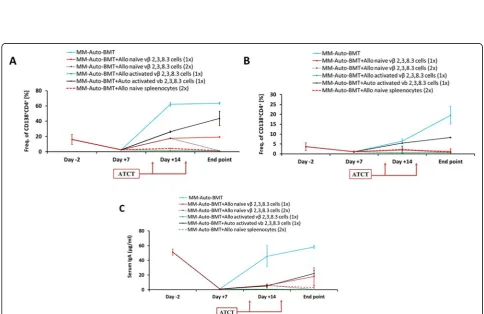
At sacrifice, all ATCT treated groups who received ei-ther naïve or activated Vbeta T cells or naïve unselected spleenocytes, had significantly lower myeloma cell infil-tration in the spleen compared to the control group (Fig. 2a, *p= 0.0006, **p= 0.0018, ***p= 0.0001 respect-ively) and accordingly they produced less serum para-protein (Fig. 2b). Percentages of activated CD4+ and CD8+ T cells were significantly higher in the BM and spleen of mice that received MT cells (Fig.2c), suggest-ing that these cells might be responsible for the observed GvM effect. These data indicate that infusion of donor myeloma-reactive T cells can provoke a potent GvM ef-fect, without GvHD, leading to extended, but nonethe-less limited, overall survival.
Improved activation of B10.D2 Vβ2, 3 and 8.3 T cells
We questioned whether a more clinically effective GvM (no GvHD) response might be obtained by improving the ex vivo activation protocol of the Allo-MT cells. Therefore, spleenocytes from B10.D2 or Balb/c mice were stimulated by Mitomycin-C-treated MOPC315.BM cells for 2 days in medium containing 50 U/mL rIL-2 and anti-CD3/anti-CD28 antibodies (referred to as IL-2/Ab) [24]. This protocol resulted in an expansion of CD4+T cells and a significant expansion of CD8+ T cells (2-fold) in B10.D2 spleenocyte cultures (Fig. 3). In Balb/c spleenocyte cultures, only CD8+ T cells expanded. There was a strong activation induced CD25 expression on MT cell families in both B10.D2 and Balb/c spleenocyte cultures. The cytotoxic capacity of these activated lymphocytes was validated by co-culturing them in different ratios with CFSE-labeled fresh MOPC315.BM. The degree of target cell killing was depended on the effector:tar-get cell ratio with the best specific lysis (24% for B10.D2 and 19% for Balb/c) achieved at the highest E/T ratio tested (20: 1) (Additional file1: Figure S3).
Enhanced MT cell activation leads to long-term survival without GvHD
The effect of the IL-2/Ab activated MT cells was then tested in vivo. On day 10 after auto-BMT, MM-Auto-BMT mice received 2.5 × 10⁶of IL-2/Ab activated Allo- or Auto-MT cells (The equivalent dose of these cells found in healthy B10.D2 and Balb/c mouse spleens as deter-mined by flow cytometry). As shown in Fig.4, 88% of mice who received IL-2/Ab activated Allo-MT cells survived at least 109 days post auto-BMT. Significantly, none of these animals developed symptoms of GvHD. Infusion of IL-2/ Ab activated Auto-MT cells also provided a significant, al-beit short-term GvM effect (MST = 44 d versus MST = 19 d, respectively;*p< 0.0001), although 100% of these mice eventually succumbed to myeloma progression.
We also tested whether an additional dose of naïve Allo-MT cells might circumvent the need for pre-activation. As shown in Fig.4, mice who received an additional infusion of these cells on day 17 displayed no symptoms of GvHD and 80% of them had survived by the end of the experiment (109 days). Mice who received unselected B10.D2 spleeno-cytes displayed the typical signs of chronic GvHD and suc-cumbed to the disease with a MST of 35 days.
can be also be obtained with repeated infusion of naïve MM-specific donor B10.D2 T cell families.
Discussion
Allogeneic immunotherapy remains the only potentially curable treatment for MM but the frequent co-development of GvHD after this type of therapy severely limits its clinical application. Unfortunately, the clinical success of strategies to reduce GvHD while retaining the GvT response have been limited [3,10,25].
Korngold and colleagues demonstrated that CDR3-size spectratyping of the TCRVβ-chain can characterize and dif-ferentiate alloreactive from GvT-specific T-cell repertoire re-sponses, highlighting the potential for tailoring the donor inoculum to target only the recipient’s malignant cells [18,26,
27]. Our aim was to apply TCRVβ-chain CDR3-sizing to
allo-immunotherapy, by positively selecting MM-specific donor T cell families and testing if their infusion could affect a clinically relevant GvM response without inducing GvHD.
We used the well-established MHC-matched/ miHA-disparate B10.D2→Balb/c BMT model [28] and induced MM in recipients by injecting MOPC315.BM MM cells [21]. MM-bearing mice were treated by total body irradiation and auto-BMT, followed by infusion of donor myeloma-reactive TCR Vβ+T cells (Vβ2, 3 and 8.3 families) identified previ-ously [19]. In vitro experiments (Additional file1: Figure S3) and the finding that the transplantation of these cells induced life-prolonging GvM effects but with no clinical (Fig.4), bio-marker (Fig.5) or histological (Fig.6) signs of GvHD indicates that these Vβ T cells families indeed respond to tumor-specific antigens expressed on MOPC315.BM cells. Similar to human MM cells, MOPC315.BM cells express and secrete
(See figure on previous page.)
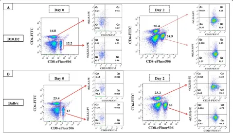
Fig. 2Involvement of vβTCR CDR3 2, 3, 8.3 T cell families in the graft-versus-myeloma effect in myeloma bearing Balb/c mice treated by irradiation, Auto-BMT and then allogeneic lymphocyte infusion. Recipient mice were sacrificed when severe GvHD symptoms, myeloma symptoms or apathy were present. Flow cytometry staining was performed on cells from spleen and bone marrow at sacrifice.aInfiltration of MOPC MM cells in the bone marrow and spleen identified as CD138+CD4+double positive cells. *p= 0.0006. **p= 0.0018 (Studentttest).b Paraprotein serum IgA quantitation (μg/ml) by ELISA before Auto-BMT, 1 week after and at sacrifice. *p= 0.0003. **p= 0.005 (Studentttest).c vβ(2 + 3 + 8.3)+T cell populations in the graft-versus-myeloma effect. Shown are percentages of activated CD4+vβ(2 + 3 + 8.3)+T cells (CD69+ within CD4+vβ(2 + 3 + 8.3)+T cells) and activated CD8+vβ(2 + 3 + 8.3)+T cells (CD69+ within CD8+vβ(2 + 3 + 8.3)+T cells) in the spleen (left panel) BM (right panel) in the MM-Auto-BMT, MM-Auto-BMT + Allo naive vβ2, 3, 8.3 (× 1) group, MM-Auto-BMT + Allo activated vβ2, 3, 8.3 (× 1) or in healthy Balb/c mice.*p< 0.0001;**p< 0.0001;***p< 0.05 (Studentttest)
Fig. 4Survival curves of myeloma bearing Balb/c recipient mice treated by irradiation, Auto-BMT and then allogeneic or autologous lymphocyte infusion. The presented results represent the average of two independent experiments. On day 10 and/or day 17 after Auto-BMT, recipient mice were injected i.v. with naïve or activated B10.D2 / Balb/c Vβ2, 3 and 8.3 T cells. Recipient mice were sacrificed when severe GvHD symptoms, myeloma symptoms or apathy were present. Statistical significance between survival curves was determined using the Log-Rank test. MM-Auto-BMT versus MM-Auto-MM-Auto-BMT + Auto activated vβ2, 3, 8.3 (× 1), *p< 0.0001; MM-Auto-BMT versus MM-Auto-BMT + Allo naive vβ2, 3, 8.3
(× 1), **p= 0.0001
an idiotypic (Id) antibody and peptides from this antibody presented in association with MHC-Class I molecules would likely be one target recognized by the donor MT cells [29]. The induction of anti-MM-Id peptide responses has been studied following the vaccination of MM patients with autologous Id-pulsed dendritic cells [30] and a recent trial (#NCT01426828) aims to evaluate whether infusion of Id-KLH primed CD3/CD28 activated autologous lym-phocytes mediates a clinically relevant Id-specific immun-ity.. Unfortunately, there is no information on other potential MOPC315.BM tumor specific molecules that might be recognized by MT cells. A search in several immunoinformatic databases (IMTG, VDJdb, McPAS-TCR) did not clearly indicate which MHC presented pep-tides might be bound by TCR bearing VβCDR3 2, 3 and 8.3 sequences. With regards to human MM there is cur-rently no information on the myeloma-specific TCR se-quence repertoire in MM patients [31].
The relative contribution of each Vβ family to the overall GvM response we observed is a subject for on-going studies. Not all families may contribute equally to the GvM effect, possibly because only some of them are presented with dominant MHC-bound peptides [32,33], or because they secrete cytokines that induce more ef-fective anti-tumor responses. In another study, the Vβ13 family by itself was shown to dominate the B10.BR CD8
T-cell response against a myeloid leukemia cell line. Transplantation of these cell induced a slight GvT re-sponse with no concomitant acute GvHD [27].
Appropriate T-cell co-stimulation is critical for induc-tion of effective anti-tumor T-cell funcinduc-tion [24, 34–37]. Porter et al. [35] and Biavati et al. [38] showed that ex vivo co-stimulation of T cells via their CD3 and CD28 recep-tors can produce activated T cells that enhance the antitu-mor effect of donor lymphocyte infusions after allogeneic hematopoietic stem cell transplantation in patients with chronic myelogenous leukemia and MM. Noonan et al. were the first to report that infusion of autologous, ex vivo activated, marrow-infiltrating T cells could induce anti-tumor reactivity and enhance progression-free survival in MM patients, although there was no difference in overall survival [39]. Our results are in line with these findings. We saw that although in vitro activation of auto-MT cells led to target cell killing (Additional file1: Figure S3) and transplantation of IL2/Ab stimulated auto-MT cells more than doubled the mean survival time (from 20 to 43 days, p< 0.0001), the mice eventually relapsed. The short-lived response after auto-MT infusion may be due to T cell ex-haustion, a topic currently under intensive study [40, 41]. While appropriately activated (IL-2/Ab) allogeneic MT cells responded aggressively to target cells in vitro and in-duced long-term survival in vivo, on the other hand,
transplantation of one dose of naïve allo-MT cells lead to only short-term clinical efficacy. Interruption of the GvM effect may have been due to development of effector T cell exhaustion because an additional infusion of naïve allo-MT cells was more effective allowing for long-term disease-free survival (Additional file1: Figure S4, Fig.4).
Another explanation for the lack of efficacy of auto-MT cells infusion may be the specificity of the Balb/c MT cells themselves. Flow cytometry clearly showed that co-culture with target cells resulted in overall expansion and activation of both CD4+ and CD8+ B10.D2 populations but only the CD4+ Balb/c population. While Balb/c T cells expressing 2, 3 and 8.3 Vβfamily containing TCRs did become activated (but did not expand) they may not be the best anti-MOPC315.BM effector T cell clones and may only induce a weaker and short-lived GvM response. Transcriptome ana-lysis of the Balb/c CD8+T cell TCRs may reveal that other subfamilies are more effective. This may also be true in pa-tients, however there is currently no data available to ad-equately address this question. A third explanation may be that the effectiveness of the allo- over auto-MT cell activity in our model is due to a miHA antigen (or antigens) recog-nized on the MOPC315.BM by B10.D2 but not Balb/c T cells (MOPC315 cells are Balb/c derived). These antigens would need to be different from the shared myeloma and allo-antigens reported by Binsfeld et al. that are recognized by TCRVβfamilies other than those used in our study [19]. A number of human leukemic restricted miHAs have been identified, including some on MM cells [42]. Some of these are capable of eliciting anti-tumor T cell responses [43] and indeed recent studies report development of engineered T cells bearing human miHA specific TCRs [44,45]. Their ac-tivity towards MM has not been demonstrated.
Conclusion
We have shown for the first time invocation of a strong and life-saving GvM response and prevention of GvHD by integrating auto-BMT with a ATCT composed only of transcriptome-identified MM reactive Vβ T cell families. With the use of new TCR sequencing technologies [46–48] it should become feasible to characterize, isolate and infuse tumor-specific donor T cell Vβfamilies into patients. This strategy is significant for MM therapy because it highlights the opportunity to develop a more effective treatment protocol combining a vigorous GvM response that elimi-nates residual MM cells in patients who have undergone pre-conditioning and auto-HSCT without inducing GvHD.
Supplementary information
Supplementary informationaccompanies this paper athttps://doi.org/10. 1186/s40425-019-0776-9.
Additional file 1 Figure S1. Survival curve of Balb/c mice injected i.v. with MOPC315.BM (1x10⁶,n= 4/group).Figure S2.In vitroreactivity of
T-cells after 4-days co-culture with MOPC315.BM T-cells.Figure S3. Target cell cytotoxicity of activated B10.D2 or Balb/c vβ2, 3 8.3 T cells.Figure S4.Monitoring of post-transplant reconstitution of spleen(A)and BM(B) T -cell subsets in normal Balb/c mice (n= 10/group) who received 6.5Gy irradiation and then autologous bone marrow transplantation (Auto-BMT).Video S1.Video of representative Balb/c mouse with hind leg paraplegia 35 days after i.v. injection with MOPC315.BM myeloma cells.
Abbreviations
ASCT:Autologous stem cell transplantation; ATCT: Adoptive Allogeneic T-cell therapy; BMT: Bone marrow transplantation; CFSE: Carboxyfluorescein succinimidyl ester; GvHD: Graft versus host disease; GvM: Graft-versus-myeloma; MM: Multiple Myeloma; MST: Median survival times; MT cells: Myeloma-specific T cells; TCR: T cell receptor
Acknowledgements
The authors are sincerely grateful to Prof. Bjarne Bogen and Peter O. Hofgaard (Institute of Immunology, Oslo, Norway) for providing the MOPC315.BM cells, Ab2.1-4 antibody, M315 protein standard and for helpful discussions.
Authors’contributions
SY designed and carried out the experiments, analyzed the results and wrote the initial draft GL advised on design of experiments and results analysis and helped write the manuscript OZ advised on the experimental concept and on the manuscript RO advised on the experimental concept and on the manuscript MF oversaw and advised on design of experiments and results analysis and helped write the manuscript. All authors read and approved the final manuscript.
Funding
SA was supported by graduate fellowships from Ariel University and from the Daon-Lang Cancer Research Fund. This research was funded by the Daon-Lang Cancer Research Fund and in part by the Ariel University Re-search Authority.
Availability of data and materials
The datasets supporting the conclusions of this article are included within the article and its additional file. For further information on original data, contact the Corresponding Author.
Ethics approval
All experimental procedures were performed in accordance with protocols approved by the Ariel University Institutional Animal Care and Use Committee. Animal welfare was assessed at least daily. After completion of experiments mice were euthanized in a CO2chamber.
Consent for publication Not applicable.
Competing interests
The authors declare that they have no competing interests.
Author details
1Chemical Engineering and Biotechnology, and Adelson School of Medicine, Ariel University, 40700 Ariel, Israel.2Ariel Center for Applied Cancer Research, Ariel University, 40700 Ariel, Israel.3Department of Bone Marrow
Transplantation and Cancer Immunotherapy, Hadassah-Hebrew University Medical Center, Jerusalem, Israel.4Adelson Medical School, Ariel University, 40700 Ariel, Israel.
Received: 25 July 2019 Accepted: 9 October 2019
References
2. Binsfeld M, Fostier K, Muller J, Baron F, Schots R, Beguin Y, et al. Cellular immunotherapy in multiple myeloma: Lessons from preclinical models. Biochim Biophys Acta - Rev Cancer. 2014;1846:392–404 Elsevier B.V. 3. Malek E, El-Jurdi N, Kröger N, de Lima M. Allograft for myeloma: examining
pieces of the jigsaw puzzle. Front Oncol. 2017;7:1–9.
4. Jing W, Gershan JA, Blitzer GC, Palen K, Weber J, McOlash L, et al. Adoptive cell therapy using PD-1+ myeloma-reactive T cells eliminates established myeloma in mice. J Immunother Cancer. 2017;5:51 Available from:http://jitc.
biomedcentral.com/articles/10.1186/s40425-017-0256-z. [cited 2018 Nov 6]. 5. Jamil MO, Mineishi S. State-of-the-art acute and chronic GVHD treatment.
Int J Hematol. 2015;101:452–66 Springer Japan.
6. Giralt S. Stem cell transplantation for multiple myeloma: current and future status. Hematology Am Soc Hematol Educ Program. 2011;2011:191–6. 7. Azar Y, Shainer R, Almogi-Hazan O, Bringer R, Compton SR, Paidas MJ, et al.
PreImplantation factor reduces graft-versus-host disease by regulating immune response and lowering oxidative stress (murine model). Biol Blood Marrow Transplant. 2013;19:519–28 Elsevier Ltd.
8. Vadakekolathu J, Rutella S. T-cell manipulation strategies to prevent graft-versus-host disease in Haploidentical stem cell transplantation. Biomedicines. 2017;5:33.
9. Li Pira G, Biagini S, Cicchetti E, Merli P, Brescia LP, Milano GM, et al. Immunoselection techniques in hematopoietic stem cell transplantation. Transfus Apher Sci. 2016;54:356–63 Elsevier Ltd.
10. Or-Geva N, Reisner Y. The evolution of T-cell depletion in haploidentical stem-cell transplantation. Br J Haematol. 2016;172:667–84.
11. Bertaina A, Merli P, Rutella S, Pagliara D, Bernardo ME, Masetti R, et al. HLA-haploidentical stem cell transplantation after removal of ab 1 T and B cells in children with nonmalignant disorders. Blood. 2015;124:822–7. 12. Bleakley M, Heimfeld S, Loeb KR, Jones LA, Chaney C, Seropian S, et al.
Outcomes of acute leukemia patients transplanted with naive T cell– depleted stem cell grafts. J Clin Invest. 2015;125:1–13.
13. Shikari H, Antin JH, Dana R. Ocular graft-versus-host disease: a review. Surv Ophthalmol. 2013;58:233–51 Elsevier Inc.
14. Moyer JS, Maine G, Mulé JJ. Early vaccination with tumor-lysate-pulsed dendritic cells after allogeneic bone marrow transplantation has antitumor effects. Biol. Blood Marrow Transplant. 2006;12:1010–9.
15. Jones SC, Friedman TM, Murphy GF, Korngold R. Specific donor V??-associated CD4+ T-cell responses correlate with severe acute graft-versus-host disease directed to multiple minor histocompatibility antigens. Biol Blood Marrow Transplant. 2004;10:91–105.
16. Friedman TM, Statton D, Jones SC, Berger MA, Murphy GF, Korngold R. Vbeta spectratype analysis reveals heterogeneity of CD4+ T-cell responses to minor histocompatibility antigens involved in graft-versus-host disease: correlations with epithelial tissue infiltrate. Biol Blood Marrow Transplant. 2001;7:2–13. 17. Friedman TM, Gilbert M, Briggs C, Korngold R. Repertoire analysis of CD8+ T
cell responses to minor histocompatibility antigens involved in graft-versus-host disease. J Immunol. 1998;161:41–8.
18. Patterson AE, Korngold R. Infusion of select leukemia-reactive TCR Vbeta+ T cells provides versus-leukemia responses with minimization of graft-versus-host disease following murine hematopoietic stem cell transplantation. Biol Blood Marrow Transplant. 2001;7:187–96. 19. Binsfeld M, Beguin Y, Belle L, Otjacques E, Hannon M, Briquet A, et al.
Establishment of a murine graft-versus-myeloma model using allogeneic stem cell transplantation. PLoS One. 2014;9:1–19.
20. Eisen HN, Simms ES, Potter M. Mouse myeloma proteins with antihapten antibody activity. Protein produced by plasma cell tumor MOPC-315. Biochemistry. 1968;7:4126–34.
21. Hofgaard PO, Jodal HC, Bommert K, Huard B, Caers J, Carlsen H, et al. A novel mouse model for multiple myeloma (MOPC315.BM) that allows noninvasive spatiotemporal detection of osteolytic disease. PLoS One. 2012;7:1–13. 22. Cooke KR, Martin TR, Kobzik L, Brewer J, Delmonte J, Ferrara JL. The roles of
Alloreactivity and endotoxin in an experimental model of idiopathic pneumonia syndrome after bone marrow transplantation.•909. Pediatr Res. 1996;39:154.
23. Riedel SS, Mottok A, Brede C, Bäuerlein CA, Jordán Garrote AL, Ritz M, et al. Non-invasive imaging provides spatiotemporal information on disease progression and response to therapy in a murine model of multiple myeloma. PLoS One. 2012;7:1–13.
24. Maus MV, Thomas AK, Leonard DGB, Allman D, Addya K, Schlienger K, et al. Ex vivo expansion of polyclonal and antigen-specific cytotoxic T lymphocytes by artificial APCs expressing ligands for the T-cell receptor, CD28 and 4-1BB. Nat
Biotechnol. 2002;20:143–8 Available from:http://www.nature.com/articles/ nbt0202-143. Nature Publishing Group. [cited 2018 Jan 15].
25. Eefting M, de Wreede LC, Von dem Borne PA, Halkes CJM, Kersting S, Marijt EWA, et al. Donor T-cell responses and disease progression patterns of multiple myeloma. Bone Marrow Transplant. 2017;52:1609–15 Nature Publishing Group.
26. Friedman TM, Goldgirsh K, Berger SA, Zilberberg J, Filicko-O’Hara J, Flomenberg N, et al. Overlap between in vitro donor antihost and in vivo posttransplantation TCR V?? Use: a new paradigm for designer allogeneic blood and marrow transplantation. Blood. 2008;112:3517–25.
27. Fanning SL, Zilberberg J, Stein J, Vazzana K, Berger SA, Korngold R, et al. Unraveling graft-versus-host disease and graft-versus-leukemia responses using TCR V Spectratype analysis in a murine bone marrow transplantation model. J Immunol. 2013;190:447–57.
28. Jaffee BD, Claman HN. Chronic graft-versus-host disease (GVHD) as a model for scleroderma. I. Description of model systems. Cell. Immunol. 1983;77:1–12. 29. Lauritzsen GF, Weiss S, Dembic Z, Bogen B. Naive idiotype-specific CD4+ T cells
and immunosurveillance of B-cell tumors. Proc Natl Acad Sci. 2006;91:5700–4. 30. Garfall AL, Stadtmauer EA. Cellular and vaccine immunotherapy for multiple
myeloma. Hematology. 2016;2016:521–7 Available from:http://www.ncbi. nlm.nih.gov/pubmed/27913524. [cited 2018 Oct 30].
31. Krejcik J, Casneuf T, Nijhof IS, Verbist B, Bald J, Plesner T, et al. Daratumumab depletes CD38+ immune regulatory cells, promotes T-cell expansion, and skews T-cell repertoire in multiple myeloma. Blood. 2016;128:384–94 Available from:http://www.bloodjournal.org/cgi/doi/10.1182/blood-2 015-12-687749. [cited 2018 Dec 25].
32. Valmori D. Enhanced generation of specific tumor-reactive CTL in vitro by Melan-A/Mart-1 immunodominant peptide analogs. Immunol Lett. 2002;56:223–4. 33. Kjer-nielsen L, Clements CS, Purcell AW, Brooks AG, Whisstock JC, Burrows
SR, et al. A Structural Basis for the Selection of Dominant. Immunity. 2003; 18:53–64.
34. Fischer K, Mackensen A. The flow cytometric PKH-26 assay for the determination of T-cell mediated cytotoxic activity. Methods. 2003;31:135–42. 35. Porter DL, Levine BL, Bunin N, Stadtmauer EA, Luger SM, Goldstein S, et al.
A phase 1 trial of donor lymphocyte infusions expanded and activated ex vivo via CD3 / CD28 costimulation. Blood. 2006;107:1325–31. 36. Klebanoff CA, Gattinoni L, Palmer DC, Muranski P, Ji Y, Hinrichs CS, et al.
Determinants of successful CD8 + T-cell adoptive immunotherapy for large established tumors in mice. Clin Cancer Res. 2011;17:5343–52.
37. Laport GG, Levine BL, Stadtmauer EA, Schuster SJ, Luger SM, Grupp S, et al. Adoptive transfer of costimulated T cells induces lymphocytosis in patients with relapsed/refractory non-Hodgkin lymphoma following CD34+-selected hematopoietic cell transplantation. Blood. 2013;102:2004–13.
38. Biavati L, Noonan K, Luznik L, Borrello I. Activated allogeneic donor-derived marrow-infiltrating lymphocytes display measurable in vitro antitumor activity. J Immunother. 2019;42:73–80.
39. Noonan KA, Huff CA, Davis J, Lemas MV, Fiorino S, Bitzan J, et al. Adoptive transfer of activated marrow-infiltrating lymphocytes induces measurable antitumor immunity in the bone marrow in multiple myeloma. Sci Transl Med. 2015;7:288ra78 Available from:http://stm.sciencemag.org/lookup/doi/1 0.1126/scitranslmed.aaa7014. [cited 2018 Oct 30].
40. Wang JC, Xu Y, Huang ZM, Lu XJ. T cell exhaustion in cancer: mechanisms and clinical implications. J Cell Biochem. 2018;119:4279–86.
41. Suen H, Brown R, Yang S, Weatherburn C, Ho PJ, Woodland N, et al. Multiple myeloma causes clonal T-cell immunosenescence: identification of potential novel targets for promoting tumour immunity and implications for checkpoint blockade. Leukemia. 2016;30:1716–24.
42. Spaapen R, Mutis T. Targeting haematopoietic-specific minor
histocompatibility antigens to distinguish graft-versus-tumour effects from graft-versus-host disease. Best Pract Res Clin Haematol. 2008;21:543–57 Available from:https://linkinghub.elsevier.com/retrieve/pii/S15216926 08000522. [cited 2019 Sep 3].
43. van Bergen CAM, Kester MGD, Jedema I, Heemskerk MHM, van Luxemburg-Heijs SAP, Kloosterboer FM, et al. Multiple myeloma-reactive T cells recognize an activation-induced minor histocompatibility antigen encoded by the ATP-dependent interferon-responsive (ADIR) gene. Blood. 2007;109: 4089–96 Available from:http://www.ncbi.nlm.nih.gov/pubmed/17234742. American Society of Hematology. [cited 2019 Sep 3].
44. Dossa RG, Cunningham T, Sommermeyer D, Medina-Rodriguez I, Biernacki MA, Foster K, et al. Development of T cell immunotherapy for
relapse. Blood. 2017;131 blood-2017-07-791608. Available from:http://www. ncbi.nlm.nih.gov/pubmed/29051183. [cited 2019 Sep 3].
45. Pilunov AM, Kuchmiy AA, Sheetikov SA, Filkin SY, Romaniuk DS, Rosov FN, et al. Modification of Cytotoxic Lymphocytes with T Cell Receptor Specific for Minor Histocompatibility Antigen ACC-1Y. Mol Biol (Mosk). 2019;53:456– 66 Available from:http://elibrary.ru/item.asp?doi=10.1134/S002689841903 0145. [cited 2019 Sep 3].
46. Ogishi M, Yotsuyanagi H. Quantitative prediction of the landscape of T cell epitope immunogenicity in sequence space. Front Immunol. 2019;10:1–20. 47. Bentzen AK, Such L, Jensen KK, Marquard AM, Jessen LE, Miller NJ, et al. T
cell receptor fingerprinting enables in-depth characterization of the interactions governing recognition of peptide–MHC complexes. Nat Biotechnol. 2018;36:1191–6.
48. Zhang SQ, Ma KY, Schonnesen AA, Zhang M, He C, Sun E, et al. High-throughput determination of the antigen specificities of T cell receptors in single cells. Nat Biotechnol. 2018;36:1156–9.
Publisher’s Note