R E V I E W
Open Access
Radiation therapy and PD-1/PD-L1
blockade: the clinical development of an
evolving anticancer combination
Jun Gong
1, Thang Q. Le
2, Erminia Massarelli
1, Andrew E. Hendifar
3and Richard Tuli
4*Abstract
Several inhibitors of programmed cell death-1 (PD-1) and programmed death ligand-1 (PD-L1) have been approved
as a form of immunotherapy for multiple cancers. Ionizing radiation therapy (RT) has been shown to enhance the
priming and effector phases of the antitumor T-cell response rendering it an attractive therapy to combine with
PD-1/PD-L1 inhibitors. Preclinical data support the rational combination of the 2 modalities and has paved way for
the clinical development of the combination across a spectrum of cancers. In this review, we highlight the preclinical
and clinical development of combined RT and PD-1/PD-L1 blockade to date. In addition to a comprehensive
evaluation of available safety and efficacy data, we discuss important points of consideration in clinical trial design for
this promising combination.
Keywords:
Radiation therapy, PD-1, PD-L1, Clinical trials, Preclinical, Antitumor, Immune response, Checkpoint inhibitor
Background
Early preclinical evidence demonstrated that activation
of the programmed cell death 1 (PD-1) and programmed
death ligand 1 (PD-L1) axis suppressed the activation
and proliferation of tumor antigen-specific T-cells and
promoted tumorigenesis [
1
,
2
]. These processes were
reversed with PD-1/PD-L1 blockade and supported
the concept of PD-1/PD-L1 blockade as a potential
form of anti-cancer immunotherapy. The first agents
in the family of PD-1/PD-L1 inhibitors to be
ap-proved by the Food and Drug Administration (FDA)
were the humanized monoclonal IgG4 antibodies,
pembrolizumab and nivolumab, that targeted PD-1 in
unresectable or advanced melanoma [
3–10
]. There are
currently 5 PD-1/PD-L1 inhibitors approved by the
FDA for the treatment of a number of solid tumors
and hematologic malignancies [
11–43
].
Ionizing radiation therapy (RT) is widely used in the
definitive and metastatic setting for local tumor control;
however, the ability of radiation to elicit a systemic
tumor response with associated regression of untreated
metastases outside of the radiation field has been
reported and was first described as the abscopal effect
[
44
]. Increasing evidence supports that the abscopal
ef-fect is likely immune-mediated
–
largely, in a T-cell
dependent manner with a complex interplay between
proimmunogenic and proinflammatory factors [
45–53
].
Over time, recognition of the immunomodulatory
prop-erties of radiation has led to the integration of RT with
immune-modulating agents including immune
check-point inhibitors to potentially develop a combination
therapy with enhanced or synergistic anticancer activity
(Fig.
1
).
Indeed, an initial preclinical study showed that
com-bining RT (1
–
2 fractions of 12 Gray (Gy) to the primary
tumor) with an anti-cytotoxic T lymphocyte-associated
antigen-4 (CTLA-4) monoclonal antibody resulted in
synergistic antitumor activity in a poorly immunogenic
metastatic mammary carcinoma mouse model when
CTLA-4
blockade
by
itself
was
ineffective
[
54
].
Enhanced antitumor responses have also been observed
across several preclinical animal models treated with
combined RT and CTLA-4 blockade [
55–58
]. Since the
first preclinical studies that highlighted the synergistic
antitumor activity of combination RT and CTLA-4
* Correspondence:[email protected]4Departments of Radiation Oncology and Biomedical Sciences, Samuel Oschin Comprehensive Cancer Institute, Cedars-Sinai Medical Center, 8700 Beverly Blvd, AC 1023, Los Angeles, CA 90048, USA
Full list of author information is available at the end of the article
blockade, several prospective clinical trials have
re-ported on the activity of RT and ipilimumab in
ad-vanced solid tumors [
59–66
]. Similarly, there are
numerous ongoing clinical trials investigating the
combination of RT and CTLA-4 blockade that have
been extensively reviewed and are beyond the scope
of this manuscript [
67
,
68
]. Herein, we review in
detail the preclinical and clinical development of the
combination of RT and PD-1/PD-L1 inhibitors in
can-cer therapy.
Preclinical studies
The efficacy of combination RT and checkpoint blockade
is associated with modulation of immune parameters
within the tumor microenvironment
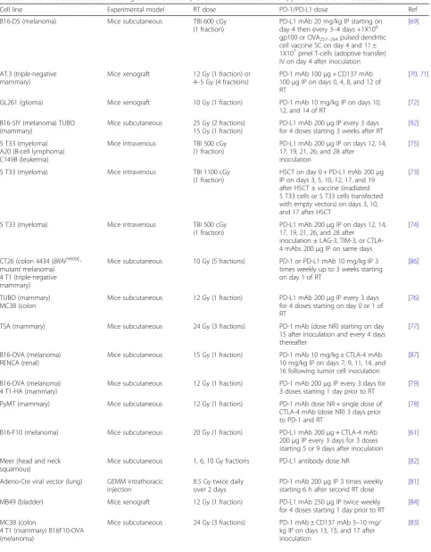
Early investigations in mouse models of solid and
hematologic malignancies showed enhanced antitumor
effects when treated with PD-1 or PD-L1 blockade in
combination with in-field RT, sublethal total body
irradiation (TBI), or stereotactic radiosurgery (SRS)
compared to single modality treatment (Table
1
) [
69–85
].
Table 1
Preclinical studies demonstrating antitumor activity of combined radiation therapy and PD-1/PD-L1 blockade
Cell line Experimental model RT dose PD-1/PD-L1 dose Ref.
B16-D5 (melanoma) Mice subcutaneous TBI 600 cGy (1 fraction)
PD-L1 mAb 20 mg/kg IP starting on day 4 then every 3–4 days +1X10⁶ gp100 or OVA257–264pulsed dendritic cell vaccine SC on day 4 and 11 ± 1X10⁷pmel T-cells (adoptive transfer) IV on day 4 after inoculation
[69]
AT.3 (triple-negative mammary)
Mice xenograft 12 Gy (1 fraction) or 4–5 Gy (4 fractions)
PD-1 mAb 100μg + CD137 mAb 100μg IP on days 0, 4, 8, and 12 of RT
[70,71]
GL261 (glioma) Mice xenograft 10 Gy (1 fraction) PD-1 mAb 10 mg/kg IP on days 10, 12, and 14 of RT
[72]
B16-SIY (melanoma) TUBO (mammary)
Mice subcutaneous 25 Gy (2 fractions) 15 Gy (1 fraction)
PD-L1 mAb 200μg IP every 3 days for 4 doses starting 3 weeks after RT
[92]
5 T33 (myeloma) A20 (B-cell lymphoma) C1498 (leukemia)
Mice intravenous TBI 500 cGy (1 fraction)
PD-L1 mAb 200μg IP on days 12, 14, 17, 19, 21, 26, and 28 after
inoculation
[75]
5 T33 (myeloma) Mice intravenous TBI 1100 cGy (1 fraction)
HSCT on day 0 + PD-L1 mAb 200μg IP on days 3, 5, 10, 12, 17, and 19 after HSCT ± vaccine (irradiated 5 T33 cells or 5 T33 cells transfected with empty vectors) on days 3, 10, and 17 after HSCT
[73]
5 T33 (myeloma) Mice intravenous TBI 500 cGy
(1 fraction)
PD-L1 mAb 200μg IP on days 12, 14, 17, 19, 21, 26, and 28 after
inoculation ± LAG-3, TIM-3, or CTLA-4 mAbs 200μg IP on same days
[74]
CT26 (colon 4434 (BRAFV600E -mutant melanoma) 4 T1 (triple-negative mammary)
Mice subcutaneous 10 Gy (5 fractions) PD-1 or PD-L1 mAb 10 mg/kg IP 3 times weekly up to 3 weeks starting on day 1 of RT
[86]
TUBO (mammary) MC38 (colon
Mice subcutaneous 12 Gy (1 fraction) PD-L1 mAb 200μg IP every 3 days for 4 doses starting on day 0 or 1 of RT
[76]
TSA (mammary) Mice subcutaneous 24 Gy (3 fractions) PD-1 mAb (dose NR) starting on day 15 after inoculation and every 4 days thereafter
[77]
B16-OVA (melanoma) RENCA (renal)
Mice subcutaneous 15 Gy (1 fraction) PD-1 mAb 10 mg/kg ± CTLA-4 mAb 10 mg/kg IP on days 7, 9, 11, 14, and 16 following tumor cell inoculation
[87]
B16-OVA (melanoma) 4 T1-HA (mammary)
Mice subcutaneous 12 Gy (1 fraction) PD-1 mAb 200μg IP every 3 days for 3 doses starting 1 day prior to RT
[79]
PyMT (mammary) Mice subcutaneous 12 Gy (1 fraction) PD-1 mAb dose NR + single dose of CTLA-4 mAb (dose NR) 3 days prior to PD-1 and RT
[78]
B16-F10 (melanoma) Mice subcutaneous 20 Gy (1 fraction) PD-L1 mAb 200μg + CTLA-4 mAb 200μg IP every 3 days for 3 doses starting 5 or 9 days after inoculation
[61]
Meer (head and neck squamous)
Mice subcutaneous 1, 6, 10 Gy fractions PD-L1 antibody dose NR [82]
Adeno-Cre viral vector (lung) GEMM intrathoracic injection
8.5 Gy twice daily over 2 days
PD-1 mAb 200μg IP 3 times weekly starting 6 h after second RT dose
[81]
MB49 (bladder) Mice xenograft 12 Gy (1 fraction) PD-L1 mAb 250μg IP twice weekly for 4 doses starting 1 day prior to RT
[84]
MC38 (colon
4 T1 (mammary) B16F10-OVA (melanoma)
Mice subcutaneous 24 Gy (3 fractions) PD-1 mAb ± CD137 mAb 5–10 mg/ kg IP on days 13, 15, and 17 after inoculation
Combined modality therapy was associated with higher
levels of CD8+/interferon-
γ
(IFN
γ
)+/tumor necrosis
factor-
α
(TNF
α
) + cytotoxic T-cells, increased PD-1, T-cell
immunoglobulin mucin-3 (TIM-3), lymphocyte-activation
gene 3 (LAG-3), and 2B4 (immune checkpoints)
expres-sion on CD8+ T-cells, decreased numbers of CD4
+/FOXP3+ regulatory T-cells (Tregs) and myeloid-derived
suppressor cells (MDSCs), upregulation of PD-L1 on
dendritic cells and tumor cells in irradiated tumors,
RT-induced upregulation of major histocompatibility
complex
(MHC)
class
I
tumor-associated
antigen
complexes, and enhanced antigen cross-presentation in
draining lymph nodes compared to single modality
arms [
71
,
72
,
74
,
76–79
].
Combination modality-induced immune profile changes
may be time-dependent
Early syngeneic mouse tumor models demonstrating
significant improvements in survival and tumor volume
reduction with the combination of RT and PD-1 or
PD-L1 blockade compared to single modality and
con-trol arms identified elevations in tumor cell PD-L1
ex-pression that were CD8+ T-cell and IFN
γ
-dependent
following irradiation (10 Gy over 5 daily fractions)
com-pared to non-irradiated mice with peak levels occurring
72 h after last dose of RT [
86
]. RT-induced increases in
the CD8+/Treg ratio and PD-L1 expression occurred
24
–
96 h post-RT in a separate mouse model [
81
]. In
colon
carcinoma
tumors,
the addition
of
PD-L1
Table 1
Preclinical studies demonstrating antitumor activity of combined radiation therapy and PD-1/PD-L1 blockade
(Continued)
Cell line Experimental model RT dose PD-1/PD-L1 dose Ref.
4-hydroxytamoxifen induction (BRAF-mutant, PTEN-deficient melanoma)
GEMM topical induction
14 Gy (1 fraction) PD-1 + CD137 or PD-1 + CTLA-4 mAb 100μg IP twice weekly for 4 doses on day 1 of RT
[99]
344SQ (lung) Mice subcutaneous 36 Gy (3 fractions) PD-1 mAb 10 mg/kg IP starting on day 1 of RT and continued for additional 3–4 doses
[91]
ARK (esophageal squamous) Mice subcutaneous 20 Gy (10 fractions) PD-1 mAb (dose NR) starting 2 days before RT and every 3 days thereafter ± carboplatin and paclitaxel IP (dose NR) on day 1 of RT and every 3 fractions
[85]
GL261 (glioma) Mice xenograft 10 Gy (1 fraction) PD-1 mAb 200μg IP on days 10, 12, and 14 of RT ± TIM-3 mAb 250μg IP days 7, 11, and 15 of RT
[90]
CT26 (colon 4434 (BRAFV600E -mutant melanoma)
Mice subcutaneous 10 Gy (5 fractions) PD-1 or PD-L1 mAb 10 mg/kg IP 3 times weekly for 1 week starting on day 1 of RT
[88]
TSA (mammary) Mice subcutaneous 24 Gy (3 fractions) on days 12, 13 and 14 after inoculation
PD-1 mAb 200μg IP on days 12, 15, 19, 22 and 26 after inoculation
[89]
Hep-55.1c (hepatocellular) Mice orthotopic 30 Gy (3 fractions) PD-1 mAb 250μg IP on days 7, 14, and 21 after inoculation
[96]
KPC and Pan02 (pancreatic) Mice subcutaneous 6, 12, or 20 Gy (1 fraction) 10 Gy (5 fractions) 15 Gy (5 fractions)
PD-L1 mAb 10 mg/kg IP on days 4, 7, 10, and 13 after inoculation + gemcitabine 100 mg/kg IP on days 0 and 3 of inoculation
[95]
HCa-1 (hepatocellular) Mice intramuscular 10 Gy (1 fraction) PD-L1 mAb 10 mg/kg IP every 3 days for 4 doses starting on day 1 of RT
[97]
LM8 (osteosarcoma) Mice subcutaneous 10 Gy (1 fraction) PD-L1 mAb 150μg + CTLA-4 mAb 150μg IP every 3 days for 3 doses starting on days 9, 12, and 15 after inoculation
[98]
CT26 (colon Mice intradermal RFA 17-gauge single
ablation electrode for 3.5–4.5 min at target temperature of 70 degrees C
PD-1 mAb 200μg IP every 3 days for 4 doses
[94]
RTradiation therapy,TBItotal body irradiation,cGycentigraymAbmonoclonal antibody,IPintraperitoneal,SCsubcutaneous,IVintravenous,GyGray,
blockade on day 1 of RT (schedule A), day 5 of RT
(schedule B), or 7 days after RT (schedule C) showed
that there was no significant difference in overall
sur-vival (OS) between schedule A and B (
p
> 0.05) though
sequential therapy (schedule C) was ineffective in
enhan-cing OS compared to RT alone (median OS 30 days vs.
35 days,
p
> 0.05) [
86
]. Notably, PD-1 expression was
sig-nificantly decreased on CD8+ T-cells 7 days after RT
compared to time-matched controls (
p
< 0.05).
Abscopal effects and systemic immunity
On
subcutaneous
tumor
flank
rechallenge
of
treatment-naïve mice and mice cured by combination
RT and checkpoint blockade, immunologic memory was
established in cured mice but not in treatment-naïve
mice suggesting that the immune system in cured mice
retained the ability to recognize tumor-associated
antigens and mount an immune response of greater
magnitude and speed upon rechallenge, i.e., systemic
im-munity [
71
,
72
]. Abscopal effects have been shown to be
mediated, in part, by PD-1 as administration of a single
fraction of 15 Gy by stereotactic ablative radiotherapy
(SABR) to the primary tumor in a melanoma
subcutane-ous msubcutane-ouse model resulted in significant reduction in
tumor volumes of secondary nonirradiated tumors in
PD-1-knockout mice compared to PD-1-wild-type (WT)
mice [
87
]. Addition of a PD-1 inhibitor to SABR resulted
in synergistic antitumor activity on the primary tumor
compared to PD-1 inhibitor or SABR alone and
recapit-ulated abscopal effects on secondary nonirradiated
tu-mors in PD-1-WT mice when treatment alone with
anti-PD-1 or SABR did not reduce secondary tumor
growth. Furthermore, following RT, higher levels of
PD-1+ CD11a
highCD8+ T-cells were seen in primary
tu-mors compared to secondary tutu-mors and higher levels
in irradiated compared to nonirradiated tumors; this
population of cells appeared to comprise the principal
tumor-specific reactive phenotype. This latter finding
has been confirmed in another study where RT
in-creased T-cell receptor (TCR) repertoire clonality and
diversity of the TCR repertoire in irradiated tumors
compared to controls, however, the addition of PD-1
in-hibition to RT increased TCR diversity both in irradiated
and out-of-field sites [
88
]. Further analysis revealed that
most of these TCR clones arose from progenitor clones
that were established in tumors prior to therapy, and it
is the influx of tumor-infiltrating lymphocytes (TILs)
from outside the tumor along with resident-tumor
infiltrating T-cells that contribute to the enhanced
tumor responses seen with combination therapy.
Recently, durable regression of irradiated tumors and
abscopal responses observed in mammary tumor-bearing
mouse models treated with combination RT and
check-point blockade were shown to be dependent on cancer
cell-intrinsic activation of the type I IFN pathway as
medi-ated by cyclic GMP-AMP (cGAMP) synthase (cGAS) and
stimulator of interferon genes (STING) signaling [
89
].
RT-induced abscopal responses with PD-1 blockade were
additionally shown to be regulated by
Trex1
where
induc-tion of Trex1 expression in cancer cells resulted in loss of
abscopal responses in mice treated with the combination.
Combined modality therapy reverses T-cell exhaustion
and resistance to RT and anti-PD-1 therapy
Murine tumor xenografts have shown that increasing
levels of PD-1 and TIM-3 co-expression in CD4+
T-cells, CD8+ T-cells, and Tregs over time contribute to
an exhausted or impaired T-cell phenotype [
90
].
Furthermore,
resistance
to
anti-PD-1
therapy
in
RT-refractory tumors has been characterized by
signifi-cant elevations in expression of genes associated with
T-cell exhaustion, increased levels of checkpoints
includ-ing LAG-3, TIM3, and CTLA-4 on CD4+ T-cells, and
decreased number of CD11c + tumor-associated
macro-phages (TAMs) [
81
]. The addition of immune
check-point inhibitors to RT has been shown to enhance
tumor response compared to controls across several
mouse
tumor
models
through
reinvigoration
of
exhausted CD8+ TILs characterized by increased Ki67+
GzmB+ T-cells within the exhausted PD-1+ Eomes+
T-cell pool, increased CD8+ CD44+ TILs, and increased
CD8+/Treg ratio [
61
,
77
,
85
].
Moreover, an anti-PD-1-resistant murine lung cancer
model established through sequential in vivo passage of
nonresponsive tumors to ongoing anti-PD-1 therapy was
characterized by significant downregulation of MHC
class I and II genes including
β
2-microglobulin and
reduction in CD4+/CD8+ TILs and IFN-
γ
production in
resistant tumors compared to parental tumors [
91
].
Addition of RT induced IFN-
γ
production and MHC
class I expression and ultimately restored response to
PD-1 blockade in resistant tumors. Addition of a PD-L1
inhibitor has been shown to reverse RT-induced tumor
equilibrium in favor of tumor regression in mice
sub-cutaneously injected with melanoma and breast tumors
demonstrating RT-induced stable disease (SD, defined as
≥
3 weeks) characterized by a transient rise and fall in
levels of tumor-infiltrating CD8+ T-cells and IFN
γ
[
92
].
Extrinsic RT resistance has been recently shown to be
contributed by RT-induced host STING activation
resulting in immunosuppressive MDSC recruitment that
is mediated by chemokine receptor type 2 (CCR2) in a
syngeneic mouse model of colon carcinoma [
93
].
A growing body of preclinical evidence supports the
combination of other immunotherapeutic agents with
RT or radiofrequency ablation (RFA), immune
check-point blockade, and/or chemotherapy to enhance
tumor growth control (and often systemic control)in
preclinical mouse models; synergistic antitumor
activ-ity with multimodalactiv-ity therapy was characterized by
tumor cell PD-L1 expression in a
JAK/Stat1-depen-dent manner and reduced numbers of CD11b + Gr1+
cells (MDSCs) [
90
,
94–99
].
Toxicities
Several preclinical studies have investigated the toxicity
of combined RT and checkpoint blockade. Notably, one
investigation of lung-irradiated (20 Gy) C57bl/6-WT
mice receiving anti-PD-1 antibody (10 mg/kg
intraperi-toneal twice per week for 5 doses) showed more findings
of abnormal alveoli, inflammatory changes, and exudates
in the alveolar septa associated with a 2.1-fold increase
in CD8+ T-cells in the irradiated lung tissues of mice in
the RT and PD-1 blockade arm though post-RT
mortal-ity up to 120 days was not significantly different in the
RT alone vs. RT and PD-1 blockade arm (
p
= 0.657)
[
100
]. A separate study, however, using a similar dose of
20 Gy of thoracic RT (designed to induce mortality) to
C57bl/6 mice identified worse survival with RT and
PD-1 blockade (36% survived) than RT alone (70%
survived,
p
= 0.0169) at 21 days post-RT and increased
T-cell infiltrates in lung and cardiac tissues (both
in-and out-of-field) of mice treated with RT in-and PD-1
blockade compared to RT alone putatively due to
enhanced healthy tissue damage by T-cell activation with
the addition of PD-1 blockade to thoracic RT [
101
].
Incorporating PD-1 blockade to cardiac RT in mice
has also shown to decrease survival and exacerbate
cardiac dysfunction and myocarditis that are CD8+
T-cell-mediated [
102
].
Clinical studies
Retrospective studies
Numerous case reports and case series have documented
clinically significant, and often durable, tumor responses
to the combination of RT and PD-1/PD-L1 blockade in
advanced or metastatic melanoma, NSCLC, Hodgkin
lymphoma, RCC, and cervical cancer [
103–112
]. Initial
retrospective series of patients with melanoma brain
metastases treated with SRS or fractionated RT within
3
–
6 months of receiving anti-PD-1 therapy produced
promising 1-year OS rates and significantly improved
6-and 12-month distant brain metastasis control 6-and OS
rates in those treated with SRS and anti-PD-1 therapy
vs. SRS and chemotherapy (Table
2
) [
113
,
114
]. In 24
patients with brain metastases from melanoma (54%)
and NSCLC (46%), treatment with SRS before, during,
or after PD-1 blockade produced 6- and 12-month
median OS rates of 85 and 78%, respectively [
115
]. One
retrospective study investigated 53 patients with
meta-static melanoma treated with RT sequential or
concur-rent to anti-PD-1 therapy or as salvage therapy in the
setting of progression on anti-PD-1 therapy (35 patients
received extracranial RT or intracranial SRS and 21
pa-tients received whole brain radiotherapy (WBRT)) and
showed that median OS and ORR were not significantly
different between concurrent and sequential RT/SRS
cohorts (Table
2
) [
116
].
A single-institute retrospective trial analyzed the
effi-cacy of concurrent SRS and anti-PD-1 or anti-CTLA-4
therapy (defined as SRS within 4 weeks of administration
of checkpoint inhibitors) in 75 patients with melanoma
brain metastases and identified significantly improved
median percent reduction in lesion volume with
concur-rent compared to nonconcurconcur-rent arms and with
anti-PD-1 compared to anti-CTLA-4 arms at 3 months
and 6 months [
117
]. However, when both anti-PD-1 and
anti-CTLA-4 therapies were combined there was no
sig-nificant difference in median OS between nonconcurrent
(9.0 months, range 2.1
–
61.8) and concurrent arms
(19.1 months, range 2.7
–
64.2,
p
= 0.0691). In solely
metastatic NSCLC patients (
n
= 21), combined RT to
oli-goprogressive sites along with PD-1/PD-L1 blockade or
other immune therapies resulted in excellent local
con-trol, median time to systemic progression of 2.3 months
(95% confidence interval (CI) 1.0
–
4.5), and median OS
of 7.2 months (95% CI 4.2
–
11.1) [
118
]. Among 25
pa-tients with unresectable melanoma, abscopal responses
(CR or PR) were observed in 56% of patients with the
addition of late RT (> 3 months of insufficient response
to anti-PD-1 monotherapy) [
119
].
A group of 137 patients with metastatic melanoma,
NSCLC, and RCC treated with WBRT, SRS, or
extracra-nial RT before or after initiation of PD-1 blockade
expe-rienced a median OS 249 days (8 months; interquartile
range (IQR) 90
–
689) following the start of anti-PD-1
therapy though OS was 25.7 months in the cohort
re-ceiving brain RT as first form of palliative RT [
120
]. On
multivariate analysis, melanoma patients fared best as
the hazard ratio (HR) for death was 3.1 (95% CI 1.7
–
5.9)
for NSCLC and HR of 3.2 (95% CI 1.2
–
7.9) for RCC
compared to melanoma (
p
= 0.0008) possibly due to
im-proved responses to checkpoint inhibitors in melanoma
with the incorporation of both PD-1 and CTLA-4
inhib-itors into standard care.
Table 2
Retrospective clinical studies with available results on the antitumor activity of combined radiation therapy and PD-1/PD-L1
blockade
Study n Design Outcomes Toxicities Ref.
RS 26 Melanoma BMs treated with SRS or FSRT (16–30 Gy X 1–5 fractions) within 6 mo of nivolumab (1, 3, or 10 mg/kg every 2 weeks for 12 doses then every 12 weeks for 8 doses)
Median OS 11.8 mo (range 0.5–33.9) and 1-year OS 55% in unresected BMs; median OS not reached and 1-year OS 100% in resected BMs
1 grade 2 headache relieved with steroids
[114]
RS 96 Melanoma BMs treated with SRS (majority 24 Gy X 1 fraction) within 3 mo of nivolumab 3 mg/kg every 2 weeks, pembrolizumab 2 mg/kg every 3 weeks, or other systemic therapies
6- and 12-mo distant BM control rate 61%/38% anti-PD-1, 26%/21% anti-CTLA-4, 53%/20% BRAF/MEK inhibitor, 15%/5% chemotherapy (p= 0.008); 6- and 12-mo OS 81%/ 66% anti-PD-1, 84%/50% anti-CTLA-4, 83%/75% BRAF/MEK inhibitor, 70%/15% chemotherapy (p= 0.004)
For anti-PD-1 therapy: 1 grade 2 headache managed with steroids
[113]
RS 24 Melanoma and NSCLC BMs treated with SRS (median 20 Gy/fraction, IQR 16–21) within median 19 weeks (range 0–107) of nivolumab or pembrolizumab (median 5 cycles, IQR 3–6)
6- and 12-mo OS 85 and 78%; median OS not reached; 6- and 12-mo distant brain progression rate 37 and 65%
2 patients grade≥3 CNS toxicity: 1 seizure and 1 symptomatic radionecrosis requiring surgery
[115]
RS 53 Metastatic melanoma treated with extracranial RT/intracranial SRS (8– 30 Gy X 1–10 fractions) or WBRT (median 30 Gy X10 fractions) and pembrolizumab 2 mg/kg every 3 weeks or nivolumab 3 mg/kg every 2 weeks as concurrent, sequential, or salvage (following progression on anti-PD-1 therapy) therapy
Medians OS 6.4 vs. 8.6 mo (p= 0.7672) for concurrent vs. sequential RT/SRS; ORR 31% vs. 36% (p= 1) for concurrent vs. sequential RT/SRS; lesional response rate 45% for 30 progressing lesions treated with salvage RT/SRS
For RT arm: 3 patients grade≥3 rash, 1 grade≥3 diarrhea, 2 grade≥ 3 radiation dermatitis, 1 grade≥3 radionecrosis; for WBRT arm: 1 grade≥3 nausea, 1 grade≥3 cognitive changes, 2 grade≥3 rash
[116]
RS 75 Melanoma BMs treated with SRS (median 20 Gy, range 12–24 Gy) within ±4 weeks (concurrent) of pembrolizumab 2 or 10 mg/kg every 2–3 weeks or nivolumab 3 mg/kg every 2–3 weeks or ipilimumab
Median % lesion volume reduction at 3 mo (−83.0% vs. -52.8%, p< 0.0001) and 6 mo (−94.9% vs. -66.2%,p< 0.0001) for concurrent vs. noncurrent; median % lesion volume reduction at 3 mo (−89.3% vs. -66.2%,p< 0.0001) and 6 mo (−95.1% vs. -75.9%,p= 0.0004) for anti-PD-1 vs. anti-CTLA-4
NR [117]
RS 21 Metastatic NSCLC treated with RT (8–30 Gy X 1–10 fractions) while receiving anti-PD-1, anti-PD-L1, and/or anti-CTLA-4, or other immune therapy
6- and 12-mo local control rates 91.7 and 85.2%; median time to systemic progression 2.3 mo (95% CI 1.0–4.5); median OS 7.2 mo (95% CI 4.2–11.1)
1 grade 4 cerebral edema (WBRT) and 1 grade 3 pneumonitis
[118]
RS 25 Unresectable melanoma treated with hypofractionated RT (1 weekly fraction over 4–5 weeks (84%) or 1 gammaknife RT for BMs (16%)) within 3 mo of anti-PD-1 (early) or > 3 mo after anti-PD-1 therapy (late)
CR, PR, SD, and PD rates for radiated sites 24, 8, 44, and 28% and for nonirradiated sites 29, 19, 19, and 33%, respectively; abscopal responses (CR or PR) in 56% for addition of late RT
No unusual AEs reported [119]
RS 15 Metastatic melanoma, RCC, NSCLC treated with palliative RT (total 8–36 Gy via 3–8 Gy fractions) within ±75 days of PD-1 inhibitor
Safety analysis All-grade immune-related AEs in 3 patients (20%) and 1 RT-related AE (7%) of moderate mucositis; no cases of pneumonitis
[123]
RS 84 Metastatic melanoma, NSCLC, and other solid tumors treated with thoracic RT (median total dose 3000 cGy (range 600–7400 X 10 fractions) within 1 month (concurrent) or up 6 months (sequential) of PD-1/PD-L1 and/or CTLA-4 blockade
No significant differences in toxicity rates between PD-1/PD-L1 and CTLA-4 inhibitors or concurrent and sequential treatment
For all-grade AEs: 6 patients with pneumonitis (7.2%, 1 grade≥3); for grade≥2 AEs: 14 fatigue, 9 rash, 10 GI toxicities, 12 infections, 8 thyroid dysfunction, 7 renal injury, and 9 other
not receive RT, respectively [
121
]. In spite of these
inter-esting clinical results, no data are provided on the type,
dose, schedule of radiotherapy or the tumor burden of
patients receiving therapy making the results hard to
in-terpret. Interestingly, one retrospective series of 108
pa-tients with melanoma brain metastases treated with SRS
and/or WBRT concurrently with various contemporary
systemic therapies highlighted that RT in combination
with anti-PD-1 therapy produced among the best OS in
the cohort without clinically significant increases in
neurotoxicity [
122
].
Safety analyses
Retrospective safety analyses in patients with
ad-vanced solid tumors receiving RT and PD-1/PD-L1
and/or CTLA-4 blockade have generally not
demon-strated increased risk of toxicity with the combination
beyond those expected with each modality
independ-ently [
123
,
124
]. There were no significant differences
in toxicity rates between choice of PD-1/PD-L1 and
CTLA-4 inhibitor or concurrent and sequential
treat-ment with RT [
124
]. However, another series of 29
metastatic NSCLC patients given thoracic RT and
Table 2
Retrospective clinical studies with available results on the antitumor activity of combined radiation therapy and PD-1/PD-L1
blockade
(Continued)
Study n Design Outcomes Toxicities Ref.
RS 29 Metastatic NSCLC treated with thoracic RT (10–70 Gy X 1–35 fractions) within 6 mo of PD-1/PD-L1 and/or CTLA-4 blockade
Median PFS and OS of 3.8 mo (95% CI 1.9–8) and 9.2 mo (95% CI 5.1-not reached)
Possible treatment-related AEs: 1 grade 5 pneumonitis and 2 grade 3 pneumonitis
[125]
RS 133 Metastatic NSCLC, melanoma, and RCC treated with palliative RT (8–37.5 Gy X 1–15 fractions) within 180 days of PD-1 or CTLA-4 inhibitor
No significant difference in immune-related AEs between those receiving RT during/after checkpoint inhibitors and before checkpoint inhibitors (p= 0.053), receiving RT within 14 days or outside 14 days of checkpoint blockade (p= 0.06), and of site of irradiation
All-grade immune-related AEs: 20% dermatitis, 8% colitis, 5% transaminitis; grade≥3 immune-related AEs: 4% colitis, 2% transaminitis, 2% hypophysitis
[127]
RS 137 Metastatic NSCLC, melanoma, and RCC treated with WBRT (12–39 Gy), SRS (15–30 Gy), or extracranial RT (8–66 Gy) within a median 85 days (IQR 34–181) of anti-PD-1 therapy
Median OS 249 days (IQR 90–689) following PD-1 blockade; on multivariate analysis HR for death 3.1 (95% CI 1.7–5.9) for NSCLC and HR 3.2 (95% CI 1.2–7.9) for RCC vs. melanoma (p= 0.0008)
No grade 4–5 immune-related AEs [120]
RS 17 NSCLC BMs treated with SRS or FSRT (18–25 Gy X 1–5 fractions) within ±6 mo of nivolumab or durvalumab
Distant brain control rate 57% (RT during or before PD-1/PD-L1 blockade) vs. 0% (RT after, p = 0.05); median OS for SRS during/before PD-1/PD-L1 blockade vs. SRS after (HR 3.6, 95% CI 0.74–26.9,p= 0.11) on multivariate analysis
No neurologic/ cutaneous AEs with SRS and anti-PD-1/PD-L1 therapy (41% received prophylactic dexamethasone before SRS); 1 patient each discontinued PD-1/PD-L1 inhibitor due to colitis and pneumonitis
[128]
RS 137 Melanoma BMs treated with SRS or WBRT (median 20 Gy, range 12–30) within 1 year of PD-1 or CTLA-4 blockade
Median OS 16.9 mo; for radionecrosis: 37 patients (27%); no difference in risk between ipilimumab and pembrolizumab (p= 0.549) or CTLA-4 and PD-1 (p= 0.86); 1-year OS 78.4% vs. 55.06% (without radionecrosis,p= 0.341)
See outcomes [129]
RS 98 Advanced NSCLC treated with palliative RT any time point before (median 9.5 mo, range 1–106) first cycle of pembrolizumab 2 or 10 mg/kg every 2–3 weeks
Any previous RT vs. no previous RT: median PFS 4.4 mo (95% CI 2.1–8.6) vs. 2.1 mo (95% CI 1.6–2.3, HR 0.56, 95% CI 0.34–0.91,p= 0.019); median OS 10.7 mo (95% CI 6.5–18.9) vs. 5.3 mo (95% CI 2.7–7.7, HR 0.58, 95% CI 0.36–0.94,p= 0.026)
All-grade treatment-related pulmon-ary toxicity in 3 patients (13%, with RT) vs. 1 (1% without RT,p= 0.046); grade≥3 treatment-related pulmonary toxicity similar in both arms (1 each,p= 0.44)
[121]
RS 108 Melanoma BMs treated with SRS and/or WBRT (dose NR) within ±6 weeks of various systemic therapies
In combination with RT: median OS 7.5 mo with CTLA-4 (95% CI 4.4– 15.6), 20.4 mo PD-1 (95% CI 8.8-NA), and 17.8 mo BRAF ± MEK inhibitor (95% CI 11.8-NA)
2 radiation necrosis (SRS + anti-PD-1) treated with surgery, steroids, and bevacizumab
[122]
RSretrospective study,BMsbrain metastases,SRSstereotactic radiosurgery,FSRTfractionated stereotactic RT,GyGray,OSoverall survival,NSCLCnon-small cell lung cancer,IQRinterquartile range,CNScentral nervous system,RTradiotherapy,WBRTwhole brain radiation therapy,ORRoverall response rate,NRnot reported,
CIconfidence interval,CRcomplete response,PRpartial response,SDstable disease,PDprogressive disease,AEsadverse events,RCCrenal cell carcinoma,
PD-1/PD-L1 and/or CTLA-4 inhibitors identified 1
case of possibly treatment-related grade 5 pneumonitis in
a patient who received 20 Gy over 5 fractions of thoracic
RT initiated 1 month after the last dose of anti-PD-1
ther-apy [
125
]. Interestingly, case reports have documented the
existence of PD-1 inhibitor-induced radiation recall
pneu-monitis even after 2 years of RT [
126
].
A multicenter safety analysis demonstrated no
signifi-cant differences in immune-related AEs regardless of site
of irradiation, between those receiving RT during/after
checkpoint inhibitors and before checkpoint inhibitors
(
p
= 0.053), and between those receiving RT within 14 days
or outside 14 days of checkpoint blockade (
p
= 0.06) [
127
].
One retrospective series demonstrated that brain RT and
PD-1/PD-L1 blockade was relatively well-tolerated in
pa-tients with NSCLC brain metastases as toxicity rates were
consistent with those seen with checkpoint inhibitors
alone [
128
]. Interestingly, the distant brain control
(out--of-field) rate for RT during/before PD-1/PD-L1 blockade
was 57% compared to 0% (RT after,
p
= 0.05). Another
retrospective series of 137 patients with melanoma brain
metastases identified 37 patients (27%) who developed
radionecrosis following SRS or WBRT and anti-CTLA-4
or anti-PD-1 therapy with a median time of onset of
6 months (range 1.3
–
31.4 months), which is comparable
to rates seen in other series though prospective studies are
limited [
129–132
]. Notably, 1-year OS did not
signifi-cantly differ between those that developed
radionecro-sis vs. those without (Table
2
). However, risk of
radionecrosis was significantly associated with
concur-rent use of chemotherapy within 6 months of SRS
(HR 2.20, 95% CI 1.22
–
3.97,
p
= 0.009) and increased
number of lesions treated (HR 1.09, 95% CI 1.03
–
1.15,
p
= 0.002). The lack of significant difference in
OS between presence and absence of radionecrosis
conflicts with the results of other studies though the
number of patients treated with brain RT and PD-1
blockade were likely much smaller [
130
,
133
].
Prospective studies
A combined preclinical and phase I study was among the
first to provide preliminary results for the efficacy of
com-bined RT and checkpoint blockade in the prospective
set-ting [
134
]. In the phase I dose-finding cohort of 5 patients
given local RT for mixed response or asymptomatic
pro-gression to atezolizumab, dual RT and anti-PD-L1 therapy
was well-tolerated without any dose-limiting toxicities
(DLTs) or severe immune-mediated AEs and all 5 patients
experienced at least SD (Table
3
).
In another phase I trial, 9 patients with advanced
mel-anoma received RT during induction, between induction
and maintenance, or during maintenance therapy with
ipilimumab and/or nivolumab [
135
]. Combined RT and
checkpoint inhibition resulted in SD or response by first
assessment at all irradiated sites and the best ORR was
44% (4 patients with partial responses (PRs)) by World
Health Organization (WHO) criteria (Table
3
). A phase
I/II study investigated the safety and efficacy of
concur-rent local palliative RT and durvalumab (PD-L1
inhibi-tor) in 10 patients with unresectable or metastatic
advanced solid tumors [
136
]. When RT (to 15 localized
lesions) was given a median of 8.5 days (range 1
–
35)
from the last dose of durvalumab, the combination was
generally tolerated with no grade
≥
3 RT-related AEs
(Table
3
). The 1-year OS and progression-free survival
(PFS) rates were 44% (95% CI 12
–
77) and 30% (95% CI
2
–
58), respectively.
Preliminary results from a phase I dose-finding study
of stereotactic body RT (SBRT; 8 Gy X 1 or 5 Gy X 5)
and durvalumab or the CTLA-4 inhibitor tremelimumab
(or
combination
of
all
3)
was
administered
as
second-line therapy to 24 metastatic pancreatic
adeno-carcinoma patients. No DLTs have been observed so far
[
137
]. The best response was SD in 5 patients (21%) with
rapid progression within 4 weeks in an additional 5
pa-tients. A phase II trial involving locally advanced NSCLC
patients recently reported preliminary results from part I
of the study [
138
]. Out of 10 enrolled patients, 7 have
received atezolizumab added to consolidation
carbopla-tin and paclitaxel following weekly carboplacarbopla-tin/paclitaxel
and RT and 2 patients have demonstrated PD after 6 and
8 doses of the PD-L1 inhibitor. Given the safety and
tolerability of patients in part I, criteria were met for
ad-vancement to part II of the study where atezolizumab
will be added to the chemoradiation portion followed by
consolidation atezolizumab, carboplatin, and paclitaxel.
Recently, the PD-L1 inhibitor durvalumab was granted
FDA approval based on superior PFS but similar safety
compared to placebo following platinum-based
chemo-radiation in locally advanced, unresectable NSCLC in
the phase III PACIFIC trial [
139
]. Patients who did not
demonstrate PD after
≥
2 cycles of platinum-based
chemotherapy concurrent with definitive RT were
ad-ministered durvalumab or placebo within 1
–
42 days for
up to 1 year (Table
3
). Improved outcomes were
ob-served in the experimental arm irrespective of PD-L1
status or histology.
Discussion
Table 3
Prospective clinical studies with available results on the antitumor activity of combined radiation therapy and PD-1/PD-L1
blockade
Study n Design Outcomes Toxicities Ref.
Phase I 4 solid tumors, 1 hematologic malignancy
Atezolizumab 0.01–20 mg/kg every 3 weeks (dose-finding cohort) + local fractionated RT (dose NR) for mixed responses or asymptomatic PD
Stabilization of systemic progression in all 5 patients (PR at systemic site in 1 patient)
Transient grade 1–2 inflammatory AEs (fevers, flu-like symptoms) observed but no DLTs or serious immune-related AEs
[134]
Phase I 9 advanced melanoma
Nivolumab 0.3–10 mg/kg every 3 weeks X 21 weeks (induction) then every 12 weeks X 84 weeks (maintenance) ± ipilimumab 3 or 10 mg/kg every 3 weeks X 9 weeks (induction) then every 12 weeks X 84 weeks (maintenance) or combined nivolumab 1 mg/kg and ipilimumab 3 mg/kg every 3 weeks X 12 weeks then nivolumab 3 mg/kg every 2 weeks up to 96 weeks + RT (median 30 Gy X 5 fractions, range 21–37.5 Gy X 1–15 fractions) during induction or maintenance
ORR 44% (4 PRs) as best response by WHO criteria; median OS 27 mo; 1- and 2-year OS rates of 89 and 78%, respectively
5 patients with non-laboratory grade≥3 AEs, 2 RT-related grade≥3 AEs (intracranial hemorrhage, diarrhea)
[135]
Phase I/II 10 unresectable or metastatic solid tumors (≥5% PD-L1 expression)
Durvalumab 10 mg/kg every 2 weeks + local RT (median 20 Gy, range 6–33 X median 5 fractions, range 1–10) given a median of 8.5 days (range 1–35) of last dose of durvalumab
In-field ORR 60% (2/10 CRs, 4/10 PRs); median OS 11.5 mo (95% CI 8.8–13.7); median PSF 6.2 months (95% CI 4.5–12.4); out-of-field 10/14 SD, no responses or abscopal effects were seen
5 cases of (50%) RT-related grade 2 AEs (3 mucositis, 1 diarrhea, 1 vomiting)
[136]
Phase I 24 metastatic pancreatic adenocarcinoma
SBRT (8 Gy X 1 fraction or 25 Gy X 25 fractions) + durvalumab (dose NR) every 2 weeks or tremelimumab (dose NR) every 4 weeks X 6 doses then every 12 weeks for 3 doses or triple therapy
SD as best ORR in 5 patients (21%)
No DLTs observed; most common AE was grade 1–2 fatigue at dose level 2
[137]
Phase II 10 locally advanced NSCLC
Weekly carboplatin (AUC 2) and weekly paclitaxel 50 mg/m2+ RT 5 days/week for 6–7 weeks (60–66 Gy over 30–33 fractions) followed by atezolizumab 1200 mg every 3 weeks + consolidation carboplatin (AUC 6) and paclitaxel 200 mg/m2on days 1 and 22 for 2 cycles then atezolizumab alone up to 1 year
Out of 7 patients receiving atezolizumab, 2 patients developed PD after 6 and 8 doses of atezolizumab
3 patients with potential immune-related AEs (1 grade 3 arthralgia, 1 grade 2 pneumonitis resolved with steroids, 1 grade 3 dyspnea)
[138]
Phase III 709 stage III, locally advanced, unresectable NSCLC
2 or more cycles of platinum-based chemotherapy (defined by local practice) + concurrent definitive RT (54–66 Gy with mean dose to the lung < 20 Gy or volume of lung parenchyma receiving≥20 Gy < 35%) followed by (within 1–42 days) durvalumab 10 mg/kg every 2 weeks up to 1 year or placebo if no PD during chemoradiation
Median PFS 16.8 months (95% CI 13.0–18.1) vs. 5.6 months (95% CI 4.6–7.8) with placebo (HR 0.52, 95% CI 0.42–0.65,p< 0.001); median TTD or distant metastasis 23.2 months (95% CI 23.2-NE) vs. 14.6 months (95% CI 10.6–18.6) with placebo (HR 0.52, 95% CI 0.39–0.69, p< 0.001); ORR 28.4% vs. 16.0% with placebo (p< 0.001)
Grade 3–4 AEs 29.9% vs. 26.1% (placebo); most common grade 3–4 AEs pneumonia (4.4% vs. 3.8%), pneumonitis (3.4% vs. 2.6%), and anemia (2.9% vs. 3.4%) in durvalumab vs. placebo arms
[139]
tumors [
79
,
141
]. Local RT appears necessary in eliciting
abscopal effects, but RT alone remains insufficient in
complete eradication of local and distant tumors likely,
in part, due to activation of negative T-cell regulatory
pathways including the PD-1/PD-L1 axis and immune
checkpoints such as CTLA-4 [
76
,
86
,
87
]. However, RT
has been shown to upregulate expression of PD-1 and
PD-L1 on immune and tumor cells rendering it an
at-tractive modality to combine with PD-1/PD-L1 blockade
[
71
,
76
,
78
,
79
,
86
,
97
]. Activation of cGAS-STING
sig-naling has also been recognized to mediate systemic
tumor rejection by combined RT and checkpoint
block-ade given that knockdown of cGAS and STING in
can-cer cells abrogated priming of CD8+ T-cells in
tumor-draining sites and infiltration of abscopal tumors
by CD8+ T-cells [
89
].
In efforts to characterize the synergistic antitumor
ac-tivity of combined RT and PD-1/PD-L1 blockade,
nu-merous studies have identified significant elevations in
CD8+ IFN
γ
+ TNF
α
+ T-cells but decreases in CD4+
FOXP3+ Tregs leading to an increased CD8+/Treg ratio,
increases in tumor-antigen specific CD8+ TILs with a
CD44+ effector memory phenotype, decreases in
im-munosuppressive MDSCs, reinvigoration of CD8+ TILs
with an exhausted phenotype, and increases in TCR
rep-ertoire clonality and diversity of the TCR reprep-ertoire in
ir-radiated and out-of-field sites as a consequence of
combination radioimmunotherapy [
61
,
72
,
76
,
79
,
88
].
Furthermore, addition of anti-PD-L1 therapy to tumors
that are nonresponsive to RT has shown the ability to
reverse RT-induced tumor equilibrium in favor of tumor
regression [
92
]. Resistance to RT also appears to be
regulated by host STING activation via CCR2;
add-itional targeting of the CCR2 pathway may therefore
aid in reversing RT resistance in the context of
checkpoint blockade [
93
]. Conversely, integration of
RT to anti-PD-1-resistant tumors restores response to
PD-1 blockade highlighted by RT-induced IFN-
γ
pro-duction and MHC class I expression [
91
].
Immune modulation from immune checkpoint
in-hibitors and RT through nonredundant pathways that
altogether contribute to synergistic antitumor activity
now represents an emerging theme from ongoing
in-vestigations in combination RT and immunotherapy
[
61
,
77
,
85
,
88
,
90
,
142
]. For example, anti-CTLA-4
therapy has been shown to predominantly inhibit Tregs,
increase the CD8+ T-cell/Treg ratio, and promote T-cell
expansion. Radiation enhances the diversity of the TCR
repertoire, shapes the TCR repertoire of expanded
periph-eral T-cell clones in an antigen-driven selection manner,
and promotes tumor infiltration by antigen-specific CD8+
T-cells. Addition of PD-1/PD-L1 blockade reverses T-cell
exhaustion to offset decreases in the CD8+ T-cell/Treg
ra-tio and further enhances oligoclonal T-cell proliferara-tion.
Several points of consideration remain that could
po-tentially impact the rational combination of RT and
PD-1/PD-L1 inhibitors and their efficacy. Firstly,
im-munogenic cell death has been shown to be induced by
RT in a dose-dependent manner in vitro [
68
]. In other
preclinical studies, increasing radiation doses (single
fractions above 7.5 Gy but not 5 Gy) were
immunosti-mulatory, associated with elevated IFN-
γ
production,
and prevented increases in Tregs [
143
]. At higher doses
(single fractions
≥
15 Gy), dose-dependent increases in
Tregs were observed and associated with no
improve-ment in antitumor immune responses. Fractionation of
the 15 Gy generally resulted in superior immune
re-sponses compared to single-fraction 15 Gy. In a seminal
study of 2 preclinical mouse carcinoma models,
evalu-ation of RT (20 Gy X 1, 8 Gy X 3, or 6 Gy X 5 fractions
over
consecutive
days)
in
combination
with
an
anti-CTLA-4 antibody determined that fractionated RT
but not single-dose RT achieved significantly enhanced
tumor responses both within and outside the radiation
field (abscopal effects) when combined with CTLA-4
blockade [
55
]. It has been further corroborated that
frac-tionated RT (8 Gy X 3) with checkpoint blockade was
able to elicit abscopal effects whereas checkpoint
block-ade with RT doses
≥
20 Gy single dose were
character-ized by complete loss of abscopal responses through
induction of Trex1 and downregulation of type I IFN
signaling [
89
].
The timing of RT in relation to administration of
checkpoint inhibitors represents another issue of
discus-sion. Preclinical data support that RT-associated
in-creases in the CD8+ T-cell/Treg ratio, CD8+ T-cell
PD-1 expression, and tumor cell PD-L1 expression often
occur early with peak levels occurring within 24
–
96 h
RT and checkpoint blockade on survival as several
retro-spective studies have identified that there is no significant
difference in OS between concurrent and nonconcurrrent
radioimmunotherapy while another study demonstrated a
significant improvement in PFS and OS in patients having
ever received RT prior to PD-1 blockade compared to
those with no prior RT [
116
,
117
,
121
]. It is worthwhile to
mention that these retrospective studies were likely
lim-ited by variability in RT modality, tumor histology, patient
characteristics, and cohort size. Notably, abscopal effects
have been observed in 56% of patients with the addition of
late RT to PD-1 blockade as well (> 3 months of
insuffi-cient response to anti-PD-1 monotherapy) [
119
].
Another point of consideration in clinical trial design is
the issue of toxicity with combined RT and PD-1/PD-L1
blockade. Several preclinical studies demonstrated more
findings of abnormal alveoli, inflammatory changes,
exu-dates in the alveolar septa, and cardiac toxicity in mice
re-ceiving thoracic RT and anti-PD-1 therapy, when
compared to controls, though effects on survival have
been mixed [
100–102
]. Retrospective analyses have
gener-ally shown no increased risk of toxicity with the
combin-ation of RT and checkpoint blockade beyond those
expected with either modality alone [
121
,
124
,
127
]. For
brain RT, a study of 137 patients treated with SRS or
WBRT in combination with PD-1 or CTLA-4 blockade
identified radionecrosis in 27% though 1-year OS did not
significantly differ between those that developed
radione-crosis and those that did not [
129
]. Reassuringly,
retro-spective series of > 200 patients receiving combined RT
and immunotherapy have demonstrated that there are no
significant differences in toxicities regardless of site of
ir-radiation, choice of checkpoint inhibitor, or treatment
schedule (concurrent vs. sequential) [
124
,
127
].
Taking together the preclinical evidence on the
kinet-ics of PD-1 and PD-L1 expression in relation to RT and
the clinical data on the safety and tolerability of
radioim-munotherapy, there is growing evidence to support that
PD-1/PD-L1 blockade is optimal when synchronized
with the administration of fractionated RT to prevent
the development of immunological anergy [
144
]. Indeed,
the concept of administering PD-1/PD-L1 inhibitors
concurrently or immediately following fractionated RT
has already been employed in clinical trials with
evi-dence that the combination is generally well-tolerated
(Table
3
). However, despite our increased understanding,
preclinical and clinical data have yet to offer a consensus
on optimal dosing and modality sequencing to date [
68
].
The majority of retrospective and prospective studies on
combination RT and checkpoint blockade have
predomin-antly used fractionated dosing schemes (Tables
2
and
3
).
However, depending on the tumor type, target site, and
modality employed, total RT doses from retrospective
series have ranged widely from 8 to 74 Gy (Table
2
). Of
the limited number of larger prospective trials, PD-1 and
PD-L1 blockade have often been incorporated into
stand-ard dosing regimens of SBRT and chemoradiation
rou-tinely used in the treatment of locally advanced pancreatic
cancer and NSCLC, for example (Table
3
).
It is worthwhile to mention that the Phase III PACIFIC
trial demonstrated the superiority of chemoradiation
followed by durvalumab when the latter was included
within 1
–
42 days of chemoradiation over
chemoradia-tion followed by placebo in locally advanced NSCLC
[
139
]. On review of the study protocol and Supplementary
Appendix, the investigators emphasized the initiation of
durvalumab as close as possible to chemoradiation when
antigen release and PD-L1 expression is likely to be at its
greatest. An analysis of benefit in those receiving
durvalu-mab closer to chemoradiation compared to those treated
later relative to chemoradiation was not provided; an
ana-lysis of this nature may provide further insight on the
pro-posed synergism offered by this combination. For reasons
which are unclear, the median PFS of the placebo arm
(5.6 months) appears worse than historical standards
[
145
]. It is also unclear whether the benefit derived from
the combination arm is due to the efficacy of
immuno-therapy in settings of smaller disease volume as seen
pre-viously [
146
]. All of these are potential factors that may
contribute to the difference seen in efficacy between
ex-perimental and control arms.
Despite the promising results and feasibility of the
PA-CIFIC trial, clinical studies on an upper threshold RT
dose with checkpoint inhibition by which no further
im-provement in antitumor immunity is offered (as
foresha-dowed by preclinical evidence discussed previously) are
virtually nonexistent, yet duly warranted. Dedicated
dose-escalation studies on combination PD-1/PD-L1
in-hibitors and RT are also needed in other tumor types to
determine safety and tolerability. Early phase studies of
this nature are emerging and have demonstrated the
feasibility of this combination while recognizing the
im-portance of timing of checkpoint blockade with respect
to RT administration [
147
]. Extrapolation of RT dose
effects from animal to human studies is not
straightfor-ward and great caution is needed in applying dosing
schemes and regimens involving combination RT and
PD-1/PD-L1 blockade in human patients [
148
]. Further
Lastly, phase I trials of RT and anti-PD-1 therapy have
already provided glimpses into potential mechanisms of
failure even with the combination as 1 patient with
metastatic RCC who rapidly progressed on combined
RT and pembrolizumab had biomarker analyses showing
an absence of TILs and presence of other nonredundant
immune checkpoints in the tumor microenvironment
and periphery that may have contributed to treatment
failure [
149
]. Accordingly, future studies may seek to
target multiple checkpoints in combination with RT.
The incorporation of additional immunotherapeutic
strategies or other systemic therapies to enhance
im-mune responses with RT represents another potential
avenue of therapy. Several studies have investigated
combined RT, PD-1/PD-L1, and CTLA-4 blockade while
others have evaluated RT and immune checkpoint
ther-apy with various combinations of chemotherther-apy, vaccine
therapies, or targeted therapies across a spectrum of
cancers [
150–157
].
Abbreviations
AEs:Adverse events; CCR2: Chemokine receptor type 2; cGAS: Cyclic GMP-AMP (cGGMP-AMP) synthase; CI: Confidence interval; CR: Complete response; CTLA-4: Cytotoxic T-lymphocyte antigen 4; DLTs: Dose-limiting toxicities; FDA: Food and drug administration; GEMM: Genetically engineered mouse model; Gy: Gray; HCC: Hepatocellular carcinoma; HR: Hazard ratio; HSCT: Hematopoietic stem cell transplantation; IFNγ: Interferon-γ; IQR: Interquartile range; LAG3: Lymphocyte activation gene 3 protein; MDSCs: Myeloid-derived suppressor cells; MHC: Major histocompatibility complex; NSCLC: Non-small cell lung cancer; OS: Overall survival; PD-1: Programmed cell death 1; PD-LPD-1: Programmed death-ligand 1;
PFS: Progression-free survival; PR: Partial response; RCC: Renal cell carcinoma; RFA: Radiofrequency ablation; RT: Radiation therapy; SABR: Stereotactic ablative radiotherapy; SD: Stable disease; SRS: Stereotactic radiosurgery; STING: Stimulator of interferon genes; TAMs: Tumor-associated macrophages; TBI: Total body irradiation; TCR: T-cell receptor; TILs: Tumor-infiltrating lymphocytes; TIM-3: T-cell immunoglobulin mucin-3; TNFα: Tumor necrosis factor-α; Tregs: Regulatory T-cells; WBRT: Whole brain radiotherapy; WHO: World Health Organization; WT: Wild-type
Authors’contributions
JG, TL, EM, AH and RT–literature search and review, writing, and editing; JG and RT–conception and design and editing. All authors read and approved the final manuscript.
Ethics approval and consent to participate
Not applicable
Competing interests
The authors declare that they have no competing interests.
Publisher
’
s Note
Springer Nature remains neutral with regard to jurisdictional claims in published maps and institutional affiliations.
Author details
1
Department of Medical Oncology, City of Hope National Medical Center, Duarte, CA, USA.2Division of Angiography and Interventional Radiology, Brigham and Women’s Hospital, Harvard Medical School, Boston, MA, USA. 3Division of Medical Oncology, Department of Medicine, Samuel Oschin Comprehensive Cancer Institute, Cedars-Sinai Medical Center, Los Angeles, CA, USA.4Departments of Radiation Oncology and Biomedical Sciences, Samuel Oschin Comprehensive Cancer Institute, Cedars-Sinai Medical Center, 8700 Beverly Blvd, AC 1023, Los Angeles, CA 90048, USA.
Received: 26 December 2017 Accepted: 16 May 2018
References
1. Dong H, Strome SE, Salomao DR, Tamura H, Hirano F, Flies DB, et al. Tumor-associated B7-H1 promotes T-cell apoptosis: a potential mechanism of immune evasion. Nat Med. 2002;8:793–800.
2. Iwai Y, Ishida M, Tanaka Y, Okazaki T, Honjo T, Minato N. Involvement of PD-L1 on tumor cells in the escape from host immune system and tumor immunotherapy by PD-L1 blockade. Proc Natl Acad Sci U S A.
2002;99:12293–7.
3. Hamid O, Robert C, Daud A, Hodi FS, Hwu WJ, Kefford R, et al. Safety and tumor responses with lambrolizumab (anti-PD-1) in melanoma. N Engl J Med. 2013;369:134–44.
4. Larkin J, Chiarion-Sileni V, Gonzalez R, Grob JJ, Cowey CL, Lao CD, et al. Combined nivolumab and ipilimumab or monotherapy in untreated melanoma. N Engl J Med. 2015;373:23–34.
5. Postow MA, Chesney J, Pavlick AC, Robert C, Grossmann K, McDermott D, et al. Nivolumab and ipilimumab versus ipilimumab in untreated melanoma. N Engl J Med. 2015;372:2006–17.
6. Ribas A, Puzanov I, Dummer R, Schadendorf D, Hamid O, Robert C, et al. Pembrolizumab versus investigator-choice chemotherapy for ipilimumab-refractory melanoma (KEYNOTE-002): a randomised, controlled. phase 2 trial Lancet Oncol. 2015;16:908–18.
7. Ribas A, Wolchok JD, Robert C, Kefford R, Hamid O, Daud A, et al. P0116 Updated clinical efficacy of the anti-PD-1 monoclonal antibody pembrolizumab (MK-3475) in 411 patients with melanoma [abstract] Eur J Cancer. 2015; 51:Abstr nr P0116.
8. Robert C, Ribas A, Wolchok JD, Hodi FS, Hamid O, Kefford R, et al. Anti-programmed-death-receptor-1 treatment with pembrolizumab in ipilimumab-refractory advanced melanoma: a randomised dose-comparison cohort of a phase 1 trial. Lancet. 2014;384:1109–17.
9. Robert C, Schachter J, Long GV, Arance A, Grob JJ, Mortier L, et al. Pembrolizumab versus ipilimumab in advanced melanoma. N Engl J Med. 2015;372:2521–32.
10. Weber JS, D'Angelo SP, Minor D, Hodi FS, Gutzmer R, Neyns B, et al. Nivolumab versus chemotherapy in patients with advanced melanoma who progressed after anti-CTLA-4 treatment (CheckMate 037): a randomised, controlled, open-label, phase 3 trial. Lancet Oncol. 2015;16:375–84. 11. Borghaei H, Paz-Ares L, Horn L, Spigel DR, Steins M, Ready NE, et al.
Nivolumab versus docetaxel in advanced nonsquamous non-small-cell lung cancer. N Engl J Med. 2015;373:1627–39.
12. Brahmer J, Reckamp KL, Baas P, Crinò L, Eberhardt WE, Poddubskaya E, et al. Nivolumab versus docetaxel in advanced squamous-cell non-small-cell lung cancer. N Engl J Med. 2015;373:123–35.
13. Garon EB, Rizvi NA, Hui R, Leighl N, Balmanoukian AS, Eder JP, et al. Pembrolizumab for the treatment of non-small-cell lung cancer. N Engl J Med. 2015;372:2018–28.
14. Herbst RS, Baas P, Kim DW, Felip E, Pérez-Gracia JL, Han JY, et al. Pembrolizumab versus docetaxel for previously treated, PD-L1-positive, advanced non-small-cell lung cancer (KEYNOTE-010): a randomised controlled trial. Lancet. 2016;387:1540–50.
15. Langer CJ, Gadgeel SM, Borghaei H, Papadimitrakopoulou VA, Patnaik A, Powell SF, et al. Carboplatin and pemetrexed with or without
pembrolizumab for advanced, non-squamous non-small-cell lung cancer: a randomised, phase 2 cohort of the open-label KEYNOTE-021 study. Lancet Oncol. 2016;17:1497–508.
16. Reck M, Rodríguez-Abreu D, Robinson AG, Hui R, Csőszi T, Fülöp A, et al. Pembrolizumab versus chemotherapy for PD-L1-positive non-small-cell lung cancer. N Engl J Med. 2016;375:1823–33.
17. Ferris RL, Blumenschein G, Fayette J, Guigay J, Colevas AD, Licitra L, et al. Nivolumab for recurrent squamous-cell carcinoma of the head and neck. N Engl J Med. 2016;375:1856–67.
18. Motzer RJ, Escudier B, McDermott DF, George S, Hammers HJ, Srinivas S, et al. Nivolumab versus everolimus in advanced renal-cell carcinoma. N Engl J Med. 2015;373:1803–13.