R E S E A R C H
Open Access
Fundamentals of Presbyopia: visual
processing and binocularity in its
transformation
Olga I. Rozanova
1*, Andrey G. Shchuko
1,2and Tatyana S. Mischenko
1Abstract
Background:The accommodation has considerable interactions with the pupil response, vergence response and
binocularity. The transformation of visual reception processing and the changes of the binocular cooperation during the presbyopia development are still poorly studied. So, the regularities of visual system violation in the presbyopia formation need to be characterized. This study aims to reveal the transformation of visual reception processing and to determine the role of disturbances in binocular interactions in presbyopia formation.
Methods:This study included 60 people with emmetropic refraction, uncorrected distance visual acuity 1.0 or higher (decimal scale), normal color perception, without concomitant ophthalmopathology. The first group consisted of 30 people (from 18 to 27 years old) without presbyopia, the second cohort comprised 30 patients (from 45 to 55 years old) with presbyopia. The eyeball anatomy and optics were evaluated using ultrasound biomicroscopy, aberrometry, and pupillometry. The functional state of the visual system was investigated under monocular and binocular conditions. The registration of the disparate fusional reflex limits was performed by the original technic using a diploptic device which facilitated investigation of the binocular interaction under natural conditions without the accommodation response, but with the different vergence load. The disparate fusional reflex was analyzed using the proximal and distal fusion borders, and the convergence and divergence fusion borders. The calculation of the area of binocularity field was performed in cm2.
Results:The presbyopia formation is characterized by a change in an intraocular anatomy, optics, visual processing, and binocularity. The processes of binocular interaction inhibition make a significant contribution to the misalignment of the visual perception. The modification of the proximal, distal and convergence fusion borders was determined. It was revealed that 87% of the presbyopic patients had binocularity shortage, whereas the reduction of binocularity field area in extreme grade was seen in 6% of cases.
Conclusions:The presbyopia formation is accompanied by a significant reorganization of the visual system activity and by the creation of the new visual processing interactions. These data may be useful in presbyopia surgery. Keywords:Presbyopia, Binocularity, Visual processing, Visual reception
* Correspondence:[email protected]
1Irkutsk branch of S. Fyodorov Eye Microsurgery Federal State Institution,
Irkutsk, Russian Federation
Full list of author information is available at the end of the article
Background
According to the International Classification of Diseases (ICD-10, version: 2010) presbyopia belongs to the class of refraction and accommodation disturbance and is slow, age-related and irreversible accommodation de-cline. Currently, over 1.7 billion people in the world are afflicted with presbyopia [1]. The problem appears in people at the age of 40–45 years, which is the period of maximal professional and creative activity involving ana-lyses of a significant volume of visual information [2–4].
The problem of presbyopia correction is an actual task, but in spite of the active introduction of advanced methods, the question about adequate presbyopia correction remains open and unsettled [5]. For the compensation of accommo-dation deficit, different variants of a multifocal optical sys-tem creation have been described [1, 5–12].
But the creation of a multifocal monocular optical sys-tem or anisometropic binocular optical syssys-tem is closely associated with the adaptation processes, which are very complicated in some patients. The multifocal monocular optical system does not have any physiological analogs [13–15]. Regardless of the multifocal IOL model, there is always a proportion of patients who complain of the visual dysphotopsia as blurring, misting, “holographic” view, 3D - view. In 5% of cases, this dysphotopsia syn-drome becomes rigid and is an indication for IOL explantation [16]. The reasons for the visual dysphotop-sia are not clear enough. The explantation of multifocal intraocular lens is debated as an “army method of re-fractive surgery”[16] and is about 3 to 10% according to the data by different authors [16–20].
Most of the presbyopia theories consider the intraocu-lar changes purely. The presbyopia is mainly viewed as an accommodation decrease determined by the reduc-tion of lenticular elasticity and changing lens suspension apparatus [21–24]. At the same time, the accommoda-tive response is a part of the near synkinetic reflex. The accommodation has significant interactions with the pupil response, vergence response, and binocularity.
However, based on the theory of functional systems, the loss of useful component in the body’s activity is accom-panied by the measures to compensate or to adapt for it [25–27]. Therefore, the reduction of the accommodative response that underlies the presbyopia development must be inevitably accompanied by the imbalance among the components of the near synkinetic reflex. How does the accommodation decrease influence the ocular motor forces which are responsible for the stable constant visual image? This problem has not been solved yet.
The changes of the binocular cooperation during the presbyopia development are still poorly studied. Accord-ing to our previous studies, there is some deficit of binocular cooperation in age-related accommodation loss [28]. Besides, the aging-induced accommodation
decrease is strongly linked with other physiological aging processes and the degradational changes of sensory neu-rons. The normal aging is accompanied by the visual acuity decrease, contrast sensibility decrease, color per-ception changing, stereoacuity decrease [29–32]. The age-related reduction of stereovision is marginally corlated with fusional ability decrease [33]. But what re-mains unclear is the role of binocular disorders among mechanisms underlying presbyopia pathogenesis. There-fore, comprehending the binocular interaction changes in people with presbyopia is a critical issue, and the re-gularities of visual system violation in the presbyopia formation need to be characterized.
The purpose of this study is to examine the transformation of visual reception processing and to determine the role of binocular interactions disturbance in presbyopia formation.
Methods
Subjects
The study adhered to the tenets of Helsinki Declaration and was approved by the Institution Research and Ethics Committee (protocol number 8/13 from 25/11/2013). All patients were adequately informed and signed a con-sent form. Of the 60 people examined, the first group consisted of 30 people (from 18 to 27 years old) without presbyopia, and the second group comprised 30 patients (from 45 to 55 years old) with presbyopia.
The criteria for inclusion in this study comprise the presence of emmetropic refraction (i.e., the spherical equivalent of cycloplegic measurements from +0.25 D to −0.25 D), uncorrected distance visual acuity of each eye 1.0 (decimal scale) or higher, normal color perception, ophthalmopathology absence. All patients of the second group complained of the insufficient near vision, and they made use of the glasses.
Considering that binocularity correlates the hetero-phoria value and interpupillary distance [34, 35], the ex-clusion criteria comprised a heterophoria degree greater than five prism diopter, the pupillary distance less than 62 mm and greater than 64 mm.
Measurements
All patients had a full ophthalmological examination in-cluding the evaluation of 80 parameters of eye anatomy, visual processing, and binocularity.
Assessment of the eye anatomy and the physiological optics
The refractive error was the average spherical equivalent of five cycloplegic measurements taken with an autorefractor/ keratometer (KR8800, Topcon, Japan). All cycloplegic mea-surements were made 25 min after the administration of 1% tropicamide (2 drops during 5 min interval twice).
The amplitude of accommodation (AA) was measured using a minus lens method. The subjects were asked to fixate N8 target at a distance of 40 cm. Then minus lenses were introduced in 0.25 D steps until the patient reported the first sustained blur that could not be cleared by the further conscious effort. This procedure was done for each eye first monocularly and then binocularly. The total AA was estimated as the endpoint minus lens which was pos-sible to see the target at 40 cm under binocular condi-tions. The AA measurement in people with presbyopia was done using the near addition lens.
The axial length, lens thickness, anterior chamber (AC) measurements were made with the help of an ultrasound biometer (AL-3000, Tomey, Japan). An aver-age of three measurements for each parameter was used. Ultrasound biomicroscopy (UBM) measurements were made using HiScan 2000 (Optikon, Italy), and UBM was done in the supine position as described by C. Pavlin and F. Foster [36]. Images from the iris root to the pars plicata zone were obtained in 12 o’clock direction. The ciliary body thickness in cross section and the length of anterior portion Zinn’s ligament were examined. The ocular wavefront aberration across a 3 mm zone in the pupil and the pupil diameter (under photopic and meso-pic conditions) were obtained using the principle of au-tomated retinoscopy (OPD-Scan, NIDEK, Japan). The anterior chamber volume, iris and lens configuration, corneal aberrations, lens light transmission, were fixed using Pentacam (Oculus Optikgeräte GmbH, Germany).
Assessment of visual processing under monocular conditions
The distance visual acuity and the near visual acuity were measured with logical geometric scale Bailey-Lovie (logMAR). Other examinations included evaluation of the contrast sensitivity at spatial frequencies 3, 6, 12, 18 cycles per degree (CSV-10000E, VectorVision, USA), the threshold of light sensitivity (EP3000, Tomey, Japan), the flicker fusion threshold, the amplitude and implicit time of maximal electric retinal response, and the ampli-tude and implicit time of visual evoked potentials (EP1000, Tomey, Japan).
Binocularity assessment
For this purpose, the data reflecting the different levels of binocular interaction were systematized. These in-clude the near physiological diplopia, the stereovision test Lang I&II, and the binocularity field (spatial limits of disparate fusional reflex).
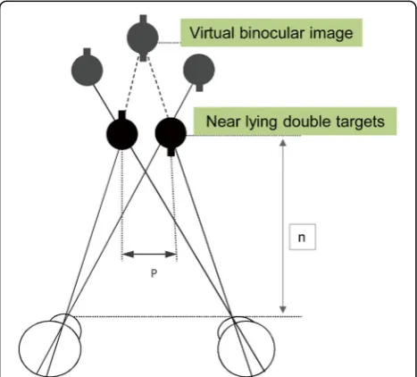
To induce the physiological diplopia, each patient was asked to look into the far distance (5 m). Then a near-lying target (in the distance 10 cm, i.e., not in the horop-ter curve) was presented. The presence (yes/no) of double virtual objects was fixed.
In the case of physiological diplopia, the registration of the disparate fusional reflex limits was performed. For this, a diploptic device (AVIS 01, Krasnogvardeec, Russia) was used, which facilitated investigation of the binocular interaction under natural conditions without the accommodation response, but with the different vergence load [37–41]. The first point of the meas-urement is 40 cm. Then the double targets were moved inwards until the virtual stereo image was ob-served (Fig. 1). It was a point of binocularity without vergence load.
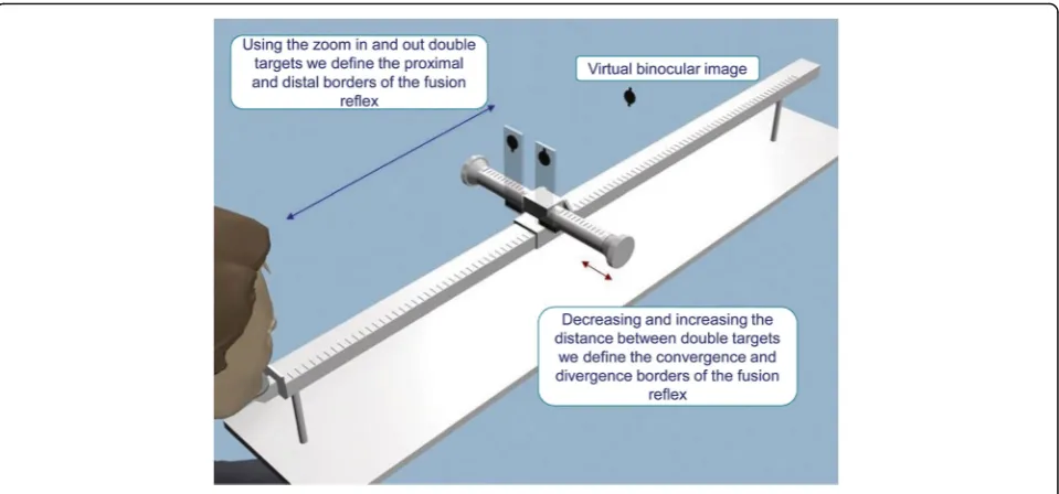
The change in the distance between the centers of the double near lying tests (p) and the distance from the eyes to the targets (n) while maintaining of the virtual binocular image perception makes it possible to define the fusional reflex limits in space. Then the targets were moved increasingly inward and outward to force the ver-gence response (Fig. 2). There were some points of max-imum convergence and divergence. The patient reported all the visual images which were recorded.
The disparate fusional reflex was analyzed using the following parameters:
1. The proximal fusion border (PF) and distal fusion border (DF) were determined while the double targets were approaching and moving away (Fig.2). The difference between these parameters
corresponds to the length of the binocularity field. 2. The convergence fusion border (CF) and the
divergence fusion border (DivF) were determined with the help of the decrease and the increase of the distance between the double targets (the point of the measurement is 40 cm from the eyes). The
difference between these parameters corresponds to the width of the binocularity field.
3. Finally, we performed the calculation of the binocularity field area (A) in cm2(Fig.3).
Statistical analysis
All data were analyzed with a spreadsheet application (Statistica ver. 8.0; StatSoft Inc., USA). The data were represented as the mean value ± standard deviation (Mean ± SD). The Shapiro-Wilk test was used for asses-sing of normality distribution. The statistical differences in measured values were analyzed using a t-test. The critical level of significance (p) upon the examination of statistical hypotheses was 0.05. The comparison analysis, the Pearson correlation analysis, and the logistic regres-sion models were done. The Pearson correlation coeffi-cient with absolute values equal to or greater than 0.7 with p< 0.001 was accepted as the close relation. The
discriminant function analysis (DFA) was used for the selection of the analytes that maximally discriminated the studied groups. The DFA was built in a step-wise manner after direct standardization. The final discrimin-atory power of each analyte was characterized by a par-tial Wilk’s Lambda coefficient; 1.0 (no discriminatory power) to 0.0 (perfect discriminatory power). The Maha-lanobis distance D2 between centroid values for each group was measured.
Results
The baseline features of the study population were sum-marized in Table 1. The mean patient age was 22.3 ± 3.2 years in the first group, and 52.4 ± 2.2 years in the second group. The groups did not differ in gender, spherical refraction equivalent, eye globe axial length, oculomotor status. In the first (control) group, the mean accommodation amplitude was 6.93 ± 1.12 D (minimum
Fig. 2Method of Assessing the Disparate Fusion Reflex Borders
Fig. 3Calculation of the Binocularity Field Area
Table 1Studied Groups Descriptive Statistics (M ± SD)
Characteristics Control Presbyopia P-value
Age, years 22.3 ± 3.2 52.4 ± 2.2 0.0001
Female: Male 15:15 15:15 –
Mean spherical equivalent of refraction, D
0.2 ± 0.1 0.2 ± 0.2 1.00
Axial length, mm 23.5 ± 0.5 23.5 ± 0.4 1.00
Keratometry, ax 90°, D 43.2 ± 1.3 43.3 ± 1.1 0.61
Keratometry, ax 180°, D 43.0 ± 1.1 43.1 ± 1.1 0.62
Amplitude of accommodation, D 6.93 ± 1.12 1.99 ± 0.89 0.0001
Mean prism equivalent of distance heterophoria, PD
−1.2 ± 0.2 −1.1 ± 0.3 0.88
5.75 D, maximum 10.0 D). All patients with presbyopia had a decreased accommodation: the mean accommoda-tion amplitude was 1.99 ± 0.89 D (minimum 0.5 D, maximum 4.0 D).
Intraocular anatomy and optics
The presbyopia formation is characterized by a change in an intraocular anatomy. Significant differences were de-tected in the anterior-posterior size of the lens – from 3.73 ± 0.23 to 4.41 ± 0.21 (p= 0.0001). The optical density (light transmission coefficient) of the lens increased in the nuclear area from 15.5 ± 1.2 to 26.6 ± 3.4% (p = 0.0001), and in the cortical layers –from 9.1 ± 0.9 to 10.8 ± 1.3% (p= 0.03). The change of the intraocular anatomy was expressed as: the decrease of the ciliary body thickness (in the inner top projection) from 0.82 ± 0.10 to 0.63 ± 0.11 mm (p= 0.001), the increase of the distance between the trabecula and the ciliary processes from 0.79 ± 0.10 to 1.02 ± 0.11 mm (p = 0.001), and the shortage of the front portion Zinn ligament length from 1.23 ± 0.31 to 1.04 ± 0.26 mm (p= 0.002).
The variation of both static and dynamic components of the optical physiological system was established in the eyes of presbyopic patients. This was evidenced by a sig-nificant increase of the total root mean square wavefront errors (from 0.13 ± 0.04 to 0.17 ± 0.05μm in pupil diam-eter 3 mm,p= 0.0001) and the corneal root mean square wavefront aberrations. In patients with presbyopia, an increase in the Zernike coefficients of the corneal spherical aberration Z40from 0.17 ± 0.05 to 0.23 ± 0.06μm
(p= 0.0001) was observed. Also, a significant decrease of the pupil excursion was found. The photopic pupil diameter decreased from 3.81 ± 0.76 to 3.35 ± 0.78 mm (p = 0.0001) whereas the mesopic pupil diameter de-creased from 6.47 ± 0.56 to 5.50 ± 0.94 mm (p = 0.0001).
Monocular visual characteristics
In the next step, a comprehensive study of the sensory activity of the visual system was carried out. The patients with presbyopia had a decrease not only in accommoda-tion amplitude and uncorrected near visual acuity but also in the most of the visual reception parameters (Table 2).
A significant decrease of the contrast sensitivity in low and high spatial frequency ranges, the average values of the b-wave ERG maximum amplitude, the flicker fusion threshold and an increase in the a-wave and b-wave ERG maximum latency were found in patients with presbyopia.
Binocularity
The change in the perception Lang stereo tests was not significant.
A study of binocular cooperation showed an inhibition of the near physiological diplopia in 20% of the patients with presbyopia.
There were multiple changes. These include a signifi-cant distortion of the binocular interaction zone with the reduction of total binocularity area, shift in the space towards the near focal point, and the fusion neutralization in the convergence zone. The changes in the proximal, distal and convergence fusion borders were determined (Table 3).
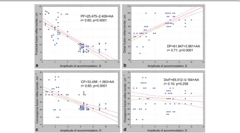
The regressions between disparate fusion reflex limits and accommodation amplitude are shown in Fig. 4. In the first (control) group, the mean area of binocularity field was 365.6 ± 45.1 cm2 (minimum 280 cm2, maximum
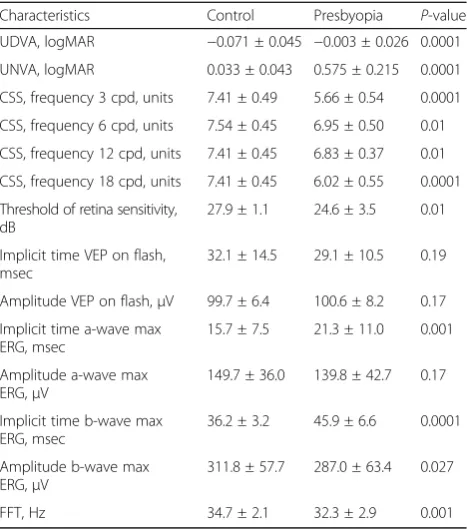
Table 2Comparisons of visual characteristics in studied groups (M ± SD)
Characteristics Control Presbyopia P-value
UDVA, logMAR −0.071 ± 0.045 −0.003 ± 0.026 0.0001
UNVA, logMAR 0.033 ± 0.043 0.575 ± 0.215 0.0001
CSS, frequency 3 cpd, units 7.41 ± 0.49 5.66 ± 0.54 0.0001
CSS, frequency 6 cpd, units 7.54 ± 0.45 6.95 ± 0.50 0.01
CSS, frequency 12 cpd, units 7.41 ± 0.45 6.83 ± 0.37 0.01
CSS, frequency 18 cpd, units 7.41 ± 0.45 6.02 ± 0.55 0.0001
Threshold of retina sensitivity, dB
27.9 ± 1.1 24.6 ± 3.5 0.01
Implicit time VEP on flash, msec
32.1 ± 14.5 29.1 ± 10.5 0.19
Amplitude VEP on flash,μV 99.7 ± 6.4 100.6 ± 8.2 0.17
Implicit time a-wave max ERG, msec
15.7 ± 7.5 21.3 ± 11.0 0.001
Amplitude a-wave max ERG,μV
149.7 ± 36.0 139.8 ± 42.7 0.17
Implicit time b-wave max ERG, msec
36.2 ± 3.2 45.9 ± 6.6 0.0001
Amplitude b-wave max ERG,μV
311.8 ± 57.7 287.0 ± 63.4 0.027
FFT, Hz 34.7 ± 2.1 32.3 ± 2.9 0.001
UDVA= uncorrected distance visual acuity;UNVA= uncorrected near visual acuity;CSS= contrast spatial sensitivity;VEP= visual evoked potentials;ERG= electroretinogram;FFT= flicker fusion threshold
Table 3Comparisons of Fusion Reflex Characteristics in Study Groups (M ± SD)
Characteristics Control Presbyopia P-value
Proximal fusion border, cm 5.23 ± 1.58 18.90 ± 6.85 0.0001
Distal fusion border, cm 90.05 ± 7.35 69.20 ± 12.21 0.0001
Convergence fusion border, 10−1cm
24.83 ± 6.14 30.31 ± 6.91 0.0001
Divergence fusion border, 10−1cm
63.38 ± 3.55 58.21 ± 5.61 0.056
Length of binocularity field, cm
84.78 ± 7.75 50.82 ± 16.05 0.0001
Width of binocularity field, 10−1cm
41.88 ± 3.35 30.39 ± 6.34 0.0001
456 cm2). All patients with presbyopia had a decreased fusion ability with the mean area of binocularity field at 174.4 ± 87.7 cm2(minimum 48 cm2, maximum 385 cm2). The regression between binocularity field area and accom-modation amplitude is shown in Fig. 5.
It is interesting to note that 77% patients with presby-opia had binocular suppression in some grade, while 6% of patients had extreme decrease in the binocularity field area (Fig. 6).
Intrasystem interactions
The multivariate kinds of statistical analyses were used for the understanding of the vision reception transform-ation in presbyopia formtransform-ation.
A Pearson correlation analysis of visual system parameters was made. The comparison of the correl-ation Pleiades (correlcorrel-ations with P-value equal or less than 0.001) within Control and Presbyopia Groups re-vealed a reduction in the strength of most relation-ships. The correlation Pleiades are represented in Fig. 7, where the positive correlations were shown as red arrows and the negative correlations as blue ones. It is evident that young people without presbyopia have a much larger number of interdependencies between structural and functional indicators of the visual system than in patients with presbyopia. In young people, 25 close relationships were established, but in patients with presby-opia, there were only 6 such relationships. Instead of destroyed relationships, there was a new correlation be-tween the width of the binocularity field and the implicit time of visual evoked potentials (r=−0,7;p= 0.001).
In the forward stepwise discriminant analysis, eight in-dices were selected for 100% discrimination of studied groups. The matrix of most informative variables for dis-crimination is represented in Table 4.
The separation between groups is not only due to the accommodation state but also to some other significant
Fig. 4Regressions between Fusion Reflex Borders and Accommodation Amplitude.arelationship between proximal fusion border and accommodation amplitude,brelationship between distal fusion border and accommodation amplitude,crelationship between convergence fusion border and accommodation amplitude,drelationship between divergence fusion border and accommodation amplitude
changes in the structural and functional parameters. The in-dicators of the tolerance show that all features are orthogonal and their contributions to the separation do not overlap.
The results of the discriminant analysis showed that the fusional ability made the high contribution in the separation of two groups. At the same time, the contri-bution of other sensory parameters in the division was less expressed.
Discussion
This research aimed to describe the transformation of the visual system functional organization during the presbyopia formation. The results of the study broaden our understanding of the presbyopia mechanisms. It was found that the structural and functional state of the vis-ual system in middle-aged patients with presbyopia is significantly different from young people.
The reduction of the accommodation is significant, but not the only sign of visual transformation in patients with
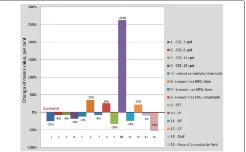
presbyopia. The increase in the number of optical errors despite the pupil tour decrease worsens the conditions for the formation of the retinal image. The formation of pres-byopia is accompanied by the misalignment of visual sen-sory processing with varying degrees of functional defect severity (Fig. 8).
The decrease of the contrast sensitivity at frequencies below four cpd reflects the amplification of the visual system internal noise (at the level of receptive fields). Whereas the contrast sensitivity at high spatial frequen-cies is limited with optical parameters (aberrations, dif-fraction phenomenon or “sampling noise” of the visual image). The patients with presbyopia had the signs both internal noise and sampling noise (noise of signal pro-cessing). The change of temporal parameters of the vis-ual system at middle age patients indicates the initial deficiency of interactions between neurons and the signs of the central nervous system fatigue. These changes can be viewed as signs of aging.
Fig. 6Distribution of Patients Depending on the value of Binocularity Field Area
The new data about binocular visual system activity was established. The processes of binocular interaction inhibition make a significant contribution to the mis-alignment of the visual perception. The area of binocu-larity field, where the disparate fusion is possible, was reduced twice. The variation of proximal fusion limit and a decrease of the binocularity field area are more serious than the variation in other sensor parameters.
On the one hand, this phenomenon may be a result of age-related changes in the neurons. On the other hand, the inhibition process can be motivated by the desire to
liberate the body from the excessive flow of the visual information under the shortage of accommodation. The process of efferent synthesis is an active selection of in-formation aimed to release biologically significant flows and is formed in such integral efferent excitations that are required by the body in a given situation.
Our results correspond to the relevant studies. Granger-Donetti revealed that majority of presbyopes had disorders of binocular cooperation in some degree due to a decrease in the slow convergence [42]. The accommodation ampli-tude decrease is accompanied by the increase of vergence
Table 4Matrix of most informative variables for discrimination studied groups
Discriminant Function Analysis Summary
Step 8, Wilks’Lambda:0.02771, approx. F (9.74) = 288.46 Mahalanobis D2= 139.84,p< 0,0001
Variables Wilks’Lambda F-remove (1.74) p-level Tolerance
UNVA 0.068917 110.0151 0.000001 0.621721
Proximal border of fusion field 0.049343 57.7521 0.000001 0.667574
Amplitude of accommodation 0.040528 34.2152 0.000001 0.619955
Binocularity area 0.038421 28.5871 0.000001 0.567583
Coefficient corneal spherical aberration Z40 0.037609 26.4206 0.000002 0.545072
Pupil diameter in photopic conditions 0.033739 16.0866 0.000143 0.718888
Implicit time b-wave max ERG, msec 0.030783 8.1942 0.005462 0.803496
CSS, frequency 3 cpd, units 0.029672 5.2271 0.025101 0.850332
movement latency, the reduction of the vergence fusion and the speed of the fast vergence [43].
In this study, we analyzed data in people with the vis-ual system that mostly meets the ideal. Even in this situ-ation, 77% of presbyopia patients had binocularity shortage. In 6% of cases, there were profound signs of the deep inhibition processes. These data are likely use-ful in presbyopia surgery. The analysis of the surgery re-sults using monovision or multifocal optical strategies in patients with an extreme deficit of binocularity is the next step of research.
Conclusions
The presbyopia formation is accompanied by a significant reorganization of the visual system activity and the creation of the new visual processing interactions. It was revealed that 77% of the presbyopia patients had binocu-larity shortage. The overall reduction of binocubinocu-larity field area in extreme grade was seen in 6% of cases. These data may have implications for presbyopia surgery.
Acknowledgements
The authors are grateful to Isay M. Mikhalevich and Vladimir V. Malyshev for their help and advice.
Funding
The authors have no financial support and sponsorship.
Authors’contributions
OIR made substantial contributions to conception and design, analysis, and interpretation of data, total statistical analysis, AGS revised it critically for important intellectual content and supervision, TSM participated in data acquisition, drafted the manuscript. All authors read and approved the final manuscript.
Competing interests
The authors have no proprietary or commercial interests in the medical devices that are involved in this manuscript.
Author details
1Irkutsk branch of S. Fyodorov Eye Microsurgery Federal State Institution,
Irkutsk, Russian Federation.2Irkutsk State Medical University, Irkutsk, Russian Federation.
Received: 31 August 2016 Accepted: 4 January 2018
References
1. Belville JK, Smith RJ. Presbyopia surgery. New York: SLACK; 2006. 2. Hashemi H, Khabazkhoob M, Jafarzadehpur E, Mehravaran S, Emamian MH,
Yekta A, et al. Population-based study of presbyopia in Shahroud, Iran. Clin Exp Ophthalmol. 2012;40:863–8.
3. Hudson C. How to succeed with multifocal contact lenses. Optometry Today. 2011;51:2.
4. Varma R, Wang MY, Ying-Lai M, Donofrio J, Azen SP; Los Angeles Latino eye study group. The prevalence and risk indicators of uncorrected refractive error and unmet refractive need in Latinos: the Los Angeles Latino Eye Study. Invest Ophthalmol Vis Sci. 2008;49:5264–73.
5. Pallikaris IG. Presbyopia surgery. In: Pallikaris I, Plainis S, Charman WN, editors. Presbyopia: origins, effects, and treatment. Danvers: Slack; 2012. p. 141–2. 6. Nijkamp MD, Dolders MG, de Brabander J, van den Borne B, Hendrikse F,
Nuijts RM. Effectiveness of multifocal intraocular lenses to correct presbyopia after cataract surgery: a randomized controlled trial. Ophthalmology. 2004;111:1832–9.
7. Bellucci R. Multifocal intraocular lenses. Curr Opin Ophthalmol. 2005;16:33–7.
8. Chang DF. Mastering refractive IOLs. The art and science. Thorofare, NJ: SLACK; 2008.
9. Gierek-Ciaciura S, Cwalina L, Bednarski L, Mrukwa-Kominek E. A comparative clinical study of the visual results between three types of multifocal lenses. Graefes Arch Clin Exp Ophthalmol. 2010;248:133–40.
10. Cochener B, Lafuma A, Khoshnood B, Courouve L, Berdeaux G. Comparison of outcomes with multifocal intraocular lenses: a meta-analysis. Clin Ophthalmol. 2011;5:45–56.
11. Friedrich R. Intraocular lens multifocality combined with the compensation for corneal spherical aberration: a new concept of presbyopia-correcting intraocular lens. Case Rep Ophthalmol. 2012;3:375–83.
12. Lichtinger A, Rootman DS. Intraocular lenses for presbyopia correction: past, present, and future. Curr Opin Ophthalmol. 2012;23:40–6.
13. Calladine D, Evans JR, Shah S, Leyland M. Multifocal versus monofocal intraocular lenses after cataract extraction. Cochrane Database Syst Rev. 2012;9:CD003169.
14. Javitt JC, Steinert RF. Cataract extraction with multifocal intraocular lens implantation: a multinational clinical trial evaluating clinical, functional, and quality-of-life outcomes. Ophthalmology. 2000;107:2040–8.
15. Tan N, Zheng D, Ye J. Comparison of visual performance after implantation of 3 types of intraocular lenses: accommodative, multifocal, and monofocal. Eur J Ophthalmol. 2014;24:693–8.
16. Bassam A, Donnenfeld E. Bonus feature: IOL Explantation: indications and strategies for multifocal IOL explantation. Cataract and Refractive Surgery Today. 2011;4:18-20.
17. Bucci FA Jr. "Vaseline Vision Dysphotopsia" and Explantation of the ReSTOR Multifocal Implant. Invest Ophthalmol Vis Sci. 2007;48(13):3119.
18. Fernández-Buenaga R, Alio JL, Muñoz-Negrete FJ, Barraquer Compte RI, Alio-del Barrio JL. Causes of IOL explantation in Spain. Eur J Ophthalmol. 2012;22:762–8.
19. Shimizu K, Ito M. Dissatisfaction after bilateral multifocal intraocular lens implantation: an electrophysiology study. J Refract Surg. 2011;27:309–12. 20. Woodward MA, Randleman JB, Stulting RD. Dissatisfaction after multifocal
intraocular lens implantation. J Cataract Refract Surg. 2009;35:992–7. 21. Timiras PS. Physiological basis of aging and geriatrics. 4th ed. Berkeley:
Informa Healthcare; 2007.
22. Glasser A, Campbell MC. Biometric, optical and physical changes in the isolated human crystalline lens with age in relation to presbyopia. Vision Res. 1999;39:1991–2015.
23. Croft MA, McDonald JP, Katz A, Lin TL, Lütjen-Drecoll E, Kaufman PL. Extralenticular and lenticular aspects of accommodation and presbyopia in human versus monkey eyes. Invest Ophthalmol Vis Sci. 2013;54:5035–48. 24. Croft MA, Kaufman PL. Role of the ciliary muscle and zonula in
accommodation and presbyopia. In: Pallikaris I, Plainis S, Charman WN, editors. Presbyopia: origins, effects, and treatment. Danvers: Slack; 2012. p. 69–76. 25. Anokhin PK. Biology and neurophysiology of the conditioned reflex and its
role in adaptive behavior. New York: Pergamon Press; 1974.
26. Meerson FZ. Adaptation, stress and prophylaxis. Berlin: Springer-Verlag; 1984. 27. Sudakov KV. The theory of functional systems: general postulates and
principles of dynamic organization (dedicated to the Anokhin Centenary). Integr Physiol Behav Sci. 1997;32:392–414.
28. Rozanova OI, Shchuko AG, Mikhalevich IM, Malyshev VV. [Regularities and mechanisms of visual perception transformation in presbyopia development]. Vestn Oftalmol. 2011;127:17–20.
29. Owsley C. Aging and vision. Vis Res. 2011;51:1610–22.
30. Ciuffreda KJ, Thiagarajan P. Presbyopia and the vergence system. In: Pallikaris I, Plainis S, Charman WN, editors. Presbyopia: origins, effects, and treatment. Danvers: Slack; 2012. p. 103–9.
31. Werner JS, Schefrin BE, Bradley A. Optics and vision of the aging eye. In: Bass M, Enoch M, Lakshminarayanan V, editors. Handbook of Optics. Vol. III. New York: McGraw Hill Inc; 2010.
32. Andersen GJ. Aging and vision: changes in function and performance from optics to perception. Wiley Interdiscip Rev Cogn Sci. 2012;3:403–10. 33. Laframboise S, De Guise D, Faubert J. Effect of aging on stereoscopic
interocular correlation. Optom Vis Sci. 2006;83:589–93.
34. Shafiee D, Jafari AR, Shafie AA. Correlation between Interpupillary Distance and stereo acuity. Bull Env Pharmacol Life Sci. 2014;3(12):26–33.
36. Pavlin CJ, Foster FS. Ultrasound biomicroscopy of the eye. New York: Springer-Verlag; 1995.
37. Mogilev LN. Mechanisms of spatial vision. Leningrad: Nauka; 1982. (in Russian)
38. Rabitchev IE. The mechanisms of binocular function correction at different forms of strabismus. J Fr d’Orthoptique. 1998;30:153–9. (in French) 39. Hofstetter HW. Dictionary of Visual Science and Related Clinical Terms.
Boston: Butterworth-Heinemann; 2000.
40. Rychkova SI, Ninio J. Paradoxical fusion of two images and depth perception with a squinting eye. Vision Res. 2009;49:530–5. 41. By Shchuko AG, Malyshev VV. Theoretical and clinical binarimetry.
Novosibirsk: Nauka; 2006. (in Russian)
42. Granger-Donetti B. Central suppression at near vision in presbyopic subjects. Perception. 2006;35(Suppl 1):172.
43. Ciuffreda KJ. Accommodation, pupil, and presbyopia. In: Benjamin WJ, editor. Borish’s clinical refraction. St. Louis: Buterworth Heinemann; 2006. p. 93–144.
• We accept pre-submission inquiries
• Our selector tool helps you to find the most relevant journal
• We provide round the clock customer support
• Convenient online submission
• Thorough peer review
• Inclusion in PubMed and all major indexing services
• Maximum visibility for your research
Submit your manuscript at www.biomedcentral.com/submit