O
RIGINAL
A
RTICLES
1182
Emergence of enteropathogenic
Escherichia coli
Escherichiacoli is named after Theodor Escherich (1857 - 1911), a German paediatrician, who identified this bacterium in the stools of healthy children and named it Bacterium coli commune. Escherich showed that E. coli could cause infection when inoculated parenterally into rabbits. On the other hand, the concept that E. coli could also cause disease in the gastrointestinal tract took much longer to gain acceptance.
Investigations during the early part of the 20th century in France, Denmark and the USA provided sound epidemiological evidence that E. coli was associated with diarrhoea in children.1
These observations were supported by reports from Adam and Goldschmidt in Germany who showed that certain strains of E. coli which were antigenically related to each other were linked to outbreaks of severe infantile gastroenteritis.1 Despite
this body of evidence, it was only after Bray and colleagues at the Hillingdon Hospital near London reported that a particular strain of E. coli, Bacterium coli neapolitanum, was significantly associated with diarrhoea in children, that the notion of diarrhoea induced by E. coli gained credence (Fig. 1).2
Bray’s seminal studies were followed by similar reports from Aberdeen and London which confirmed the epidemiological link between particular strains of E. coli and diarrhoea.1
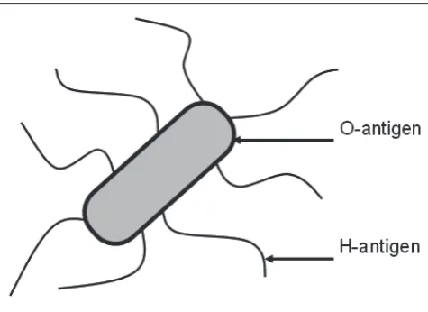
A major breakthrough in this area stemmed from the work of Kaufmann and colleagues, who established a serotyping scheme for E. coli based on the bacterium’s somatic (O) and flagellar (H) antigens (Fig. 2). This important work made it possible to compare the relatedness of E. coli isolates from different outbreaks and showed that diarrhoea-associated isolates of E. coli from Germany, England and Scotland that had been reported previously, belonged to a limited number of O-serogroups, in particular O111 and O55.3 These early
reports were followed by a series of studies, mainly in the USA and Japan, which showed that strains of E. coli that had been associated with outbreaks of severe diarrhoea in children caused diarrhoea when ingested by adult volunteers.1 In 1955,
Neter et al. coined the term ‘enteropathogenic’ E. coli (EPEC) to indicate those strains of E. coli that had been associated with outbreaks of childhood diarrhoea.4 Some years later,
Neter summarised the evidence favouring the pathogenicity of EPEC as follows: (i) during institutional outbreaks, a single O-serogroup has been found in the majority of affected infants,
The rise and rise of enteropathogenic
Escherichia
coli
Roy M Robins-Browne
Department of Microbiology and Immunology, University of Melbourne, and Mur-doch Children’s Research Institute, Royal Children's Hospital, Victoria, Australia Roy M Robins-Browne, MB BCh, PhD, DTM&H, FCPath (SA), FRCPath, FRCPA
First described in 1885, Escherichiacoli gradually achieved recognition as a cause of diarrhoea. Strains of E. coli
which belonged to a limited number of O-serogroups and had been associated with outbreaks of diarrhoea in hospitalised children were designated ‘enteropathogenic’
E. coli (EPEC) to distinguish them from E. coli strains that cause other types of infection. The discovery that some strains of E. coli can produce toxins or invade epithelial cells in a similar fashion to established pathogens, such as Vibrio cholerae and Shigella species, shed new light on
E. coli virulence. As EPEC do not exhibit either of these properties, however, their pathogenicity was brought into question. Further studies revealed that EPEC constitute a distinctive group of pathogenic bacteria that display characteristic adherence to cultured epithelial cells and produce distinctive histopathological changes, termed ‘attaching-effacing lesions’, in the intestinal epithelium. The ability to evoke these lesions, which are essential for virulence, is associated with a series of linked genes, known as the locus for enterocyte effacement, on the bacterial chromosome. In addition, the pathogenicity of some EPEC strains is associated with the presence of a plasmid-encoded, bundle-forming pilus. Naturally occurring strains of EPEC which lack this pilus are known as atypical EPEC and are an emerging cause of diarrhoea throughout the world.
[image:1.637.327.568.603.771.2]O
RIGINAL
A
RTICLES
and the serogroup involved has usually been present as the predominant aerobic bacterium cultured from the stools of sick infants; (ii) EPEC strains have been recovered far less frequently from the stools of healthy infants, older children and adults than from those of infants with diarrhoea; (iii) volunteers develop diarrhoea when fed strains identified by serogroup alone; (iv) serogroup-specific antibodies may be detected in the serum of a proportion of affected patients and volunteers; (v) antibiotics that eliminate EPEC from the stool bring relief from illness in affected infants and prevent the development of illness in susceptible infants; and (vi) extensive bacteriological and limited virological investigations of outbreaks of E. coli diarrhoea have failed to show a strong association with any other microbial agent.5
Emergence of other varieties of
diarrhoeagenic
E.
coli
In the years that followed Neter’s review, interest in EPEC and infantile diarrhoea in general appeared to wane, probably because of a reduction in the number and impact of community and nursery outbreaks of life-threatening diarrhoea that had been a prominent feature of paediatric practice in industrialised countries during the 1930s, 1940s and early 1950s.1 The role
of E. coli as an enteropathogen became firmly established, however, by the discovery that some E. coli strains secreted a novel heat-stable enterotoxin and/or a heat-labile enterotoxin resembling cholera toxin, which accounted for their virulence. This variety of E. coli came to be known as enterotoxigenic
E. coli (ETEC) and was shown to cause watery diarrhoea in volunteers.6 Also, around this time, yet another pathogenic
variety of E. coli was identified. This type of E. coli is referred to as enteroinvasive E. coli (EIEC) and causes dysentery that
is indistinguishable from that due to Shigella species, with which it shares a large number of virulence determinants. Also, during the early 1970s, Ruth Bishop, Ian Holmes and colleagues at Melbourne’s Royal Children’s Hospital and the University of Melbourne identified rotavirus, which was the first virus demonstrated to cause infantile gastroenteritis.7
At this time, I was a registrar in the School of Pathology studying microbiology under the tutelage of Hendrik Koornhof. Hendrik thought that we should look for these newly described varieties of E. coli and rotavirus, as well as traditional pathogens, in children with acute gastroenteritis in Soweto, where infantile diarrhoea was still a major health concern. In collaboration with Barry Schoub, we established the extremely laborious assays used at the time to identify rotaviruses, ETEC and EIEC.8 We then applied these assays
to an examination of faecal samples obtained from patients attending Baragwanath Hospital for management of acute gastroenteritis. To our surprise, we found that the most frequent pathogen identified in the stools of children with gastroenteritis was not ETEC or EIEC, but EPEC.9 These
findings were subsequently confirmed in a second study.10
At this time, no virulence factors of EPEC were known, and they had to be identified by determination of O-serogroup and their failure to produce known enterotoxins and to exhibit
Shigella-like invasiveness. Because EPEC as a group lacks these virulence determinants, some authorities expressed doubts as to their pathogenicity.11 These views were supported by
studies which showed that even those EPEC strains which had been associated with outbreaks of diarrhoea and had caused diarrhoea in volunteers lacked known virulence determinants, causing some investigators to suggest that these strains were in fact ETEC or EIEC which had lost the capacity to produce enterotoxins or invade epithelial cells during storage.1 Even
an elegantly designed, volunteer study reported by Levine et al.12 around this time,which confirmed the pathogenicity of
outbreak-associated strains of EPEC that had been identified by O-serogroup alone and which lacked the virulence determinants of ETEC and EIEC, failed to convince many of the sceptics.13
Identification of EPEC-specific
virulence determinants
The observation that EPEC must colonise the small intestine in order to cause diarrhoea14 suggested that adhesive factors may
contribute to the virulence of these bacteria. The suggestion was supported by studies which showed that EPEC adhere to tissue culture cells in vitro to a greater extent than other varieties of E. coli.15 In addition, most EPEC strains in
[image:2.637.72.310.147.320.2]O-serogroups, which had been classified as containing EPEC because of their association with epidemics of diarrhoea, adhered to epithelial cells in a distinctive pattern termed
O
RIGINAL
A
RTICLES
localised adherence (Fig. 3A).16 Investigations into the
mechanisms of this type of adherence revealed that adhesion is mediated by a distinctive fimbrial adhesin, termed bundle-forming pili (Bfp; Fig. 3B),17 which is encoded by plasmids
known as the EPEC adhesive factor (EAF).18 EPEC employ Bfp
to bind to epithelial cells and to each other, thus accounting for the localised adherence pattern of EPEC strains which produce these pili. Studies in volunteers have demonstrated that the EAF plasmid and Bfp themselves are essential virulence determinants of EPEC, and that bacteria which lack either of these are markedly attenuated.19,20
Studies of the pathology of EPEC infection in animal models and naturally infected children have shown that EPEC adhere to the intestinal epithelium in a distinctive manner that is characterised by bacteria intimately attached to the plasma membrane of enterocytes where the microvilli are absent (or effaced).21,22 In addition, at the sites where bacteria attach there
is evidence of cytoskeletal rearrangements resulting in changes to the morphology of the cell surface, giving rise to formations termed cups and pedestals (Fig. 4). Collectively, these changes are referred to as attaching-effacing lesions.21 Interestingly,
EPEC strains which lack Bfp or the entire EAF plasmid retain the ability to induce the formation of these lesions indicating that the EAF is not required for attaching-effacing capacity.23 A
series of elegant experiments, from Jim Kaper’s group, showed that the factors required for attaching-effacing capacity are encoded by genes co-located within a pathogenicity island, known as the locus for enterocyte effacement (LEE), which is completely absent from E. coli strains that are unable to produce attaching-effacing lesions, including ETEC and EIEC.24
The products of the approximately 40 genes which comprise the LEE act in concert to secrete proteins directly from the bacterial cytoplasm through a narrow protein channel, resembling a microsyringe, through the plasma membrane of the host cell into its cytoplasm (Fig. 5).25 One of the proteins
secreted in this way is called the translocated intimin receptor or Tir, because it acts as a receptor for a surface adhesive protein of EPEC, known as intimin. Once Tir has bound to intimin it acts as an anchor for host cytoskeletal proteins, thus completing the formation of the attaching-effacing lesions.26
A series of volunteer studies and animal experiments using deletion mutants of human EPEC and analogous animal pathogens (such as rabbit-specific EPEC and Citrobacter rodentium, an attaching-effacing pathogen of mice) have shown that intimin, Tir and almost all other LEE-encoded proteins are required for virulence.27-30
Implications and applications of
research findings
[image:3.637.327.569.145.506.2]Recent advances in our understanding of EPEC biology and virulence have led to EPEC being re-defined. The revised definition is based primarily on the presence of the LEE
Fig. 3. A: Light micrograph showing E. coli attached to HeLa cells in a localised pattern (arrows) (Giemsa stain). B: Electron micrograph showing bundle-forming pili aggregated together into rope-like bundles (arrow).
[image:3.637.326.571.556.734.2]O
RIGINAL
A
RTICLES
pathogenicity island, which is a marker of attaching-effacing capacity. Only one other pathogenic variety of E. coli may carry LEE. This is enterohaemorrhagic E. coli or EHEC, which was first reported from the USA in 1982 and has subsequently been identified in a large number of countries worldwide.31
Apart from diarrhoea, EHEC also cause haemorrhagic colitis and the haemolytic uraemic syndrome.31 In the laboratory,
EHEC can be distinguished from EPEC by its ability to secrete one or more Shiga toxins (also known as Shiga-like toxins and Verotoxins).32 Given the recent emergence of EHEC and its
close genetic relatedness to EPEC it seems likely that EHEC evolved from EPEC in part by acquiring bacteriophages which carry the structural genes for the production of Shiga toxins.33
The re-definition of EPEC means that we no longer have to rely on O-serogrouping to identify these bacteria, although serotyping remains a useful epidemiological tool to track outbreaks of infection. Currently, identification of EPEC relies upon the demonstration of attaching-effacing capacity using tissue culture cells,34 or more commonly by detection of the eae
gene, which encodes intimin and is a marker of the presence of the LEE. Distinguishing EPEC from EHEC, which can also carry intimin, requires a demonstration of the absence of Shiga toxin production and/or the genes which encode these toxins. Alternatively, one can look for the presence of bundle forming pili or their genes. We and others now routinely use these assays, usually in the form of multiplex polymerase chain reactions, to identify EPEC-specific genes and those of other
varieties of diarrhoeagenic E. coli in patients with diarrhoea.35,36
We have also used these assays to determine if E. coli strains which were isolated during the 1940s and 1950s that were labelled ‘enteropathogenic’ were truly EPEC, as defined currently, and showed that most were.37
One drawback of using Bfp as an identifying characteristic of EPEC is that there is a subtype of EPEC, known as atypical EPEC, which is naturally deficient in Bfp.38 Although the role of
atypical EPEC as a cause of diarrhoea is controversial, evidence is accumulating from widespread geographical regions that these bacteria are indeed pathogens which are particularly associated with the development of post-infectious persistent diarrhoea, i.e. diarrhoea lasting more than 14 days.35,39
Summary and conclusions
The suggestion that E. coli, a normal inhabitant of the intestinal tract of healthy humans, could also cause diarrhoea took several decades to gain credence. At the time, these varieties of diarrhoea-associated E. coli were designated ‘enteropathogenic’ to distinguish them from E. coli strains that were responsible for urinary tract infection, wound infection, meningitis, septicaemia, etc. Initially, EPEC were identified by serotyping. The emergence of ETEC and EIEC, which had readily identifiable virulence determinants at a time when EPEC did not, brought the pathogenicity of EPEC into question. Nevertheless, epidemiological studies including some in South Africa performed under the guidance of Hendrik Koornhof, suggested that EPEC identified by serotyping alone was significantly associated with diarrhoea.
Today, a considerable amount is known about the molecular pathogenesis of EPEC infections, although the mechanism by which EPEC cause diarrhoea is still unclear. EPEC are now recognised as a major cause of acute and persistent diarrhoea in children in less developed countries and a significant contributor to premature childhood mortality. EPEC are also the likely progenitor of EHEC, an important food-borne pathogen in many industrialised countries worldwide. The recent identification of a new subtype of EPEC, termed atypical EPEC, as a cause of persistent diarrhoea in children, has added another chapter to the EPEC story. Bioinformatic analysis of the genome of different E. coli strains points to their genetic plasticity and propensity to transfer genetic material between individual strains and other infectious agents, suggesting that new pathogenic varieties of E. coli will continue to arise in future.40
The author wishes to acknowledge that work in his laboratory is supported by research grants from the Australian National Health and Medical Research Council and the Australian Research Council.
[image:4.637.68.312.144.393.2]The author has no conflict of interest with respect to the research reported here.
Fig. 5. Schematic representation of the process employed by
O
RIGINAL
A
RTICLES
References
1. Robins-Browne RM. Traditional enteropathogenic Escherichiacoli of infantile diarrhea. Rev Infect Dis 1987; 9: 28-53.
2. Anonymous. Bray’s discovery of pathogenic Esch. coli as a cause of infantile gastroenteritis. Arch Dis Child 1973; 48: 923-926.
3. Kauffmann F, Dupont A. Escherichiacoli strains from infantile epidemic gastro-enteritis. Acta Pathol Microbiol Scand 1950; 27: 552-564.
4. Neter E, Westphal O, Luderitz O, Gino RM, Gorzynski EA. Demonstration of antibodies against enteropathogenic Escherichiacoli in sera of children of various ages. Pediatrics 1955; 16: 801-808.
5. Neter E. Enteritis due to enteropathogenic Escherichiacoli. Am J Dig Dis 1965; 10: 883-886. 6. DuPont HL, Formal SB, Hornick RB, et al. Pathogenesis of Escherichiacoli diarrhea. N Engl J
Med 1971; 285: 1-9.
7. Bishop RF, Davidson GP, Holmes IH, Ruck BJ. Virus particles in epithelial cells of duodenal mucosa from children with acute non-bacterial gastroenteritis. Lancet 1973; 2: 1281-1283. 8. Schoub BD, Jacobs YR, Robins-Browne RM, Koornhof HJ, Lecatsas G, Prozesky OW.
Experimental techniques in the determination of aetiology of acute infantile gastroenteritis. S Afr J Med Sci 1976; 41: 213-219.
9. Freiman I, Hartman E, Kassel H, et al. A microbiological study of gastro-enteritis in Black infants. S Afr Med J 1977; 52: 261-265.
10. Robins-Browne RM, Still CS, Miliotis MD, et al. Summer diarrhoea in African infants and children. Arch Dis Child 1980; 55: 923-928.
11. Gangarosa EJ, Merson MH. Epidemiologic assessment of the relevance of the so-called enteropathogenic serogroups of Escherichiacoli in diarrhoea. N Engl J Med 1977; 296: 1210-1213.
12. Levine MM, Berquist EJ, Nalin DR, et al. Escherichiacoli strains that cause diarrhoea but do not produce heat-labile or heat-stable enterotoxins and are non-invasive. Lancet 1978; 1: 119-122.
13. Levine MM. Escherichiacoli that cause diarrhea: enterotoxigenic, enteropathogenic, enteroinvasive, enterohemorrhagic, and enteroadherent. J Infect Dis 1987; 155: 377-389. 14. Koya G, Kosakai N, Kono M, Mori M, Fukasawa Y. Observations on the multiplication of
Escherichiacoli O-111 B4 in the intestinal tract of adult volunteers in feeding experiments: the intubation study with Miller-Abbott’s double lumen tube. Jpn J Med Sci Biol 1954; 7: 197-203. 15. Cravioto A, Cross RJ, Scotland SM, Rowe B. An adhesive factor found in strains of Escherichia
coli belonging to the traditional infantile enteropathogenic serotypes. Curr Microbiol 1979; 3: 95-99.
16. Scaletsky ICA, Silva MLM, Trabulsi LR. Distinctive patterns of adherence of enteropathogenic Escherichiacoli to HeLa cells. Infect Immun 1984; 45: 534-536. 17. Girón JA, Ho ASY, Schoolnik GK. An inducible bundle-forming pilus of enteropathogenic
Escherichiacoli. Science 1991; 254: 710-713.
18. Baldini MM, Kaper JB, Levine MM, Candy DCA, Moon HW. Plasmid mediated adhesion in enteropathogenic Escherichiacoli. J Pediatr Gastroenterol Nutr 1983; 2: 534-538.
19. Bieber D, Ramer SW, Wu CY, et al. Type IV pili, transient bacterial aggregates, and virulence of enteropathogenic Escherichiacoli. Science 1998; 280: 2114-2118.
20. Levine MM, Nataro JP, Karch H, et al. The diarrheal response of humans to some classic serotypes of enteropathogenic Escherichiacoli is dependent on a plasmid encoding an enteroadhesiveness factor. J Infect Dis 1985; 152: 550-559.
21. Moon HW, Whipp SC, Argenzio RA, Levine MM, Giannella RA. Attaching and effacing activities of rabbit and human enteropathogenic Escherichiacoli in pig and rabbit intestines. Infect Immun 1983; 41: 1340-1351.
22. Rothbaum RJ, Partin JC, Saalfield K, McAdams AJ. An ultrastructural study of enteropathogenic Escherichiacoli infection in human infants. Ultrastruct Pathol 1983; 4: 291-304.
23. Knutton S, Lloyd DR, McNeish AS. Adhesion of enteropathogenic Escherichiacoli to human intestinal enterocytes and cultured human intestinal mucosa. Infect Immun 1987; 55: 69-77. 24. McDaniel TK, Jarvis KG, Donnenberg MS, Kaper JB. A genetic locus of enterocyte effacement
conserved among diverse enterobacterial pathogens. Proc Natl Acad Sci USA 1995; 92: 1664-1668.
25. Frankel G, Phillips AD, Rosenshine I, Dougan G, Kaper JB, Knutton S. Enteropathogenic and enterohaemorrhagic Escherichiacoli: more subversive elements. Mol Microbiol 1998; 30: 911-921.
26. Campellone KG, Leong JM. Tails of two Tirs: actin pedestal formation by enteropathogenic E. coli and enterohemorrhagic E. coli O157:H7. Curr Opin Microbiol 2003; 6: 82-90.
27. Deng W, Puente JL, Gruenheid S, et al. Dissecting virulence: systematic and functional analyses of a pathogenicity island. Proc Natl Acad Sci USA 2004; 101: 3597-3602. 28. Donnenberg MS, Tacket CO, James SP, et al. Role of the eaeA gene in experimental
enteropathogenic Escherichiacoli infection. J Clin Invest 1993; 92: 1412-1417.
29. Marchès O, Nougayrede JP, Boullier S, et al. Role of Tir and intimin in the virulence of rabbit enteropathogenic Escherichiacoli serotype O103:H2. Infect Immun 2000; 68: 2171-2182. 30. Tacket CO, Sztein MB, Losonsky G, et al. Role of EspB in experimental human
enteropathogenic Escherichiacoli infection. Infect Immun 2000; 68: 3689-3695.
31. Karmali MA. Infection by Shiga toxin-producing Escherichiacoli: an overview. Mol Biotechnol 2004; 26: 117-122.
32. Robins-Browne RM, Hartland EL. Escherichiacoli as a cause of diarrhea. J Gastroenterol Hepatol 2002; 17: 467-475.
33. Feng P, Lampel KA, Karch H, Whittam TS. Genotypic and phenotypic changes in the emergence of Escherichiacoli O157:H7. J Infect Dis 1998; 177: 1750-1753.
34. Knutton S, Baldwin T, Williams PH, McNeish AS. Actin accumulation at sites of bacterial adhesion to tissue culture cells: basis of a new diagnostic test for enteropathogenic and enterohemorrhagic Escherichiacoli. Infect Immun 1989; 57: 1290-1298.
35. Nguyen RN, Taylor LS, Tauschek M, Robins-Browne RM. Atypical enteropathogenic Escherichiacoli infection and prolonged diarrhea in children. Emerg Infect Dis 2006; 12: 597-603.
36. Robins-Browne RM, Bordun A-M, Tauschek M, et al. Atypical enteropathogenic Escherichia coli: a leading cause of community-acquired gastroenteritis in Melbourne, Australia. Emerg Infect Dis 2004; 10: 1797-1805.
37. Robins-Browne RM, Yam WC, O’Gorman LE, Bettelheim KA. Examination of archetypal strains of enteropathogenic Escherichiacoli for properties associated with bacterial virulence. J Med Microbiol 1993; 38: 222-226.
38. Trabulsi LR, Keller R, Gomes TAT. Typical and atypical enteropathogenic Escherichiacoli. Emerg Infect Dis 2002; 8: 508-513.
39. Afset JE, Bevanger L, Romundstad P, Bergh K. Association of atypical enteropathogenic Escherichiacoli (EPEC) with prolonged diarrhoea. J Med Microbiol 2004; 53: 1137-1144. 40. Robins-Browne RM. The relentless evolution of pathogenic Escherichiacoli. Clin Infect Dis