Comparative effects of AT and GO sequence
selective DNA minor groove binding agents.
Submitted by Stephen M. Forrow for the degree of PhD at
the University of London.
ProQuest Number: 10017761
All rights reserved
INFORMATION TO ALL USERS
The quality of this reproduction is dependent upon the quality of the copy submitted.
In the unlikely event that the author did not send a complete manuscript and there are missing pages, these will be noted. Also, if material had to be removed,
a note will indicate the deletion.
uest.
ProQuest 10017761
Published by ProQuest LLC(2016). Copyright of the Dissertation is held by the Author.
All rights reserved.
This work is protected against unauthorized copying under Title 17, United States Code. Microform Edition © ProQuest LLC.
ProQuest LLC
789 East Eisenhower Parkway P.O. Box 1346
Abstract.
The DNA sequence preference of a series of DNA minor groove binding analogues of distamycin, containing from one to four imidazole moieties is described. Only the di- and the triimidazole analogues produced strong footprints and a detailed densitometric analysis revealed that the preferred binding site of the diimidazole compound ARI 114 was at 5’-(G.C)3(A.T), whereas the strongest footprinting sites for the triimidazole compound, ARI 144, was within two occurrences of the sequence 5’-TCGGGCT-3’. The analysis also showed that these two compounds bind to different sequences on the same DNA fragment, even though they differ by only one imidazole unit.
An attempt was made to develop an assay for the binding sites of minor groove binders in a cellular environment. Although not entirely successful, the results from experiments involving a tag polymerase stop assay indicate that the DNA binding sites of some minor groove binders may be determined at doses lower than those required with existing techniques.
The G.G selective di and triimidazole compounds were compared to two A.T selective minor groove binders, distamycin and netropsin, for their ability to alter the distribution of guanine N7 monoalkylation products produced by three representative nitrogen mustards. All four of the ligands were capable of producing quantitative and/or qualitative changes in the distribution of the alkylation adducts. Pretreatment of the DNA with netropsin, distamycin or ARI 114 resulted in significantly altered patterns of guanine N7 monoalkylation, with some adducts being suppressed whilst others were enhanced. In contrast, ARI 144 produced only a strong inhibition of monoalkylation, but was unable to produce any qualitative alteration in the pattern of adducts. A comparison of the binding sites of the ligands (deduced by DNA footprinting) with patterns of enhancement or suppression of adducts yielded no simple correlation.
restriction endonucleases chosen to have either a high A.T or a high G.C content at the core of their recognition sequences. As predicted, netropsin and distamycin but not ARI 114 or ARI 144 were able to inhibit DNA cleavage by
Eco RI and Eco RV. In contrast, neither ARI 114 or ARI 144 were very effective at inhibiting DNA cleavage by the G.C recognising enzymes Nru I and Bal I. Footprinting revealed that neither ARI 114 and ARI 144 bound at these enzyme recognition sequences under the restriction enzyme buffer conditions. DNA footprinting experiments showed that ARI 114 bound to two of three recognition sequences for the enzyme Fnu 4H1 in the fragment studied, and enzyme cleavage was shown to be inhibited only at these sites. ARI 144 bound strongly to two 5'-GGGCTC-3' sequences which are recognition sites for the enzyme
Ban II. In this case a clear stimulation of DNA cleavage by Ban II was observed over a wide dose range of ARI 144.
Index,
Title Page... 1
Abstract... 2
Index... 4
List of Figures and Tables... 9
Abbreviations... 14
Acknowledgements... 16
CHAPTER 1. Introduction. 1.1. DNA as a target for anticancer drug design... 17
1.2. Mechanisms of DNA interaction and covalent modification... 19
1.2.1. DNA base modifications and other covalent lesions... 20
DNA cross-links... 21
DNA strand breaks... 22
1.2.2. Non-covalent mechanisms of ligand interactions with DNA 23 Intercalation... 23
Groove binding... 23
1.3. Classes and properties of anticancer agents targeted against nucleic acids... 24
1.3.1. The alkylating agents... 24
Nitrogen mustards... 24
Nitrosoureas... 27
Methane sulphonate esters... 29
Aziridines... 29
Triazenes... 30
Pyrimidine analogues...33
Purine analogues... 34
1.3.4. Topoisomerase poisons... 35
epipodophyllotoxins...35
DNA intercalators... 36
1.3.5. Antitumour antibiotics... 38
Actinomycin D... 38
Mitomycin C... 39
Bleomycin... 39
1.4. Drug toxicity, drug resistance and the need for more selective antitumour agents...40
1.5. Alternative strategies for the therapeutic targeting of nucleic acids... 44
1.5.1. Antisense oligonucleotides... 44
1.5.2. Triple helix-forming oligonucleotides... 47
1.5.3. Ribozymes... 49
1.5.4. Peptide-nucleic acid chimeras... 50
1.6. Minor groove binders as potential anticancer agents... 51
1.6.1. The pyrrolo(1,4)benzodiazepines...54
1.6.2. Calicheamicin... 55
1.6.3. (+) CC-1065... 56
1.6.4. Berenil... 57
1.7 Pyrrole-amidine oligopeptide DNA minor groove binders and the development of the lexitropsins... 58
1.7.1. The mechanism of binding and the origin of the sequence specificity of netropsin and distamycin... 60
bound in the minor groove of DNA...72
1.9. Aims of the research... 74
CHAPTER 2. Materials and methods. 2.1. Materials... 76
DNA... 76
Enzymes... 76
Radioisotope... 78
Chemicals... 78
Buffers... 79
2.2. Methods... 81
2.2.1. Preparation of end labelled DNA... 81
Dephosphorylation of linearised DNA... 81
5'-end labelling of linearised dephosphorylated DNA... 82
Preparation and purification of singly end labelled DNA restriction fragments... 82
2.2.2. Generation of a purine specific marker lane... 84
2.2.3. DNA footprinting reactions... 84
2.2.4. Agarose gel electrophoresis... 85
2.2.5. Denaturing polyacrylamide gel electrophoresis of samples... 87
2.2.6. Autoradiography... 87
2.2.7. Densitometry... 87
2.2.8. Cell culture... 87
3.2. Materials and methods... 93
3.2.1. Minor groove binding ligands... 93
3.2.2. DNA... 93
3.2.3. Taq polymerase stop assay... 94
3.3. Results... 96
3.3.1. DNA footprinting results... 96
3.3.2. Taq polymerase stop assay... 102
3.4. Discussion... 105
CHAPTER 4. The effect of minor groove binders on major groove alkylation by nitrogen mustards... 4.1. Introduction... 108
4.2. Materials and methods... 112
4.2.2. Buffers... 112
4.2.3. DNA... 112
4.2.4. Guanine N7 alkylation reactions... 112
4.2.5. Piperidine treatment of alkylated DNA... 113
4.2.6. Growth inhibition studies... 113
4.3. Results... 114
4.3.1. Guanine N7 alkylation by nitrogen mustards... 114
4.3.2. The effect of minor groove binders and other DNA interacting agents on guanine N7 alkylation by nitrogen mustards... 117
Alkylation by L-phenylalanine mustard... 117
Alkylation by uracil mustard... 123
Alkylation of quinacrine mustard... 128
4.3.3. Growth inhibitory effects of nitrogen mustards in the presence of minor groove binders... 137
CHAPTER 5. The effect of minor groove binders on DNA cleavage by restriction endonucleases.
5.1. Introduction... 148
5.2. Materials and methods... 151
5.2.1. Materials... 151
5.2.2. Methods... 151
DNA cleavage inhibition assay... 151
DNA footprinting reactions... 154
5.3. Results... 155
5.3.1. Inhibition of Eco RV, Eco Rl, Bal\ and Nru I activities by minor groove binders... 155
5.3.2. DNA footprinting of minor groove binding ligands at Eco RV, Eco Rl, Bal I and Nru I sites... 163
5.3.3. Effects on restriction endonucleases of DNA binding by AR1114 and AR1144 at preferential sites... 168
Effects of AR1114 on DNA cleavage by Fnu 4HI... 168
Effects of ARI 144 on DNA cleavage by Ban II... 170
5.4. Discussion... 175
CHAPTER 6. General discussion... 183
Appendix; Base sequence of the 1-650 base pair region of pBR322 DNA... 192
List of Figures and Tables,
Figures.Chapter 1.
1.1. Illustration of the main mechanisms of drug:DNA
interactions... 18
1.2. Reactive centres within DNA... 21
1.3. The structures of four representative nitrogen mustards... 25
1.4. Reaction mechanism of the nitrogen mustards... 26
1.5. The structures of two representative nitrosoureas... 27
1.6. Proposed reaction mechanism of the nitrosoureas... 28
1.7. Busulphan... 29
1.8. The structures of two representative aziridines... 30
1.9. The structures of two representative triazines... 31
1.10. The structure of cisplatin and two of its' analogues... 32
1.11. The pyrimidine analogues 5 FU and Ara 0 ...33
1.12. The purine analogues 6-mercaptopurine and 6-thioguanine.. 34
1.13. The structures of the podophyllotoxin derivatives etoposide (VP 16) and teniposide (VM26)... 36
1.14 The structure of the DNA intercalating topoisomerase II poisons doxorubicin and daunomycin... 37
1.15. Actinomycin D... 38
1.16. Mitomycin 0 ...39
1.17. Bleomycin A2... 40
1.18. Common modifications of antisense oligonucleotide structure... 46
1.19. Comparative structures of DNA and the PNA's... 51
1.20. The hydrogen bond donating and accepting sites in the major and minor grooves of DNA... 52
1.22. The structure of the oligopeptide minor groove binders
netropsin and distamycin...59
1.23. First generation lexitropsins... 66
1.24. Proposed binding modes of netropsin and a first generation lexitropsin... 67
1.25. The structure of FCE 24517 (Tallimustine)...71
Chapter 2. 2.1. Diagrammatic representation of the MPE.Fe(ll) footprinting technique... 86
Chapter 3. 3.1. Structures of Lex 371, AR II 68, ARI 114, ARI 144, AR II 62 and MW 97-8-13... 90
3.2. Footprinting patterns produced by AR II 68, ARI 114 AR1144 and ARM 62... 97
3.3. Footprints generated by ARI 114 and ARI 144... 98
3.4. DNA binding sites of AR1114... 100
3.5. DNA binding sites of AR1144... 100
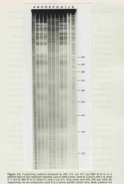
3.6. Footprints produced by ARI 114, Lex 371 and MW 97-8-13... 101
3.7. Comparison of DNA footprints with sites of inhibition of taq polymerase by netropsin... 103
Chapter 4. 4.1. Structures of the four nitrogen mustards used in chapter 4... 109
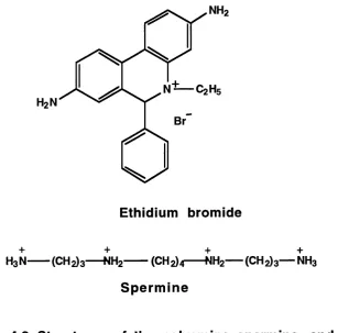
4.2. Structures of ethidium bromide and spermine... 110
4.4. Densitometric scans of figure 4.3... 116 4.5. The effect of various DNA affinity binders on guanine
N7 alkylation by L-PAM... 118 4.6. Densitometric scans of figure 4.5... 119 4.7. The effects of A.T and G.C selective minor groove
binders on the patterns of guanine N7 alkylation by
L-PAM... 121 4.8. Densitometric scans of figure 4.7... 122 4.9. The effect of various DNA affinity binders on guanine
N7 alkylation by uracil mustard... 124 4.10. Densitometric scans of figure 4.9... 125 4.11. The effects of A.T and G.G selective minor groove
binders on the patterns of guanine N7 alkylation by
uracil mustard... 126 4.12. Densitometric scans of figure 4.11... 127 4.13. The effect of various DNA affinity binders on guanine
N7 alkylation by quinacrine mustard... 129 4.14. Densitometric scans of figure 4.13... 130 4.15. The effects of A.T and G.C selective minor groove
binders on the patterns of guanine N7 alkylation by
quinacrine mustard... 131 4.16. Densitometric scans of figure 4.15... 132 4.17. Schematic representation of the patterns of guanine N7
monoalkylations produced by L-PAM in relation to minor
groove binding sites... 134 4.18. Schematic representation of the patterns of guanine N7
monoalkylations produced by uracil mustard in relation to
minor groove binding sites... 135 4.19. Schematic representation of the patterns of guanine N7
monoalkylations produced by quinacrine mustard in relation to minor groove binding sites... 136 4.20. Growth inhibitory properties of L-PAM on K562 cells in the
presence of minor groove binders... 138 4.21. Growth inhibitory properties of uracil mustard on K562 cells
4.22. Growth inhibitory properties of quinacrine mustard on K562
cells in the presence of minor groove binders... 139
Chapter 5.
5.1. Schematic representation of the restriction
endonuclease-DNA cleavage inhibition assay... 152 5.2. Map of pBR322 showing enzyme recognition sites and
restriction fragments used in the study... 153 5.3. The effect of minor groove binders on the cleavage of DNA
by Eco Rl... 157 5.4. The effect of minor groove binders on the cleavage of DNA
by Eco RV... 158 5.5. The effect of minor groove binders on the cleavage of DNA
by Ball... 159 5.6. The effect of minor groove binders on the cleavage of DNA
by Nru\... 160 5.7. Graphs showing the inhibition of restriction endonuclease
by minor grove binding agents... 162 5.8. Footprinting by minor groove binders at the Eco Rl site 164 5.9. Footprinting by minor groove binders at the Eco RV site 165 5.10. Footprinting by minor groove binders at the Bal I site... 166 5.11. Footprinting by minor groove binders at the Nru I site... 167 5.12. Representation of the restriction fragment containing the
four Fnu 4HI sites... 169 5.13. Footprinting by ARI 114 and ARI 144 at the Fnu 4H\ sites 170 5.14. Cleavage patterns produced by Fnu 4H\ on DNA
preincubated with minor groove binders... 171 5.15. Footprints generated by ARI 114 and ARI 144 at the Ban II
sites... 172 5.16. Relationship of ARI 144 binding sites to Ban II recognition
preincubated with ARI 114 and AR1144... 174 5.18. ARI 144 mediated stimulation of DNA cleavage by Ban II 173
T a b le s .
Chapter 5.
5.1. The four restriction endonucleases used in the initial part
of the study, along with their recognition sequences... 155 5.2. Concentrations of minor groove binders required to inhibit
Abbreviations.
A Adenine.
AP Apurinic/Apyrimidinic.
ATP Adenosine triphosphate.
BAP Bacterial alkaline phosphatase. BSA Bovine serum albumin.
C Cytosine.
CD Circular dichroism.
DAPI 4',6' diamidine-2-phenylindole. DHFR Dihydrofolate reductase.
DMSO Dimethylsulphoxide.
DNA Deoxyribonucleic acid.
dNTP Deoxynucleoside triphosphates.
DTT Dithiothreitol.
EDTA Ethylenediamine tetra-acetic acid. PCS Foetal calf serum.
FU Fluorouracil.
G Guanine.
HPLC High-performance liquid chromatography.
M Molar.
mg Milligram.
mM Millimolar.
MPE Methidium propyl EDTA.
M IT 3-(4,5-dimethylthiazol-2-yl)-2,5 diphenyltetrazolium bromide.
mRNA Messenger RNA.
NMR Nuclear resonance spectroscopy. PGR Polymerase chain reaction.
PNA Peptide-nucleic acid chimera.
Pu Purine.
Py Pyrimidine.
RNA Ribonucleic acid.
TAE Tris acetate-EDTA.
TeOA Tris-ethanolamine.
nM Nanomolar.
nm Nanometer.
T Thymine.
tRNA Transfer RNA.
U Uracil.
Microlitre.
|xM Micromolar.
Acknowledgements,
Chapter 1
Introduction.
1.1. DNA as a target for anticancer drug design.
Compounds which interact with DNA and covalently or non-covalently alter its structure, or its function as a template, have long been recognised to have cytotoxic or mutagenic effects for the cell (Hemminki, 1983). In some cases these mutagenic lesions may lead to the emergence of a transformed cellular phenotype.
Somewhat paradoxically, many DNA modifying and interacting agents have been found to have important and useful anticancer effects, and many are also useful as antimicrobial and antiprotazoal agents.
Single strand break
Intrastrand cross-link
DNA-protein cross-link
Interstrand cross-link
\ ^ Single-base modification
- bis-intercalation
Minor groove binding
Intercalation
V
Polymerase complex
affinity.
Many DNA directed anticancer agents and naturally occurring antibiotics do indeed show a degree of preference for reaction at specific sites or sequences on the target molecule (Warpehoski & Hurley, 1988; Thurston & Thompson 1990; Nielsen, 1991 ; Hartley & Souhami, 1993). For example, the antibiotic mitomycin C binds to the N2 position of guanine, in the minor groove, and prefers 5'-C-G-3' sequences (Tomasz et. al., 1987). The DNA intercalator actinomycin D prefers the sequences 5'-T-G-C-3' and 5'-C-G-C-3' (Rehfuss et. a!., 1990). Even relatively simple molecules such as the nitrogen mustards and the chloroethylnitrosourea family of compounds show a preference for reaction at specific DNA sequences (reviewed in Hartley, 1990; and Hartley & Souhami, 1993). These sequence preferences exhibited by some antitumour agents may in some way contribute to the antitumour efficacy of these agents, however, this has yet to be established.
Numerous types of drug induced DNA lesions have been characterised, including base modifications (Hemminki & Ludlum, 1984), interstrand or intrastrand DNA-DNA cross-links, protein associated DNA cleavage and DNA single or double strand breaks. All of these lesion types are cytotoxic to some degree, and most DNA interacting agents are capable of producing more then one type of damage (Hemminki & Ludlum, 1984).
1.2. Mechanisms of DNA interaction and covalent modification.
The DNA polymer is subject to a very high rate of endogenous damage. Four main mechanisms are responsible for this damage, namely méthylation, deamination, depurination (Saul & Ames, 1986), and, most importantly, oxidation (Ames, 1983; Saul & Ames, 1986, Lindahl, 1993). These spontaneous mechanisms of DNA damage are reviewed in Ames et. a!., (1993) and Lindahl, (1993).
react with DNA in a manner able to covalently and/or non-covalently alter its' structure. Figure 1.1. illustrates the many modes of interaction of a wide variety of chemical and physical agents. The lesions resulting from the actions of these exogenous agents are reviewed below.
1.2.1. DNA base modifications and other covaient iesions.
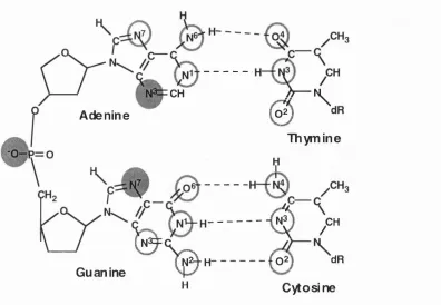
Numerous potential reaction sites have been identified in all four of the bases making up the double helix. These include the following; The N1, N3, N6 and N7 positions of adenine, N1, N2, N3 0 6 and N7 positions of guanine, N3, N4 and 0 2 positions of cytosine and the N2, 0 2 and 0 4 positions of thymine. The most reactive sites are at the N7 position of guanine and the N3 position of adenine.
Additionally, the oxygen in the phosphodiester linkage may react with electrophiles to form a phosphotriester. This is illustrated in figure 1.2 which shows the reactive centres in the four bases.
CH h— N ^
Adenine @
Thymine
u V
" 0 / "
0 = v ^
( N H H --- ( o2) dR
Guanine N -^
H Cytosine
Figure 1.2. Reactive centres in DNA. The most reactive sites are shown by shaded circles, whilst less reactive sites are circled.
DNA cross-links
DNA-DNA cross-links can occur between bases in the same strand (DNA
intrastrand cross-links), or between bases in opposite strands (DNA interstrand
cross-links). Several chemical agents such as cis-diam m inedichloroplatinum
(R oberts & Pascoe, 1972), psoralens (Cole, 1970), nitrogen and sulphur
mustards (Kohn eX. a/., 1966), as well as physical agents such as UV irradiation
at 254nm (Marmur & Grossman, 1961), and ionising radiation (Ward, 1988), are
all capable of inducing cross-links within DNA. Several different DNA cross-
linked structures have been identified, including those form ed between two
guanine N7 positions com m only formed by bifunctional mustards (Brookes &
Lawley, 1961), between guanine N1 and cytosine N3, formed, for example, by
ch lo ro e th yln itro so u re a s (Ludlum , 1990), and betw een tw o guanine N2
positions in the minor groove, as formed by mitomycin 0 (Tomasz eX. a/., 1987).
Interstrand cross-link formation has long been considered to be a particularly
important mechanism of cytotoxicity and the degree of cross-link formation has
been shown to correlate with the cytotoxic activities for nitrogen m ustards
(Garcia e t al., 1988; Sunters e t al., 1992) and the nitrosoureas (Erikson e t al.,
1980a). Cellular resistance to bifunctional alkylating agents is often associated with enhanced repair of cross-links (Burt e t at, 1991).
The second type of DNA-DNA cross-link, intrastrand cross-links, are commonly found in DNA after treatment with cisplatin and are an important modulator of the cytotoxicity of this compound. Intrastrand cross-links have been found to inhibit DNA polymerase I (Pinto & Lippard, 1985; Gralla e t al.,
1987).
An additional form of cross-links are those formed between DNA and proteins. These lesions have been found in cells treated with the topoisomerase poison etoposide (Wozniak & Ross, 1983). However the contribution of DNA-protein cross-links to the cytotoxic efficacy of other chemical and physical cross-linking agents is not certain. There is evidence that the formation of these lesions does not correlate with cytotoxic efficacy in cells treated with cisplatin (Zwelling e t al., 1979), nitrosoureas (Erickson e t al.,
1980a), or nitrogen mustards (O'Connor & Kohn, 1990).
DNA Strand-breaks.
DNA damage by some chemicals and ionising radiation can result in breaks in the sugar-phosphate backbone. Double strand-breaks may occur if two single strand-breaks occur in close proximity on opposite strands. Double strand-breaks have been identified as the lesion most likely to cause the lethal effects of ionising radiation (Ward, 1988).
Linn, 1988). Strand-breaks can also be formed via mechanisms not involving oxygen radicals. Many alkylating agents are able to attack the phosphate groups of DNA resulting in the production of a phosphotriester, which although stable at neutral pH, will hydrolyse under basic conditions. Alkylation at bases may also result in strand breaks. Base alkylation may result in depurination or depyrimidination to produce an apurinic/apyrimidinic (AP) site, which may then result in strand cleavage at the AP site (Garret and Metha, 1972).
1.2.2. Non-covalent mechanisms of ligand interactions with DNA.
Intercalation.
Many planar or near planar aromatic compounds are able to fit between the base pairs of DNA in a process called intercalation. The binding of intercalators causes significant structural modification of the double helix causing the helix to extend and unwind, and this may have detrimental effects on the template activity of DNA. The degree of unwinding of the helix varies depending on the structure of the intercalator. Bisintercalators are m olecules with two intercalating rings attached by a linker. The process of intercalation is reviewed in Saenger, (1983) and Wilson, (1990).
Groove binding.
1.3. Classes and properties of anticancer agents targeted against nucleic acids.
Many of the anticancer agents in use at the present time have DNA as their presumed major cellular target. Their modes of action are, however, very diverse, and this underlines the fact that DNA has many functionalities that can act as receptors for drugs. The major classes of nucleic acid targeted anticancer drugs are reviewed below.
1.3.1. The alkylating agents.
These compounds are generally highly reactive compounds that react so that an alkyl group or a substituted alkyl group becomes covalently linked to a nucleophilic group on a cellular constituent. Alkylation damage can inhibit DNA transcription (Pieper et. al., 1989) and DNA replication (Gralla et. a!., 1987).
The alkylating agents can be divided into five major groups; nitrogen mustards, nitrosoureas, triazenes, methane sulphonic acid esters and aziridines.
Nitrogen mustards.
The nitrogen mustards are the oldest effective cancer chemotherapeutics, and many members of this group of compounds are still in clinical use. The first truly effective anticancer agent, mechlorethamine went into clinical trials in 1942, when a patient suffering from lymphosarcoma was treated with the drug (Gilman & Phillips, 1946). Four representative nitrogen mustards are shown in figure 1.3. These compounds have, as a general feature, two chloroethyl groups attached to the nitrogen, thus imparting bifunctionality.
a — CH2CH2 a — CH2CH2 __
^ n- ch3 \
a —
CH2CH2^ Cl — CH2CH2^Nitrogen mustard M e ip h alan
a — CH2CH2
a — CH2CH2
o,
N~~~^ CH2CH2CH2CO2H N— y
a — ch2CH2^ a — ch2CH2^ °
C hloram bucil C yclophosph am ide
Figure 1.3. The structures of four representative nitrogen mustards.
three membered ring is very unstable and highly reactive and will attack nucleophilic centres. It is possible that the aziridinium ion may ring-open to form a carbonium ion intermediate. Figure 1.4. illustrates this reaction mechanism.
The major site of nucleophilic attack on DNA is at the N7 position of guanine. Bifunctional nitrogen mustards may go on to form cross-links, after the initial monoalkylation, either to a protein or to the opposite or the same strand of DNA, via the second chloroethyl group of the drug. These cross-links are thought to be the mechanism by which nitrogen mustards exert their cytotoxicity, due to their ability to inhibit vital cellular functions such as DNA replication and transcription (Brookes & Lawley, 1961; 1963). DNA cross-linking by the bifunctional nitrogen mustards has been shown to correlate with cytotoxicity (Zwelling et. al., 1981; Garcia et. al., 1988; Sunters et. al., 1992). In comparison to the bifunctional mustards, the corresponding monofunctional mustards are much less effective as anticancer agents (Brookes & Lawley, 1961). This and the fact that enhanced cross-link removal correlates with resistance to bifunctional nitrogen mustards (Batist et. al., 1989; Burt et. al., 1991) indicates that cross-links are the major cytotoxic lesion, although cell killing by nitrogen mustards is likely to be the result of a combination of lesions.
Cl— CH2CH2
\
N— CH3
a — CH2CH/^
M echloretham ine
H2N
Guanine base in DNA
CH3
a — CH2CH2
Aziridinium ion
+CH2CH2
\
Ci— CH2CH2/
N— CH3
Carbonium ion
CH2CH2-N-CH2CH— a
H2N
“ù y '
guanine aikyiated at the N"7
positionFigure 1.4. Reaction mechanism of the nitrogen mustards.
Nitrosoureas.
These are lipid soluble drugs which have the potential to penetrate the central nervous system for treatment of intercranial tumours. Examples are shown in figure 1.5.
o II
CICH2CH2—N-C—NH—CH2CH2CI B C N U
NO
O yCH2 —CH2^
CICH2CH2-N-C— NH— CH CH2 C C N U
^ ^CH2—CH2/
Figure 1.5. The structure of two representative nitrosoureas.
Nitrosoureas were originally developed after it was observed that N-methyl N'-nitrosoguanidine had antitumour activity against L I 210 cells (Greene & Greenberg, 1960). Related compounds were synthesised and tested, and N- methyl-N-nitrosourea, which was well known as a carcinogen, was also found to have antitumour activity. This observation led directly to the development of the haloethylnitrosoureas.
This class of agents act as both alkylating and carbamoylating agents, and the mechanism of action is illustrated in figure 1.6. (reviewed in Ludlum, 1990). At physiological pH, proton abstraction by a hydroxyl ion initiates spontaneous decomposition of the molecule to yield an isocyanate compound and a diazonium hydroxide molecule. Alternatively, the haloethylnitrosoureas can decompose to produce intermediates that hydroxy late DNA directly (Brundrett, 1980; Lown & Chauhan, 1982). The chloroethyl diazonium ion or the chloroethyl carbonium ion generated may then react with biological macromolecules.
| - H C I j -Cl
N
=0
RN=<
J
-R N C O
H2C-cm N OH CICH2CH2-N-C-N-R H OH Q Ç-NRH
1
,2,3
H .q' o x a d ia z o lin e
I N O
cicH2CH2”r|^c^r^”R
N V O^H-Q'oN itro s o u re a
-R N C O
+H2O
HOCH2CH2-N N OH CH3CHO
f
CICH2CH2-N+ N2+ H2O CICH=CH2■ 4
N-OH
CICH2CH2-N=N-0H
I C arb o n iu m ion
0
=C=N-RI s o c y a n a t e
I
Protein-C-N-RH
c a r b a m o y la t e d p ro te in s
+ OH +N2 CICH2CH2+ Alkylation products (DNA, DNA-DNA, DNA-protein)
Figure 1.6. Proposed reaction mechanism of the haionitrosoureas (From Brundrett, 1980; Lown & Chauhan, 1982).
the initial monoadduct. DNA-protein cross-links can also be formed by this mechanism (Kohn, 1977). The isocyanate breakdown product is capable of reacting with amine or sulphydryl groups on proteins and inhibits various DNA and RNA processing reactions. As with the nitrogen mustards, the most prominent position of monoadduct formation by the haloethylnitrosoureas is at the N7 position of guanine. The most significant cytotoxic lesions produced by the nitrosoureas are probably cross-linked adducts formed through two N7 positions of guanine, or through the N1 position of guanine and the N3 position of cytidine (Kohn, 1977; Ludlum, 1990). Guanine 0 6 alkyltransferase has been shown to be involved in the resistance to the haionitrosoureas (Erickson et. al.,
Methane sulphonate esters.
Busulphan (figure 1.7.), is the major representative of this group of alkylating agents, and is used in the treatment of chronic myelogenous leukaemia. Its' reaction with DNA is not thought to be via a reactive intermediate, as with the nitrogen mustards (which react with Sn1 type reaction kinetics). In the reaction,
the alkyl-oxygen bond splits and the molecule is able to alkylate a variety of cellular targets. Busulphan shows greater reactivity with the thiol groups of proteins then do the nitrogen mustards (Roberts & Warwick, 1957).
9 9
CH3 “ S —O —CH2CH2CH2 CH2O— S —CH3
Figure 1.7. Busulphan.
Bedford et. al. have shown that the major mechanism of cytotoxicity and antitumour activity of the dimethanesulphonic acid esters correlates with their ability to cross-link DNA (Bedford & Fox, 1983).
Aziridines.
These are analogues of the putative ring closed intermediates of the nitrogen m ustards. Two representative structures, T hiotepa and triethylenemelamine, are shown in figure 1.8. Compounds with two or more aziridine groups have been shown to have antitumour activity similar to the nitrogen mustards (Baterman, 1955). The reactivity of the aziridinium group is increased by protonation, and is therefore enhanced at low pH. The mechanism of action of these compounds is not well understood, but is possibly via a ring opening of the arizidinium group to form a reactive intermediate. Thiotepa has been used in the treatment of breast and ovary carcinoma.
N -P —
Thio-TE P A T rieth ylen em elam in e
Figure 1.8. The structures of two representative aziridines.
tumours, or those with high levels of reducing enzymes (Khan & Driscoll, 1976). AZQ was selected for clinical trials because it was able to penetrate the central nervous system (Chou et. al., 1976). One or two electron reduction of the quinone moiety is believed to facilitate protonation of the aziridine ring leading to ring opening and formation of alkylating species (Gutierrez, 1989).
Triazenes.
9 f " " N -^ C 0 N H 2
CH3NHNHCH2— > - C - N H C H / / M
" CH3
P ro carb azin e D a c a rb a zin e
Figure 1.9. The structures of two representative triazines.
1.3.2 Piatinum compounds.
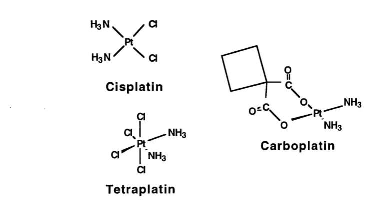
The anticancer agent cisplatin was discovered as a result of an investigation into the possible effects of electrical fields on growth processes in bacteria. Rosenburg discovered that long lived chemical species released from the platinum electrodes in the presence of ammonium and chloride ions could inhibit cell division (Rosenberg et. al., 1965). The compound responsible was identified as the platinum complex cis PtCl4(NH3)2. Later, the cisplatin complex
cis Pt(ll)(NH3)2Cl2 (Cisplatin, figure 1.10.), was found to be the most active of several platinum complexes against experimental tumours.
There has been a considerable effort to produce analogues of cisplatin with reduced nephrotoxicity, (Hamilton et. a!., 1993; Kelland, 1993), and two clinically effective platinum compounds, tetraplatin and carboplatin, are also illustrated in figure 1.10. Tetraplatin has been found to be effective in cisplatin resistant cells (Anderson et. a!., 1986). Carboplatin has a different spectrum of toxic side-effects to that of cisplatin, although its' mechanism of cytotoxicity is thought to be the same as that of the parent compound.
H aN ^ y a Pt H gN ^ \ a
C isplatin a
^ N H a
a j ^ n h 3 '
a ^ f N H 3 C arbo platin
a T e trap la tin
Figure 1.10. The structure of cisplatin and two of its analogues.
drug is capable of forming cross-links when both chloride leaving groups are replaced by nucleophilic centres within DNA. These cross-links may take the form of G-G, G-(X)-G or A-G intrastrand cross-links, or interstrand cross-links through two guanine N7 positions on opposite strands of DNA. In studies of platinated cellular DNA, the G-G and A-G intrastrand adducts make up approximately 60% and 30% respectively, of the total adducts, while the interstrand cross-links make up less than 1%. (Plooy e t al., 1985; Eastman, 1986). It is unclear which lesion is primarily responsible for the cytotoxic action of cisplatin. The transplatin isomer of cisplatin has much lower cytotoxicity than cisplatin and has been found to produce much fewer interstrand cross-links in cells (Zwelling et. a!., 1979; Zwelling et. a!., 1981). The intrastrand G-G cross links cause a marked distortion of the DNA conformation and it has been suggested that this may be responsible for the cytotoxic effect of cisplatin (Rice
et. a!., 1988).
1.3.3. Antim etabolites.
The antimetabolite class of antitumour agents consists of folic acid analogues (antifolates), purine analogues and pyrim idine analogues. Dihydrofolate reductase (DHFR) inhibitors are not thought to exert their effect by a direct interaction with DNA, although inhibition of DHFR can lead to inhibition of DNA, RNA and protein synthesis.
Pyrimidine analogues.
Analogues of both uracil and cytidine are commonly used in the treatment of cancer. Cytidine analogues, commonly called arabinose nucleosides, differ from the physiological parent compounds by the presence of a p hydroxy group
in the 2'- position of the sugar.
Several of the arabinose nucleosides have antitumour and also antiviral effects. Ara 0 (figure 1.11.), is used primarily in combination with doxorubicin or daunomycin, and has little activity as a single agent. In the cell Ara 0 is phosphorylated to the triphosphate (Ghu & Fisher, 1962), and this acts as a competitive inhibitor of DNA polymerase. Ara 0 can also be incorporated into DNA and this isj thought to be the main mechanism of its cytotoxic action (reviewed in Chabner, 1990b).
NH2
à
O ^ N CH2OH
Lf
OH '5-F lu ro u racil Cytosine Arabinoside
5-fluorouracil (5-FU), figure 1.1 1, is an analogue of the base uracil. In a situation similar to that for Ara C, 5-FU is metabolised in the cell to the active nucleotide form by the addition of sugar and phosphate moieties. The active forms, 5-FUTP and F-dUMP can be incorporated into RNA in place of UTP and this leads to inhibition of the nuclear processing of rRNA and mRNA. 5-dUMP also reversibiy inhibits thymidilate synthetase. (Grem, 1990 and references therein).
Purine analogues.
Several purine analogues have been synthesised and tested for antitumour activity. O ther purine analogues have found use as antiviral or immunosuppressive agents, for example adenosine arabinoside (Ara A) and azothioprine.
Two compounds that have found use as anticancer agents, particularly in the treatment of leukaemia, are 6-mercaptopurine and 6-thioguanine, shown in figure 1.12. These compounds are metabolised to deoxynucleotides by addition of sugar-phosphate (Elion, 1967).
6-T h io g u a n in e 6-M ercap to p u rin e
Figure 1.12. The purine analogues 6-mercaptopurine and 6- thioguanine.
cytotoxicity of the compound is undetermined. The ribonucleotide forms of the drugs are capable of inhibiting de-novo purine synthesis and purine interconversion. For a review of the purine antimetabolites see McCormack & Johns, (1990).
1.3.4. Topoisom erase poisons.
A number of DNA interacting anticancer agents exert at least a part of their cytotoxic action via a mechanism involving the ubiquitous DNA processing enzymes topoisomerase I and II (Reviewed in D'Arpa & Liu, 1989; Epstein, 1990; Lown, 1993; Pommier, 1993). These enzymes are involved in carrying out the breaking and resealing reactions of DNA necessary in such events as DNA replication and DNA repair. Drugs which poison topoisomerase II can be broadly categorised into the DNA intercalators, which include the acridines, ellipticines, actinomycins, anthracenediones and anthracyclines, and the non intercalating epipodophyllotoxins.
When cells are treated with these compounds, so called protein-associated DNA strand breaks result. This is thought to occur because topoisomerase inhibitors trap the DNA strand passage intermediates in a cleavable complex, consisting of protein-linked DNA single or double strand-breaks (Tewey et. al.,
1984), that are covalently linked to the enzyme by a phosphotyrosine residue.
Epipodophvllotoxins
figure 1.13.
OH
HO
OH
OH
Podophyllotoxin
R= CH3; Etoposide (VP16)
R= ^ " ^ ^ T e n ip o s id e (VM 26)
Figure 1.13. The structures of the podophyilotoxin derivatives etoposide (VP16) and teniposide (VM 26).
Etoposide and teniposide are generally considered to be non-DNA binding drugs (Chen e t at, 1984; Ross et. at, 1984), although this is by no means certain.
DNA intercalators.
The anthracyclines have found wide use in cancer chemotherapy and are active against epithelial tumours, lung and stomach cancer as well as lymphomas and leukaemias. The structures of two com monly used representatives of this class, doxorubicin and daunomycin, are shown in figure 1.14. Lown has recently provided a comprehensive review of the state of research into anthracyclines (Lown, 1993).
CHgO
OH OII
C -R
P O H
R=C H2 0H; Doxorubicin.
RsCHa; Daunomycin.
Figure 1.14. The structure of the DNA intercaiating topoisomerase II poisons doxorubicin and daunomycin.
intercalate into the DNA and act as topoisomerase poisons in trapping the enzyme in a cleavable complex (Bodley et. al., 1989). The anthracyclines are also able, however, to undergo one and two electron reduction with the end result being the production of a highly reactive hydroxyl radical (Bachur at. a!.,
1977). This radical is extremely reactive and is capable of causing DNA strand breaks and other cellular damage. Although the contribution of these radicals to the antitumour effects of anthracyclines is unknown, free radical formation is thought to be the cause of the dose limiting cardiotoxicity caused by the anthracyclines (reviewed in Myers & Chabner, 1990). In addition, the quinone methide product of the two electron reduction of doxorubicin is a potential alkylating agent and is capable of forming monoadducts on DNA.
Other intercalators such as mitoxantrone (an anthracenedione) are thought to exert at least a part of their antitumour activity via the formation of protein associated DNA strand breaks (Crespi at. a!., 1986).
1.3.5. Antitumour antibiotics.
A range of compounds useful in chemotherapy have been isolated by microbial fermentation. These include the anthracyclines (section 1.3.4.), bleomycins, and various unusual nucleosides.
Actinomycin D.
Actinomycin D was first isolated from streptomyces in 1940, and introduced into the clinic in 1954. The structure of this compound is given in figure 1.15. Actinomycin D is known to bind to DNA and to inhibit RNA and protein synthesis. It is thought that the chromophore intercalates between the base pairs of DNA, and the peptide lactone rings lie in the minor groove (Muller & Crothers, 1968). Actinomycin D seems to exhibit a degree of sequence selectivity in its interaction with DNA and prefers to bind at GC sequences (Kamawata & Imoniski, 1960). This early finding has been confirmed since, by footprinting studies (Scamrov & Beabealashvilli, 1983; Churchill et. al., 1990). Many analogous of actinomycin D have been isolated, but none have been found to have increased antitumour activity over the parent compound.
o
rC
-X
(CH3)2HC CH(CH3) 2
C -H H -Ç
N—CH3
H 3C -N ^
S arcosine Sarcosine L -P rilin e L -P rilin e
D -V alin e D-valine I i
o=c c=o
I I
CH
HO---I I
R -C—n
ÇH
CH3
Mitomycin C.
Mitomycin C, another fermentation product from Streptomyces, was first isolated in 1958. This compound has found use against a wide range of solid tumours, including breast cancer, non-small cell lung cancer, colorectal cancer and others. The compound is unique amongst naturally occurring compounds in that it contains an aziridine ring (Figure 1.16.).
CHoO —C— NH2
Figure 1.16. Mitomycin C.
Upon reduction (chemical or enzymatic) of the quinone moiety to either the semi- or the hydroquinone, the drug becomes activated and is able to alkylate DNA. Alkylation can be either mono- or bifunctional and the primary cytotoxic lesion is thought to be a DNA interstrand cross-link formed through two guanine N2 positions (Tomasz e t at, 1987).
Mitomycin C is considered to be a prototype 'bioreductive alkylating agent', a term used to describe a whole generation of compounds that can be converted
in-situ to their active forms. This may be achieved by enzymatic reduction, for example by DT diaphorase (reviewed in Verweij et. a/., 1990).
Bleomycin.
n NH2
T H
o
OH
I
H \^ N H^ Z is i- ç
---H
OH
OH °
O ^ NH2
Figure 1.17. Bleomycin A2.
The primary mechanism of action of bleomycin A2 is via the production of both single and double strand breaks in DNA. These strand breaks are the result of free radical production by the Fe(ll) bleomycin complex intercalated between the two strands of DNA.
Binding to DNA occurs primarily via the amino terminal tripeptide of bleomycin to guanines. The bithioazole rings of the S tripeptide bind to guanine bases in the sequences GT or GO (Umezawa et. a/., 1984). The clinical and biochemical aspects of bleomycin are extensively reviewed in Chabner, 1990a.
1.4. Drug toxicity, drug resistance and the need for more selective antitumour agents.
undiscriminating in their reactions with cellular constituents. Most have no particular affinity for DNA and will react with proteins, lipids and other macromolecules. In addition, although many of these drugs do exhibit a certain amount of DNA base sequence specificity (reviewed in Hartley & Souhami, 1993), this specificity is limited and its contribution to the antitumour efficacy of the compounds is uncertain. Because anticancer drugs exert their effects largely on cycling cells, normal tissues with a high proliferative fraction are often adversely effected. These include bone marrow, intestinal mucosa, hair follicles and gonads. Many drugs also show toxicity towards particular organs, for example doxorubicin exhibits dose-limiting cardiotoxicity.
An additional complication inherent with many conventional therapeutic agents is that of their carcinogenic properties. Because so many of the drugs are mutagens, at least some of these mutations must escape repair, and in cells that do not die these mutagenic lesions may eventually lead to the emergence of secondary tumours in later years, (reviewed in Schilsky & Erlichman, 1990 and Shulman, 1993).
drug activation and increased expression of the apoptosis inhibiting gene bcl-2
(Lotem & Sachs, 1993). There are likely to be mechanisms of resistance yet undiscovered.
To a certain extent drug resistance can be circumvented by pharmacological modulation. Modification of resistance may be achieved by the use of either chemoenhancers, to sensitise tumour cells, or alternatively by chemoprotectors which selectively protect normal tissues (Lazo & Bahnson, 1989). A well known class of chemoenhancers are the calcium channel blockers, an example of which is verapamil. These compounds are able to inhibit the function of the drug-effluxing P-glycoprotein. Attempts to modify the acquired or intrinsic resistance of tumour cells have, however, met with limited clinical success to date.
There is thus a pressing need to continually develop new drugs and treatments for cancer to further improve on therapeutic ratios and to achieve more selective kills of cancer cells over normal tissue. There are several possibilities that could be explored in the development of novel DNA acting anticancer agents. One approach would be to increase the amount of drug that is able to reach the DNA. Another would be to improve the yield of critical, cytotoxic lesions and decrease the proportion of the more mutagenic lesions. Neither of these approaches would, however, be expected to guard against the systemic toxicity seen with current antitumour compounds. An alternative approach would be to preferentially target tumour tissue. This is exemplified by the Antibody-Directed Enzyme Prodrug Therapy (ADEPT) research program in which tumour associated antigens are targeted by a specific antibody conjugated to an enzyme chosen for its drug activating properties. A prodrug is then administered which should be converted to its active form at the tumour site (Bagshawe, 1987).
the process of oncogenesis. Alternatively, the products of these genes at either the mRNA or protein level could be targeted. If these regions of DNA could be affected in such a way that their template function becomes compromised, then the protein product of the gene would become down-regulated. The consequences for the treated cell would presumably be differentiation, senescence or death.
Many genes have been implicated in the transformation of cells from a normal to a cancerous phenotype, and the list of such genes is continually expanding (Bishop, 1991). In the vast majority of cases it is found that more then one genetic change has taken place during the transformation process. Very often the products of the genes concerned are involved in the control, either negative or positive, of cellular growth and proliferation. Inhibition of the expression (or function) of myc, ras and other oncogenes have been shown to slow the replication of cells transformed by these oncogenes. For example, treatment of HL60 promyelocytic leukaemia cells, which express high levels of
c-myc and mutant ras, with c-myc antisense oligomers inhibits the replication of these cells and induces their differentiation (Holt et. al., 1988). Another example is the reduction in soft agar colony formation of ras transformed Swiss 3T3 fibroblasts when treated with antisense oligonucleotides to myc, even though
ras continued to be expressed (Sklar et. a!., 1991).
form of the tumour suppressor gene p53 may be a candidate for genetic targeting.
The situation is, of course, often considerably more complex then the simple model of producing selective therapy by inhibition of single genes. Cellular signalling mechanisms often act through multiple pathways and show a degree of redundancy. Thus, inhibition of the expression of a particular gene may result in the subsequent activation of other oncogenes in order to override this inhibition. Toxicity may also occur as a result of gene inhibition in normal tissues.
However, the two examples given above and other studies have demonstrated that the strategy of therapeutic inhibition is a distinct possibility. Moyer and Fischer have provided an excellent review and critique of the suitability of oncogenes as targets for chemotherapy, paying particular attention to ras as an example (Moyer & Fischer, 1993).
1.5. Alternative strategies for the therapeutic targeting of nucleic acids
In the quest to develop therapeutic agents with the potential to selectively modulate the expression of specific genes, a number of interesting and novel approaches are being pursued. Most of these are directed against the 'informational molecules' DNA or RNA. The most important of these strategies are reviewed below.
1.5.1. Antisense oligonucleotides.
including nuclear processing, stability, transport to the cytoplasm and translation into protein. In order to achieve a specific effect, that is interaction with only a single mRNA species, the antisense oligonucleotide needs to be between 11 and 15 bases long, depending on the relative A.T or G.C content of the molecule (Helene, 1992). The oligonucleotide has to be long enough to achieve a stable hybridisation under physiological conditions. However, overlong oligonucleotides can be subject to mismatch binding and so lose a degree of specificity of hybridisation. Because of this, the oligonucleotides are usually chosen to be between 12 and 2 0 nucleotides in length.
There are at least two mechanisms by which oligonucleotides inhibit mRNA translation (Helene & Toulme, 1990). Firstly, the oligo(dN)-mRNA hybrid is a substrate for the endogenous ribonuclease RNase H, which recognises RNA- DNA hybrids and cleaves the RNA portion of the molecule. Secondly, the oligo bound to the 5'-untranslated region of an mRNA can inhibit binding or sliding of the 408 ribosomal subunit and / or association of protein factors involved in translation initiation (Rothenberg et. al., 1989; Boiziau et. al., 1991; Helene,
1992).
not hybridise very efficiently and are unable to induce RNase H activity (Kibler
ot. a/., 1991 ).
y
y y y
0 0 0 0 0
0 = P -0 " 0=P-CH3 "S-p=0" 0 = P -0 " 0 = P “ R
y y y y y
B
Figure 1.18. Common m odifications of antisense oiigonucleotide stru ctu re. A; P hosphodiester linkage, B; M eth ylp ho sph on ate linkage, C; phosphorothioate iinkage, D; a-anom er nucieotide, E; Many substituents possible.
Phosphorothioate oligonucleotides exhibit good solubility, hybridisation efficiency, nuclease resistance and are able to induce RNase activity. However, the cellular uptake of these derivatives is poor. Phosphorothioate derivatives also show some sequence independent toxicity.
A n o th e r m a jo r type of o lig o d e o x y n u c le o tid e are the a - oligodeoxynucleotides. In these structures, the sugar-base linkage is in the a position as opposed to the (3 position seen in the naturally occurring oligonucleotides. These analogues show good solubility, stability and hybridisation efficiencies, but, as with the methylphosphonate derivatives, are unable to induce RNase activity (Carter & Lemoine, 1993).
In many of these derivatives, the internucleotide linkages are chiral centres, and thus preparations of oligonucleotides containing these linkages will consist of many stereoisomers. This is a potential source of problems because not all stereoisomers will bind to the target sequence with equal efficiency.
oligonucleotide-mRNA hybrid (reviewed in Milligan et. al., 1993). Conjugation of the oligonucleotides to polypeptides such as poly(L-lysine), membrane lipids or encapsulation in antibody-targeted liposomes enables increased cellular uptake and biological efficacy of the molecules (Leserman at. a!., 1991).
A new generation of oligonucleotides designed to covalently modify the target RNA are currently under investigation. This approach involves oligonucleotides covalently linked to alkylating, photoactivatable or free radical generating functional groups (for exam ple Saison et. al., 1991). Oligonucleotides containing intercalating moieties have also been synthesised in an attempt to further stabilise the oligonucleotide-mRNA hybrid (for example Zerial et. al., 1987; Birg et. al., 1990).
A major problem with antisense oligonucleotides seems to be their very poor ability to cross cellular membranes, and for this reason it is assumed by many workers that some form of cell permeabilization will have to be employed in the use of these agents (Wagner, 1994). Cationic liposomes have been used in some experiments to introduce the agent into cells and this method was found to greatly enhance the antisense effect of the oligonucleotides (Bennett et. al.,
1992; Colige et. al., 1993).
The effects of antisense oligonucleotides have been reported in many tissue culture experiments (Helene & Toulme, 1990; Milligan et. al., 1993; Stein & Cheng, 1993), and in several in -v iv o studies (see Wagner, 1994 and references therein). Clinical trials of antisense oligonucleotides are in progress for several diseases including acute myelogenous leukaemia (Bayever et. al.,
1993), HIV and human papillomavirus infection (Alper, 1993). The current state of research into the antisense approach is reviewed in Wagner, (1994).
1.5.2. Triple helix-forming oligonucleotides.
adversely affected. This approach is often called the anti-gene strategy (Helene & Toulme, 1990). Thymine and protonated cytosine can hydrogen bond with A.T and G.C base pairs respectively, via Hoogsteen base pairing, to generate A.T.(A) and C.G.(C+) base triplets. The oligonucleotides bind in the major groove of double stranded DNA in a parallel orientation. It is generally considered that the limitation of this approach is that at present only homopurine-homopyrimidine sequences can be targeted. The binding of the 'third strand' oligonucleotide is also sensitive to pH because it requires the protonation of the cytosine residue, i.e. a pH of approximately 4.5 to 6. Dervans' group have shown that the use of a 5-methylcytosine or a 5-bromouridine in the third strand appears to raise the apparent pK for triplex formation, thereby permitting triplex formation in the pH 6 to 7 range (Moser & Dervan, 1987). Some recent results indicate that mixed sequences may be recognised in which the guanosine residues on the antigene oligonucleotide form stable triplets with the T.A base pairs in the target sequence (Beal & Dervan, 1991). The presence of a triplex forming oligonucleotide consisting of a mixed sequence and targeted against a c-m yc upstream sequence has been demonstrated to inhibit c-myc transcription in an in vitro system (Durland et. al.,
1991 and references therein).
The goal of employing triple helix forming oligonucleotides to inhibit transcription activator binding has also been tested in vitro using recombinant S pl promoter (Maher et. al., 1989). DNase I footprinting assays were used to measure the competition between triple helix formation and S pl binding, and it was found that a homopyrimidine oligomer blocked the binding of S pl to its binding sequence.
mechanisms responsible for the observed experimental effects are indeed those assumed in the experimental design.
Antigene oligonucleotides are commonly conjugated to reactive or intercalative groups in order to stabilise the formation of the triple helix. For example, an acridine intercalator attached at the 5'- end of a homopyrimidine oligonucleotide significantly stabilised the formation of the triplex (Sun et. a/., 1989). Alternatively, the oligonucleotides have been conjugated to reactive chemical functionalities to facilitate strand specific cleavage or covalent modification. Examples of such are psoralen groups which have been shown to cross-link DNA when conjugated to an oligonucleotide and irradiated with UV light. (Takasugi et. al., 1991). Metal chelating functional groups conjugated to oligonucleotides are capable of inducing strand breaks in the tagged DNA (Moser & Dervan, 1987). For a recent review of triplex DNA see Maher (1992).
1.5.3. Ribozymes.
Ribozymes are another class of oligonucleotide that, like antisense oligonucleotides, have RNA as their target of action (Rossi & Sarver, 1990). They are short RNA molecules that consist of antisense sequences for binding to target molecules, plus a conserved sequence that allows the ribozyme-RNA complex to fold into a conformation that allows for self-cleavage. The ribozymes require that the target RNA possess a 5'-G-U-N-3' sequence in which N is any nucleoside but guanosine.
The feasibility of the ribozyme approach has been demonstrated using various target RNA's in cell free systems and ribozymes have also been shown to degrade various RNA's in cell cultures.
which GGU is converted to GUU. This then becomes a target for a ribozyme capable of cleaving GUU after the second U (Koizumi et. al. 1989).
As with the antisense and triple helix approaches, ribozymes have potential problems of delivery mechanisms if they are to be developed for therapeutic applications. An additional significant drawback of ribozymes is that they are particularly sensitive to nuclease degradation, and for this reason present strategies employing ribozymes achieve their delivery by genetic means in the form of mini-gene constructs (Cameron & Jennings, 1989). Ribozymes have also been delivered into cells in the form of liposomes, one example being the introduction of a ribozyme into K562 cells to cleave BCR/ABL mRNA resulting in a significant inhibition of proliferation of the cells (Lange et. a!., 1993).
Studies are also underway to replace as many of the ribonucleotides as possible to make the ribozymes resistant to nucleases (Smith & Dinter 1991; Herschlag, 1991; Pieken et. a!., 1991).
1.5.4. Peptide-nucleic acid chimeras.
The peptide-nucleic acid chimeras (PNA's) are oligonucleotide analogues in which the entire deoxyribose-phosphate backbone has been replaced with a structurally similar polyamide backbone consisting of (2-aminoethyl)glycine units. This is shown in figure 1.19. These compounds have proved to be very effective DNA mimics and are able to bind double stranded DNA by displacing the non-complementary strands. Evidence suggests that two PNA strands are bound to the target DNA via a mechanism in which one molecule of PNA binds to the double stranded DNA in a strand displacement complex, and the subsequent binding of a second strand of PNA 'traps' the first strand of PNA (Nielsen et. a!., 1993b).
oligonucleotides, PNA based anti-gene drugs are at present limited to targeting homopurine sequences (Nielsen et. al., 1993a).
NH2 OH
Y x " 0
O ^ N H
i
-0-p=0 o
n
n H
PNA DNA
J n
Figure 1.19. Comparative structures of DNA and the PNA's.
1.6. Minor groove binders as potential anticancer agents.
The antisense and triple helix based approaches outlined in the previous section have shown some promising results, and although they have yet to reach their full therapeutic potential, the intensity of research in this field will almost certainly lead to more applications in the future.
molecules. Therefore compounds that read DNA sequence with high fidelity do so, as a general rule, via the major groove. Figure 1.20. illustrates the informational content of the major and minor grooves.
Major groove
ÇH3 a ^ H ' N
a "
A d e n i n e T h y m in e G u a n in e C y t o s i n e
Minor groove
Figure 1.20. The hydrogen bond donating (d) and accepting (a) sites in the major and minor grooves of DNA.
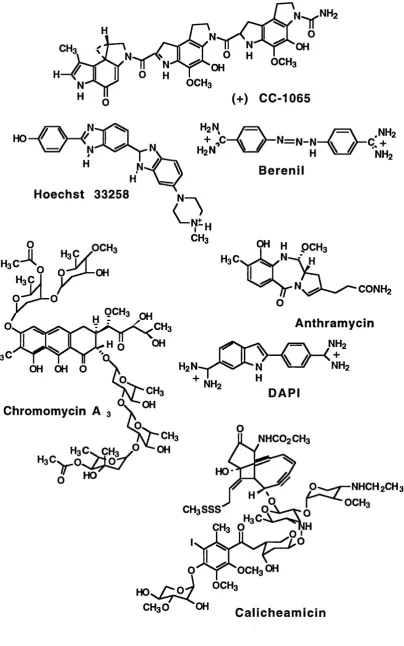
The minor groove often acts as a site of interaction for such molecules as polymerases, non-specific nucleases and many xenobiotics and antibiotics. It has been hypothesised that the minor groove, being relatively unoccupied by DNA binding proteins (in comparison to the major groove), may present a vulnerable site of attack for sequence reading, DNA directed ligands. Many naturally occurring antibiotics are DNA minor groove binders, these molecules possibly having evolved to take advantage of this facet of DNA structure.
(+) CC-1065 N ^ N H2
= \ .NHz + NHz B erenil
Hoechst 33258
i H3Ç
OH OH O
9" H PCH3
CONH2
NH2 "
Anthram ycin NH2
+ NH2
Chromomycin A 3
CH3
Jl HO
DAPI
NHCO2CH3
HO-CH3 O
NHCH2CH3 Q | ^ " ^ o c h 3
H . e S è S ^
Q - ^ "OCH3OH
CH3O OH C alich eam icin
interact non-covalently using a combination of electrostatic, hydrogen bonds and Van-der-Waals interactions.
1.6.1. The Pyrrolod .4)benzodiazepines