R E S E A R C H
Open Access
Mutually dependent degradation of Ama1p and
Cdc20p terminates APC/C ubiquitin ligase activity
at the completion of meiotic development in
yeast
Grace S Tan
1,2,4, Rebecca Lewandowski
2, Michael J Mallory
3, Randy Strich
2and Katrina F Cooper
2*Abstract
Background:The execution of meiotic nuclear divisions inS. cerevisiaeis regulated by protein degradation mediated by the anaphase promoting complex/cyclosome (APC/C) ubiquitin ligase. The correct timing of APC/C activity is essential for normal chromosome segregation. During meiosis, the APC/C is activated by the association of either Cdc20p or the meiosis-specific factor Ama1p. Both Ama1p and Cdc20p are targeted for degradation as cells exit meiosis II with Cdc20p being destroyed by APC/CAma1. In this study we investigated how Ama1p is down regulated at the completion of meiosis.
Findings:Here we show that Ama1p is a substrate of APC/CCdc20but not APC/CCdh1in meiotic cells. Cdc20p binds Ama1p in vivo and APC/CCdc20ubiquitylates Ama1p in vitro. Ama1p ubiquitylation requires one of two degradation motifs, a D-box and a“KEN-box”like motif called GxEN. Finally, Ama1p degradation does not require its association with the APC/C via its conserved APC/C binding motifs (C-box and IR) and occurs simultaneously with APC/CAma1 -mediated Cdc20p degradation.
Conclusions:Unlike the cyclical nature of mitotic cell division, meiosis is a linear pathway leading to the
production of quiescent spores. This raises the question of how the APC/C is reset prior to spore germination. This and a previous study revealed that Cdc20p and Ama1p direct each others degradation via APC/C-dependent degradation. These findings suggest a model that the APC/C is inactivated by mutual degradation of the activators. In addition, these results support a model in which Ama1p and Cdc20p relocate to the substrate address within the APC/C cavity prior to degradation.
Keywords:Cdc20p, Ama1p, Anaphase Promoting Complex, Meiosis
Background
Meiosis is a specialized developmental program during which diploid nuclei undergo two consecutive meiotic divisions to produce haploid gametes. In the budding yeast, spore wall assembly follows the second meiotic nuclear division producing four haploid spores encased in a protective ascus [1]. Similar to differentiation pro-grams in higher eukaryotes, meiotic progression is regu-lated by the transient expression of genes that are either
meiosis specific or expressed during both meiotic and mitotic divisions (reviewed in [2]). In addition, progres-sion through the meiotic diviprogres-sions is also driven by the degradation of key regulatory proteins directed by the highly conserved multi-complex ubiquitin ligase called the anaphase promoting complex/cyclosome (APC/C) (reviewed in [3-6]).
During meiosis, the APC/C is sequentially activated by two of the three known Trp-Asp activator (WD40) pro-teins, Cdc20p (reviewed in [7,8]), and Ama1p, the latter of which is only expressed during meiosis [9,10]. The Cdc20p activated APC/C (written APC/CCdc20) mediates the deg-radation of several key regulatory proteins including Pds1p * Correspondence:[email protected]
2
Department of Molecular Biology, UMDNJ-SOM, 2 Medical Center Drive, 08084, USA
Full list of author information is available at the end of the article
(securin) and the S-phase cyclin Clb5 during both meiosis I (MI) and meiosis II (MII) [8,11]. Ama1p directs the ubiquitylation of the B-type cyclin Clb1p [10], Cdc20p [12] plus other unknown substrates [13] and co-ordinates exit from MII [12]. APC/CAma1also activates Smk1p, the mei-otic MAP kinase required for spore wall morphogenesis [14] and is required for the early stages of spore wall as-sembly [11,13,15]. The third APC/C activator Cdh1p, is not required for normal meiosis [16].
It has been well documented that APC/C activator proteins recognize substrates through two conserved
degrons called the “Destruction-box” (D-box, DB) and
“KEN box” that bind the WD40 domain in the activator
[17,18]. In addition, Doc1p (Apc10), a conserved compo-nent of the APC/C complex, also recognizes these degrons. These findings have lead to the model that sub-strates are recruited to the APC/C by binding to a bi-partite substrate receptor composed of an activator protein and Doc1p ([19] and reviewed in [20]). During meiosis, Ama1p recognizes the D-box as well as variant of the KEN box called GxEN [10,12] whereas Cdc20p recognizes the D-box and the KEN box [21,22].
How-ever, in Xenopus egg extracts the APC/C recognizes
destruction motifs directly, in both a Cdc20p and Cdh1p-independent manner [23]. Similarly, much is known about how the activator proteins bind to the APC/C [5]. Structural analysis of Cdh1p has shown that a domain called the C-box interacts with Apc2p [24]. Another domain termed the IR motif promotes the asso-ciation of the activator with the TPR region of several APC/C subunits (Cdc16p, Cdc23p and Cdc27p) [25-28]. Doc1p (Apc10p), a subunit of the APC/C, also associates with the TPR subunits via its IR tail [29,30]. During mei-osis, both the C-box and IR domains are required for Ama1p and Cdc20p function [12]. However, mutational analysis revealed that the C-box in Ama1p is signifi-cantly more important for meiotic progression than the IR motif [12]. Similarly, during mitotic cell division, the IR box of Cdc20p is not required for function but con-tributes to APC/C dependent turnover [3,6].
Although much is known about how the APC/C is activated during meiotic divisions (reviewed in [8]), considerably less is known about how this ligase is inactivated as cells complete meiotic program. This is an important question as APC/C inactivation at the end of meiosis may be critical to allow the spore to reenter the mitotic cell cycle. Our previous studies have shown that both Ama1p and Cdc20p are down regulated as cells exit from meiosis II [10,12]. Furthermore, Cdc20p deg-radation is mediated by APC/CAma1[12]. In this report, we present evidence that Ama1p down regulation occurs via ubiquitin-mediated degradation directed by APC/ CCdc20. Taken together, these results indicate that the cell has solved the problem of APC/C inactivation in a
linear differentiation pathway by evolving a mutual deg-radation system for the activators.
Results
Cdc20p activates the APC/C to mediate Ama1p degradation
We have previously reported that Ama1p levels are re-duced as cells complete the second meiotic division [10]. As APC/C activators have been reported to be down-regulated by APC/C mediated proteolysis during mitotic and meiotic cell divisions (reviewed in [7,8]), we first asked if the reduction in Ama1p levels was APC/C dependent. The meiotic levels of Ama1p-T7 [12] were monitored in a strain harboring a temperature sensitive
allele of CDC16 (cdc16-1), an essential component of
the APC/C [31] that is required for meiosis [10]. To in-activate Cdc16-1p, the cells were switched to the re-strictive temperature (34.5°C) 4.5 h after meiotic entry as previously described [8,10,32]). As a control, Ama1p degradation was also examined in identically treated wild-type cells. Immunoblot analysis revealed that Ama1p-T7
levels remained elevated in the cdc16-1 strain compared
to wild type (Figure 1A, quantitated in Figure 1B). Similar results were obtained when these experiments were re-peated in acdc20-1strain (Figure 1A). Furthermore, these results are consistent with those obtained when Ama1p levels were monitored in a strain where Cdc20p was inactivated during meiosis by placing it under the control ofCLB2promoter [33]. Taken together, these results indi-cate that APC/CCdc20is required for the down regulation of Ama1p-T7 in meiosis.
A caveat to this interpretation is that Ama1p-T7 stabilization in the cdc20-1 mutant is an indirect effect of the metaphase I arrest associated with this mutation [32]. To address this issue, two approaches were taken.
First, we examined Ama1p stability in acdc20-1mutant
shifted to the restrictive temperature following meiosis II (15 h timepoint). These results show that Ama1p
re-mains stable in the cdc20-1 strain at restrictive
temperature even following 30 h in SPM (Figure 1C). To confirm that thecdc20-1cells had completed the meiotic divisions by this timepoint, the transcription profiles of meiosis-specific genes were monitored using Northern blot analysis. By 15 h in SPM, maximal transcriptional
accumulation of SPS4 was observed (Additional file 1)
which is an indicator that the meiotic divisions are
com-pleting [34]. Similarly, SPS100 mRNA induction, which
correlates with spore wall formation [35], occurs 18 h after meiotic entry.
For the second approach, we analyzed the meiotic
deg-radation of Clb5p, a known substrate of APC/CCdc20
[11]. Clb5p-HA levels were followed by immunoblot analysis in wild type andcdc20-1cultures using the same temperature shift protocol as described in panel A. The
Tanet al. Cell Division2013,8:9 Page 2 of 12
results show that, compared to wild-type cells, Clb5p was stabilized following Cdc20p-1 inactivation (Figure 1D). In
contrast, Clb1p, a known substrate of APC/CAma1[10],
is destroyed incdc20-1 cells using the same conditions
(Figure 1D). The slower induction kinetics observed for both cyclins is due to the fact that expression of early-middle, middle gene mRNAs is significantly reduced as well as delayed in this strain background [32]. Taken together, these results support a model that APC/
CCdc20 mediates the degradation of Ama1p as cells
complete the meiosis and begin spore morphogenesis.
Cdh1p is not required to mediate the degradation of Ama1p during meiosis
To determine whether Cdh1p plays a role in Ama1p prote-olysis during meiosis, Ama1p protein levels were monitored incdh1Δcells during meiosis. The results show thatcdh1Δ cells both progress through meiosis (Additional file 2: Figure S2A, S2B and S2C) and degrade Ama1p with the same kinetics as wild type (Additional file 2: Figure S2D and see Tan et al. [12] for Northern analysis). Interestingly, dissection of the resulting cdh1Δ tetrads revealed that,
different to previously published results [16],cdh1Δspores exhibit a significant reduction in their ability to form col-onies (Additional file 2: Figure S2E). These results indicate that Cdh1p does not control Ama1p stability but does play a role in promoting spore viability.
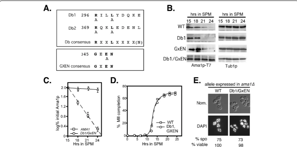
Ama1p contains functional degradation signals
Ama1p contains two motifs, the destruction box (Db)
and GxEN, that are recognized by APC/CCdc20(reviewed
in [36]), see Figure 2A). To determine if these sequences are required for Ama1p-T7 degradation, wild-type cells
expressing either Ama1pDb1Δ-T7 or Ama1pGxEN-T7
mu-tant proteins were induced to enter meiosis and their degradation profiles monitored by immunoblot analysis. These studies revealed no difference in decay kinetics for the single mutant derivatives compared to wild type (Figure 2B) indicating that individually the Db1 or GxEN motifs are not essential for Ama1p degradation. We have
recently shown that the APC/CAma1 mediates Cdc20p
degradation through more than one degron [12]. To determine if Cdc20p also recognizes multiple Ama1p degrons, wild-type cells expressing a double Db1 and
Figure 1APC/CCdc20is required for Ama1 degradation during meiosis. A: Wild-type (RSY335),cdc20-1(RSY809) and cdc16-1strains (RSY954) harboring Ama1p-T7 (pKC3036) were induced to enter the meiosis and timepoints taken as indicated. Immunoblot analysis of
GxEN AMA1 derivative were examined as just de-scribed. The results (Figure 2B, quantified in Figure 2C) show that combining the GxEN and Db1 mutations protected Ama1p-T7 from degradation similar to that
observed incdc16-1cells (compare to Figure 1A). These
results indicate that either Db1 or GxEN is sufficient to target Ama1p for degradation. No difference in the rate of meiotic progression (Figure 2D) or spore viability (Figure 2E) was noted indicating that stabilizing Ama1p did not have an adverse effect on the process.
Ama1p is a substrate of APC/CCdc20in vitro
To further confirm that APC/CCdc20 mediates the degrad-ation of Ama1p, in vitro ubiquityldegrad-ation assays were performed (see Methods for details). As Ama1p is an acti-vator of the APC/C [10], the assays were performed with an in vitro transcription coupled translation produced 35-S labeled Ama1p derivative deleted for its two APC/C bind-ing domains (C-box and IR motif). These motifs are re-quired for Ama1p function. To ensure that the added Cdc20p is the only activator in the reaction, the APC/C core complex was purified from mitotically dividingcdh1Δ cells. Furthermore, Mnd2p (Apc15p) was not present in the extracts as it inhibits meiotic APC/C activity [33]. As
pre-dicted from the in vivo studies, Ama1pCBΔ/IRΔ is
ubiquitylated by APC/CCdc20in vitro (Figure 4A, lanes 1, 2 and 3 and see Additional file 3 for input), but also that Cdc20p is required for this event (Figure 3A–lane 12).
The in vivo stability assays just described (Figure 2) in-dicated that either Db1 or the GxEN motif is sufficient to induce Ama1p degradation. Consistent with this re-sult, deletion of either of these motifs in the Ama1pCB/IR mutant still allowed ubiquitylation to occur (Figure 3A, lanes 4-6 for GxEN, 8 and 9 for Db1). However, Ama1p mutated for both Db1 and GxEN was still ubiquitylated
in vitro by APC/CCdc20 (Figure 3A, lanes 10 and 11).
This result was unexpected as this mutant is not targeted for degradation in vivo (Figure 2B). These results led us to test if the second destruction box
degron (Db2) on Ama1p can mediate
Cdc20p-dependent in vitro ubiquitylation. This was indeed the case as the mutation of Db2, in addition to Db1 and
GxEN, rendered Ama1p resistant to APC/CCdc20
-dependent ubiquitylation (Figure 3A, lane7). Taken to-gether, these results reveal that Cdc20p can recognize degrons Db1, Db2 and GxEN using in vitro assays. However, Db2 is not recognized by Cdc20p as a degron in vivo during meiosis.
The APC/C core component Doc1p forms part of the bipartite degron receptor in yeast [19,25,30]. Therefore,
Figure 2Identification of Ama1p degrons. A: Location of conserved APC/C degrons in Ama1p. The consensus sequences of destruction box and GXEN motifs are in bold face. The mutations described in the text are indicated below the consensus sequences.B: Both Db1 and GxEN degrons mediate Ama1p degradation during meiosis. Wild-type cells (RSY335) harboring plasmids expressing Ama1p-T7 or mutants as indicated were induced to enter meiosis and samples taken for immunoprecipitation and immunoblot analysis at the timepoints indicated. Tub1p levels were used as a loading control.C: Quantitation of the degradation kinetics of wild-type Ama1p-T7 and the Db1-GxEN double mutant obtained in Panel B. The mean ± s.e.m. is shown for each timepoint (n=3 independent experiments).D: The percent of tetra-nucleated cells during a meiotic timecourse inama1Δcells (RSY562) expressing either wild-type Ama1p (squares) or the DB1/GxEN double mutant (circles) plasmids.E: Fluorescence microscopy (1000X magnification) and Nomarski optics (Nom.) of DAPIDb1/GxENexpression plasmids. The percent viability of dissected spores (n=40, WT normalized to 100%) is given below.
Tanet al. Cell Division2013,8:9 Page 4 of 12
we addressed whether Doc1p is required for APC/CCd20 mediated ubiquitylation of Ama1p. The ubiquitylation as-says were repeated using Ama1pC-BoxΔ/IRΔas the substrate
and APC/C was prepared fromcdh1Δmnd2Δdoc1Δcells.
The results show a slight qualitative reduction in
Ama1pC-BoxΔ/IRΔubiquitylation when the APC/C was
pre-pared from cdh1Δ mnd2Δ doc1Δ extracts compared to
those prepared from a cdh1Δ mnd2Δ strain (Figure 3B,
compare lane 3 to 6). These results suggest that Doc1p is dispensable for Ama1p ubiquitylation in vitro.
Ama1p association with the APC/C through its C-box and IR motif is not required for its degradation
Significant structural analysis of the APC/C and its sub-strates has found two distinct locations within the cavity of the core APC/C complex that are occupied by the ac-tivator protein and the substrate. Our findings that Ama1p is both an activator and a substrate of the APC/ C raised the question of its location within the APC/C cavity before it was destroyed. To address this question, we took advantage of the observation that the conserved APC/C binding domains of Ama1p (C-box and IR
motif ) are required for APC/CAma1function and normal
association with the APC/C [12]. Therefore, we reasoned that if Ama1p was destroyed while in its activator bind-ing pocket, then disruption of this interaction should protect the protein from degradation. Immunoblot blot
analysis of ama1Δ cells harboring either wild-type
Ama1p or Ama1pCBΔ/IRΔ-T7 during meiosis revealed no
differences in the kinetic profile of Ama1p accumulation and degradation (Figure 4A). These results indicate that Ama1p association to the APC/C via the CB and IR motifs is not a prequisite for its degradation. These re-sults also suggest that the majority of Ama1p degradation
is not mediated by auto-ubiquitylation as Ama1pCBΔ/IRΔ -T7 is still degraded in the absence of a functional copy of Ama1p.
To further address this question, co-immunoprecipitation performed assays were performed between
Cdc27p-9myc and either Ama1p, Ama1pCBΔ-T7, Ama1pIRΔ-T7, or
Ama1pCBΔ/IRΔ-T7. The results showed that Ama1pCBΔ-T7
and Ama1pCBΔ/IRΔ-T7, which complemented an
ama1Δallele with 11 and <0.5% sporulation efficiency,
respectively [12], exhibited reduced Cdc27p-9myc
bind-ing (Figure 4B). Conversely, Ama1pIRΔ-T7, which
exhibited only slight reduction in activity [12], binds Cdc27p-9myc with similar affinity as wild-type Ama1p. These results were somewhat unexpected as deleting the IR and Cbox motifs in Cdh1p eliminates its ability to bind the APC/C [37]. In addition, these results suggest the presence of additional APC/C binding motif(s) in Ama1p. Consistent with this possibility, we found that a GST-Ama1p fusion construct containing the divergent amino third of Ama1p (codons 1-200) [12], can
co-immunoprecipitate with Cdc27p-9myc (Figure 4C)
whereas GST alone cannot (lanes 3 and 4). Again, we
only observe a slight reduction in Cdc27p-9myc
association when a GST-Ama1p1-200CBΔ fusion construct
(Figure 4C, lane 6). These results indicate that the amino-terminal region of Ama1p is sufficient for APC/C association and contains an uncharacterized APC/C binding motif(s).
Cdc20p and Ama1p are degraded with the same kinetics during meiosis
We have previously demonstrated that APC/CAma1directs
the degradation of meiotic Cdc20p [12]. Our results here
indicate that in a reciprocal fashion APC/CCdc20 also
Figure 3Ama1p ubiquitylation by APC/CCdc20A: in vitro ubiquitylation of Ama1p and mutant derivatives as indicated using the APC/C prepared frommnd2Δcdh1ΔCDC16::TAP strain (RSY1381, see Methods for details).In vitro transcription coupled translation produced Cdc20p was added to all extracts except for lane 12.35S labeled Ama1p harboring the following mutations:- lanes 1, 2 and 3 CB
mediates the degradation of Ama1p as cells exit meiosis
II. If Ama1p and Cdc20p are required for each other’s
degradation, one prediction of this model is that their deg-radation kinetics should be similar. To test this hypothesis, a strain was constructed harboring integrated alleles of CDC20-18myc andAMA1-3HA under the control of their own promoters. Our previous studies found that Ama1p-3HA is both functional and has the same degradation kinetics as Ama1p-T7 [10]. A meiotic timecourse was conducted and Cdc20p-18myc and Ama1p-3HA expres-sion profiles were determined by immunoblot blot ana-lysis. These studies revealed that the accumulation and subsequent degradation of both proteins were remarkably similar (Figure 4D). These results are consistent with the
model that Ama1p and Cdc20p simultaneously mediate each other’s degradation, thus terminating APC/C activity as the cells complete meiosis and form quiescent spores.
Conclusions
The APC/C ubiquitin ligase is required for the meiotic nuclear divisions in yeast. Previous studies have found that the two APC/C activators in meiosis, Ama1p and Cdc20p, are down regulated as cells complete meiosis II.
Cdc20p is targeted for degradation by APC/CAma1[12].
In this study, we demonstrate that the reverse is true in
that APC/CCdc20 is required for Ama1p degradation.
Using a combination of stability assays and in vitro ubiquitylation experiments, we show that Cdc20p, but
Figure 4Ama1p binding to the APC/C is not required for its degradation. A:ama1Δstrain (RSY562) harboring either Ama1p-T7 (pKC3036) or Ama1pCBΔ/IRΔ-T7 (pKC3048) expression plasmids were induced to enter meiosis and timepoints taken as indicated. Immunoprecipitation and immunoblot analysis of protein extracts was conducted to detect Ama1p-T7 and Ama1pCBΔ/IRΔ-T7. Immunoblot analysis of Tub1p was used as a loading control.B: Ama1p deleted for the CB and IR regions shows reduced binding to Cdc23p-9myc during meiosis. The Cdc27-9myc expressing strain (KCY328) harboring either the vector control, Ama1p-T7 or mutant versions of Ama1p as indicated were induced to enter meiosis and the cells harvested 12 h following transfer to SPM when bothCDC27andAMA1are expressed. Immunoprecipitation and immunoblot analysis was conducted to detect the presence of both proteins. The top and middle panels control for protein expression (input). The bottom panel assays co-immunoprecipitation.C: The amino-terminal region (codons 1-200) of Ama1p is sufficient for APC/C association. The Cdc27-9myc expressing strain RSY1337 harboring either GST (lanes 3 and 4), GST-Ama1p1-200(lanes 1, 2 and 5) or GST-Ama1p1-200CBΔ(lane 6) expression plasmids were grown in raffinose/galactose medium to induce the fusion genes. Immunoprecipitation and immunoblot analysis was conducted to detect the presence of both proteins. The top and middle panels control for protein expression (input). The bottom panel assays co-immunoprecipitation. [] represents the no antibody mock immunoprecipitation. The asterisk represents a background band.D: A wild-type strain (RSY750) harboring integratedAMA1-3HA andCDC20-18myc alleles were induced to enter meiosis and timepoints taken as indicated. Immunoblot analysis of immunoprecipitated protein extracts was conducted to detect Ama1p-3HA and Cdc20p-18myc. Immunoblot analysis of Tub1p was used as a loading control. In all experiments, the approximate times of meiosis I (MI) and meiosis II (MII) were determined by DAPI analysis.
Tanet al. Cell Division2013,8:9 Page 6 of 12
not Cdh1p, targets Ama1p through either one of two degrons, Db1 and GxEN. We also provide evidence to support a model in which degradation of Ama1p does not occur by auto-ubiquitylation as the non-functional
Ama1pCBΔ/IRΔ mutant is still degraded with wild-type
kinetics in ama1Δ cells. Finally, we show that the deg-radation of Ama1p and Cdc20p at MII exit occurs with similar kinetics. Taken together, these results suggest a model in which the mutually dependent degradation of Ama1p and Cdc20p terminates APC/C ubiquitin ligase activity at the completion of meiotic development in yeast.
Understanding how the APC/C is regulated during both mitotic and meiotic divisions is important as un-scheduled APC/C activity can lead to mis-segregated chromosomes and aneuploid gametes. Many studies have been devoted dissecting the precise mechanisms by which the APC/C is both activated and inactivated in mitotic cells (reviewed in [5]). These studies revealed that the complete inactivation of the APC/C late in G1 is driven by inhibition of Cdc20p and Cdh1p. This sys-tem not only resets the APC/C clock, which is critical for maintaining ploidy as it ensures that the pre-replication complex is assembled prior to S phase (reviewed in [36]). Cdh1p inactivation is achieved by phosphorylation (reviewed in [7]). However, Cdc20p regulation is more complex. Initially, it was shown that Cdc20p is inactivated by transcriptional oscillation and
turnover by APC/CCdh1 (reviewed in [4]). However,
re-cently it was shown that APC/CCdh1 only partially
con-tributes to Cdc20p degradation during anaphase [38]. Instead, Cdc20p degradation is predominantly mediated by an auto-ubiquitylation event [6,39]. Ama1p degrad-ation does not seem to take the same course as the
non-functional CBΔ/IRΔ is still degraded in ama1Δ cells
(Figure 4A).
Even less is known about how the APC/C is inactivated as cells exit meiosis II. This is an important question as APC/C inactivation is important for normal
embryonic development inDrosophila[40]. Similarly, we
find that the two APC/C activators are degraded late in meiotic development. However, we find no significant ef-fect on meiosis II fidelity or overall spore viability when either Cdc20p or Ama1p degradation is inhibited ([12] and Figure 2). These observations suggest that either APC/C inactivation is not required for the normal exe-cution of meiosis and spore formation or that this ubi-quitin ligase is disabled by redundant systems. In support of the latter possibility, several mechanisms are known to control APC/C function including inhibitory phosphorylation [41-44], APC/C specific inhibitors [45-52], or removal of the activator from the APC/C complex [53]. The roles these mechanisms play as cells exit the meiotic program are not well understood.
However, inXenopus andS. pombe, inhibitors of meiotic Cdc20p have been identified [54,55].
Model for substrate recognition by APC/C activators
Extensive studies have been devoted to understanding the molecular mechanisms of APC/C activator binding and substrate recognition (reviewed in [5]). Currently, two non-mutually exclusive models have been proposed. In the bi-partite model (outlined in model A, Figure 5), the substrate binds to both the activator and to Doc1p in the inner cavity of the APC/C. This dual association increases the affinity of the substrate enzyme complex [19,24,25,30]. However, Doc1p it is not essential for sub-strate binding in yeast [56] and its contribution to mei-osis is not well documented. In the second model, coined the allosteric model, binding of the activators to the APC/C induces a conformational change which leads to substrate recognition [57]. Currently, the bipartite model is favored but the two models can co-exist as the bi-partite model can still accommodate activator associ-ation promoting conformassoci-ational changes.
That being said, how does Ama1p fit into these models when it becomes a substrate of the APC/C? Recently, work by Foe et al. [6] has shed some light on this ques-tion. This group demonstrated that the majority of the late mitotic turnover of Cdc20p occurs while Cdc20p is bound as an activator and is driven by auto-ubiquitylation (see model in Figure 5C, cis-model). Consistent with this
model, Cdc20pIRΔ mutants show increased steady state
levels and reduced auto-ubiquitylation [3,6]. In contrast, we present evidence that Ama1p degradation is independ-ent of APC/C binding via the CB and/or IR motifs (see Figures 4 and 3A). As the CB and IR motifs associate with Cdc27p/Cdc23p and Apc2p, respectively [3], our data sup-port a model (outlined in Figure 5B, trans-model) in which Ama1p disassociates from Cdc27/23 and Apc2 be-fore it is recognized as a substrate by APC/CCdc20. Thus, the residual association that we observed between Cdc27p
and Ama1pCBΔ/IRΔ (Figure 3B and C) could be due to
Ama1p associating with the APC/C in the substrate loca-tion. This suggests a model in which C-box and IR motifs anchor Ama1p in the activator position but in their ab-sence, Ama1p switches into the substrate position binding the APC/C via as yet uncharacterized motifs. The mechanism that triggers this disassociation remains un-known but recently it has been shown that phosphoryl-ation of Cdc20p prevents its CB-dependent activphosphoryl-ation
of the APC/C in Xenopus egg extracts [44]. Lastly a
“cis-dimer” model (Figure 5D) where Ama1p remains
in the activator position and is degraded when an APC/ CCdc20 complex forms a dimer partner is also possible. This model is not favored as although yeast APC/C exist as dimers, recent work has shown that the
thus positioning the substrate binding sites in opposite directions [19,60].
Finally, the observation that Cdc20p and Ama1p both regulate each other leads to the mechanistic question of which protein is the last one to be degraded. Analysis of both proteins under the control of their own promoters in a single meiotic timecourse experiment showed that they were down regulated at the same time. These results sug-gest that it may not be critical as to which activated APC/ C molecule is the last one. To conclude, these data presented here allow us to propose a model of how APC/ C activators are recognized as substrates of the APC/C during meiosis. It remains to be seen if this model is con-served during gametogenesis in other systems.
Methods
Yeast strains and plasmids
The strains used in this study (Table 1) are isogenic to RSY335 [61] and are derived from an SK1 background
[62]. The only exception to this is RSY1337 that is iso-genic a W303a-related strain RSY10 [63]. The
Cdc27-9myc::LEU2 strains (KCY328 and RSY1337) were made
by inserting CDC27-9myc tagged allele (P. Hieter) into
RSY335 and RSY10 respectively. The mnd2Δ::KANMX
cdh1Δ::LEU2 CDC16-TAP strain (KCY1381) was made as follows. First, the TAP cassette was inserted into the
carboxyl terminus ofCDC16by recombining PCR
prod-ucts from pFA6a-TAP-kanMX6 (D. Barford) to create
KCY456. Next, the mnd2Δ::KANMX haploid (KCY419)
was created in the opposite mating type using the gene disruption [64]. These two haploids were then mated
and an mnd2Δ::KAN CDC16::TAP::KANMX haploid
(RSY1248) spore clone was identified that showed 2:2 distribution of the KANMX allele following tetrad
ana-lysis. CDH1 was deleted from RSY1248 using pWS176
(W. Seufert) to create RSY1381. Finally DOC1 was
de-leted from this strain using standard gene disruption techniques [64] to create RSY1748. The
temperature-Figure 5Possible mechanisms for mutually dependent degradation of Cdc20p and Ama1p. A:Generic APC/C model derived from genetic, biochemical and structural information (adapted from models presented in [19,24,58,59]); both the activator (green) and the substrate (red) are located in the inner cavity of the multi-subunit complex. The substrate is represented as binding between the interface of the activator (via D-box or GxEN) and Doc1p (purple, via D-box [30]). The“platform”(Apc1p, Apc4p and Apc5p) and Apc2p are shown in blue and the“arc lamp” (Cdc16p, Cdc23p and Cdc27p) in light brown. The activator is connected to the arc lamp (via Cdc27p) and to the platform (via Apc2p) by its IR and C-box motifs respectively [67]. Doc1p is also connected to Cdc27p via its IR motif and to Apc2p (reviewed in [3,5]). E2 shuttles into the complex during the course of a polyubiquitylation reaction.B: Trans-model. Ama1p and Cdc20p are destroyed when they are released from the activator binding position and move into the substrate position.C: Cis-model.Ama1p remains in the activator position and is destroyed by auto-ubiquitylation.D: Cis-dimer model. Ama1p and Cdc20p remain in the activator position. They are destroyed when they come in contact with another APC/C subunit bearing the reciprocal activator.
Tanet al. Cell Division2013,8:9 Page 8 of 12
sensitive cdc20-1 strain (RSY809) has been previously described [32].The temperature-sensitive cdc16-1strain RSY954 was made by back crossing H20c1a5 [10] into the RSY335 strain background eight times. The strain harboring integrated epitope-tagged alleles of both AMA1 and CDC20 (RSY750) was made by using
inte-grating plasmids containing functionalAMA1-3HA [10]
and CDC20-18myc (from W. Zachariae), respectively. Tables 2 and 3 list the oligonucleotides and plasmids used in this study, respectively. Details of plasmid constructions are available on request. In brief, all theAMA1-T7 tagged plasmids were derived from pKC3036 [12]. The Ama1p expressing plasmids used for ubiquitylation assays were derived from pME67 (D. Morgan). The Cdc20p plasmid used for ubiquitylation assays was pME41 (D. Morgan).
The CLB5-3HA plasmid (pKC440) was made by cloning
an Xho1-Cla1 fragment containing Clb5-3HA (from C.
Wittenberg) under the control of its own promotor and terminator into Ycplac222. The Clb1-9HA plasmid was made by first cloning aPst1-Pst1 fragment from aCLB1/
CLB6contig (from C. Wittenburg) into pRS315 and then
inserting 9 repeats of the HA epitope just upstream of the stop codon to create pKC427. The galactose inducible
GST-Ama11-200fusion construct (pKC3113) has been
pre-viously described [12]. In brief, AMA1 was introduced
into pEG[KT], which contains GST under the control of the galactose promotor (a gift from M. Solomon). Site di-rected mutagenesis was used to delete the C-box in this construct to make pKC3071. All mutations were intro-duced using the Quikchange Site-directed Mutagenesis (SDM) Kit (Stratagene) according to the manufacturer’s protocol. All introduced mutations were verified by DNA sequencing (MWG/Operon).
Meiotic and mitotic timecourse experiments
Growth and sporulation conditions were accomplished
as previously described [63]. To permit cdc20-1 and
cdc16-1 cultures to exit mitosis and enter the meiotic program, these cells were maintained at 23°C following transfer to sporulation medium for the amount of time indicated in the text before switching to the restrictive temperature (water bath). Quantitation of meiosis I and II was achieved by analyzing 4’, 6-diamidino-2-phenylindole (DAPI) stained cells as described [68]. A Nikon E800 fluorescence microscope was used for all experiments at a final magnification of 1000X. At least 200 cells were counted per timepoint. For the experiments using the gal-actose inducible GST expression constructs (Figure 4C), cells were grown to 1 × 10⁷ cells/ml in 2% raffinose, 2% galactose medium as previously described [69].
Northern blot analysis, protein extract preparation, co-immunoprecipitation and Immunoblot analysis
Northern blot analysis was executed as previously de-scribed [32]. Protein extracts for co-immunoprecipitation and Western blot analyses (referred to as Immunoblot in text) were prepared as described [12]. Immunoblot analysis and co-immunoprecipitation experiments were conducted with 100μg and 1 mg of soluble protein, respectively. Im-munoblot signals were detected using goat anti-mouse sec-ondary antibodies conjugated to alkaline phosphatase (Sigma) and the CDP-Star chemiluminescence kit (Tropix, Bedford, MA). Quantitation of Ama1p immunoblot signals from the mem brane was performed with an Image Station 4000R (Kodak Inc.) using Molecular Imaging Software (4.0.5) and standardized to tubulin. For all comparative
Table 1 Yeast strains used in this study
Strain Genotype Source
RSY335 MATa/MATαcyh2r-z ho::LYS2 leu2::hisG lys2 trp1:: hisG ura3
[63]
RSY562 ama1::KANMX4 [10]
RSY750 AMA1-3HA CDC20-18MYC::URA3 This study
RSY776 MATacdh1::LEU2 This study
RSY777 cdh1::LEU2 This study
RSY809 cdc20-1 [32]
RSY954 cdc16-1 This study
RSY1248 MATaCDC16::TAP mnd2::KANMX4 This study
RSY1337 MATαade2 ade6 can1-100 his3-11,15 leu2-3,112 trp1-1 ura3-1 CDC27-9myc::LEU2
This study
RSY1381 MATaCDC16::TAP::KAN/CDC16 mnd2::KANMX4 cdh1::LEU2
This study
RSY1748 MATaCDC16::TAP/CDC16 mnd2::KANMX4 cdh1:: LEU2 doc1:: TRP1
This study
KCY328 CDC27-9myc::LEU2 This study
KCY419 MATαmnd2::KANMX4 This study
KCY456 MATaCDC16::TAP This study
*All strains, except RSY1337 are isogenic to RSY335. All strains are diploids and all alleles are homozygous unless indicated.
Table 2 Oligonucleotides used in this study with their accompanying mutation identified
Name Gene target Mutation
Created Oligonucleotide
Db1 AMA1 RXXL-AXXA ATTGTTGGTACAAAATTTGGCGCTATTCTTGCATATGATCAAAAAGAATTTTTTCATTCC
Db2 AMA1 RXXL-AXXA TTCCCCATAAAAAACTGGAGTAAAGCACGTAAGGCCGAAGATGAAAATTTAATAGGATTGAAA
immunoblot analyses, the membranes were treated with the same probe at the same time and the resulting signals were developed to the same extent.
In vitro ubiquitylation assays
The in vitro ubiquitylation assays were performed as previously described [32,70]. In brief, the APC/C com-plex was purified from yeast extracts utilizing tandem affinity purification (TAP) tagged Cdc16p, a core com-ponent of this ubiquitin ligase. The ligase was incubated with E. coli produced ubiquitin conjugating enzyme
(made from His6-Ubc4p (from M. Solomon) and in vitro
transcription/translation produced Cdc20p. The Ama1p substrates were synthesized by in vitro transcription/trans-lation (Promega) but in the presence of 35S-methionine. As previously described [70], 1 μl of the substrate was used per reaction (see Additional file 3 for input). The ubiquitylation reactions were conducted for the times indicated with fixed Cdc20p amounts (2.5μl). The reac-tions were stopped by addition of 2X sample buffer and separated by SDS PAGE. The gels were fixed, soaked in
AmplifyW (Amersham Biosciences), then dried and
subjected to autoradiography.
Additional files
Additional file 1:Analysis ofcdc20-1during meiosis.A: Northern blot analysis ofcdc20-1cells progressing through meiosis at 23°C showing the expression of early (IME2), early middle (NDT80), middle (SPS4) and late genes (SPS100).ENO1represents the loading control.
Additional file 2:Cdh1p is not required to degrade Ama1p during meiosis. A:Fluorescence and Nomarski (Nom.) images (1000X magnification) of DAPI stained wild type (RSY335) andcdh1Δ(RSY777) diploids 24 h after transfer to sporulation medium.B:Rate of appearance of bi- and tetranucleated cells in wild type andcdh1Δcells after entry into the meiotic program. Percentage of cells in the culture executing at least one meiotic division, presented as a function of time following transfer to sporulation medium. MI, Meiosis I; MII meiosis II.C:%mono, bi and tetranucleated cells in the total population after 24 h in sporulation medium.D:cdh1Δstrain (RSY777) harboring Ama1p-T7 (pKC3036) was induced to enter meiosis and timepoints taken as indicated. Immunoblot analysis of immunoprecipitated protein extracts was conducted to detect Ama1p-T7. Immunoblot analysis of Tub1p was used as a loading control.
E:Viability of wild type (RSY335) andcdh1Δ(RSY777) tetrad spores.
Additional file 3:35S labeled Ama1p input for ubiquitylation
assays.1μl of35S labeled in vitro transcription/translation Ama1p prepared from either pKC3095 (lane 1), pKC3122 (lane 2) pKC3148 Table 3 Plasmids used in this study
Mutation Gene Epitope tag Plasmid name Promotor Type References
WT AMA1 1 T7 pKC3036 AMA1 2μ [12]
CB AMA1 1 T7 pKC3045 AMA1 2μ [12]
IR AMA1 1 T7 pKC3046 AMA1 2μ [12]
CB/IR AMA1 1 T7 pKC3048 AMA1 2μ [12]
Db1 AMA1 1 T7 pKC3126 AMA1 2μ This study
GxEN AMA1 1 T7 pKC3123 AMA1 2μ This study
Db1/GXEN AMA1 1 T7 pKC3127 AMA1 2μ This study
Db1/Db2/GXEN AMA1 1 T7 pKC3129 AMA1 2μ This study
3HA AMA1 3HA pKC2057 own Int [10]
18Myc CDC20 18Myc pCdc20-myc18 own Int [65]
Codons 1-200 AMA1 GST pKC3113 GAL CEN This study
Codons 1-200 CB AMA1 GST pKC3017 GAL CEN This study
9HA Clb1 3HA pKC427 own CEN [32]
3HA Clb5 3HA pKC440 own This study
deletion Cdh1 No tag pWS176 own Int. [66]
CbΔ/IRΔ AMA1 no tag pKC3095 T7 - This study
CbΔ/IRΔ/GXEN AMA1 no tag pKC3122 T7 - This study
CbΔ/IRΔ/GXEN/Db1 AMA1 no tag pKC3124 T7 - This study
CbΔ/IRΔ/GXEN/Db1/Db2 AMA1 no tag pKC3148 T7 - This study
9Myc Cdc27 9Myc Cdc27-9Myc own int P. Hieter
WT CDC20 no Tag pME41 T7 - David Morgan
WT UBC4 6HIS 6His-Ubc4 T7 - Mark Solomon
- GST - pEGKT GAL1 * 2μ [67]
1-200CB GST-AMA1 none pKC3113 GAL1* 2μ [12]
1-200CB GST-AMA1 none pKC3017 GAL1* 2μ This study
*CYC1promoter driven byGAL1UAS.
Tanet al. Cell Division2013,8:9 Page 10 of 12
(lane 3) or pKC3124 (lane 4) or zero DNA control was visualized by autoradiography.
Abbreviation
APC/C:Anaphase promoting complex; Db1: Destruction box (degron); GxEN: (destruction degron); CB: C-box (APC/C binding motif); IR: (APC/C binding motif); MI: Meiosis I; MII: Meiosis II; WT: Wild-type; SPM: Sporulation medium.
Competing interests
The authors declare that they have no competing interests.
Authors’contributions
GT performed the experiments outlined in Figure 1A, B and C, 2 and 4A and B. RL performed the experiments outlined in Figure 3. MM performed the experiment in Figure 1D and 4C. KFC and RS wrote the manuscript. All authors read and approved the final manuscript.
Acknowledgements
We thank D. Bradford, P. Hieter, D. Morgan, W. Seufert, M. Solomon, C. Wittenberg and W. Zachariae for plasmids. This work was supported by ACS grant # CCG106162 to K. F. C. and by Public Health Service grant #’s CA-099003 and GM086788 from the National Institutes of Health, U.S.A. to R. S.
Author details
1Current address: The Children Hospital of Philadelphia, Department of
Pathology and Laboratory Medicine, 3501 Civic Center Boulevard, CTRB RM 4300, Philadelphia, PA 19104, USA.2Department of Molecular Biology, UMDNJ-SOM, 2 Medical Center Drive, 08084, USA.3Current address: Department of Biochemistry and Biophysics, University of Pennsylvania, School of Medicine, 3700 Hamilton Walk, 19104, USA.4Current address: Division of Cancer Pathobiology, The Children's Hospital of Philadelphia, Colket Translational Research Building - RM 4300, 3500 Civic Center Blvd., Philadelphia, PA 19104, USA.
Received: 5 February 2013 Accepted: 12 June 2013 Published: 1 July 2013
References
1. Herskowitz I:Life style of the budding yeastSaccharomyces cerevisiae.
Microbiol Rev1988,52:536–553.
2. Vershon A, Pierce M:Transcriptional regulation of meiosis in yeast.
Curr Opin Cell Biol2000,12:334–339.
3. Thornton BR, Ng TM, Matyskiela ME, Carroll CW, Morgan DO, Toczyski DP:
An architectural map of the anaphase-promoting complex.Genes Dev 2006,20:449–460.
4. Yu H:Cdc20: a WD40 activator for a cell cycle degradation machine.
Mol Cell2007,27:3–16.
5. Barford D:Structural insights into anaphase-promoting complex function and mechanism.Philos Trans R Soc Lond B Biol Sci2011,366:3605–3624. 6. Foe IT, Foster SA, Cheung SK, DeLuca SZ, Morgan DO, Toczyski DP:
Ubiquitination of Cdc20 by the APC occurs through an intramolecular mechanism.Curr Biol2011,21:1870–1877.
7. Pesin JA, Orr-Weaver TL:Regulation of APC/C activators in mitosis and meiosis.Annu Rev Cell Dev Biol2008,24:475–499.
8. Cooper KF, Strich R:Meiotic control of the APC/C: similarities & differences from mitosis.Cell Div2011,6:16.
9. Chu S, DeRisi J, Eisen M, Mulholland J, Botstein D, Brown PO, Herskowitz I:
The transcriptional program of sporulation in budding yeast.Science 1998,282:699–705.
10. Cooper KF, Egeland DE, Mallory MJ, Jarnik M, Strich R:Ama1p is a Meiosis-Specific Regulator of the Anaphase Promoting Complex/Cyclosome in yeast.Proc Natl Acad Sci USA2000,97:14548–14553.
11. Diamond AE, Park JS, Inoue I, Tachikawa H, Neiman AM:The anaphase promoting complex targeting subunit Ama1 links meiotic exit to cytokinesis during sporulation in Saccharomyces cerevisiae.Mol Biol Cell 2009,20:134–145.
12. Tan GS, Magurno J, Cooper KF:Ama1p-activated anaphase-promoting complex regulates the destruction of Cdc20p during meiosis II.Mol Biol Cell2011,22:315–326.
13. Rabitsch KP, Toth A, Galova M, Schleiffer A, Schaffner G, Aigner E, Rupp C, Penkner AM, Moreno-Borchart AC, Primig M,et al:A screen for genes required for meiosis and spore formation based on whole-genome expression.Curr Biol2001,11:1001–1009.
14. McDonald CM, Cooper KF, Winter E:The Ama1-Directed Anaphase-Promoting Complex Regulates the Smk1 Mitogen-Activated Protein Kinase During Meiosis in Yeast.Genetics2005,171:901–911.
15. Coluccio A, Bogengruber E, Conrad MN, Dresser ME, Briza P, Neiman AM:
Morphogenetic pathway of spore wall assembly in Saccharomyces cerevisiae.Eukaryot Cell2004,3:1464–1475.
16. Kamieniecki RJ, Liu L, Dawson DS:FEAR but not MEN genes are required for exit from meiosis I.Cell Cycle2005,4:1093–1098.
17. Glotzer M, Murray AW, Kirschner MW:Cyclin is degraded by the ubiquitin pathway.Nature1991,349:132–138.
18. Pfleger CM, Lee E, Kirschner MW:Substrate recognition by the Cdc20 and Cdh1 components of the anaphase-promoting complex.Genes Dev2001,
15:2396–2407.
19. Buschhorn BA, Petzold G, Galova M, Dube P, Kraft C, Herzog F, Stark H, Peters JM:Substrate binding on the APC/C occurs between the coactivator Cdh1 and the processivity factor Doc1.Nat Struct Mol Biol2011,18:6–13. 20. Barford D:Structure, function and mechanism of the anaphase
promoting complex (APC/C).Q Rev Biophys2011,44:153–190. 21. King EM, van der Sar SJ, Hardwick KG:Mad3 KEN boxes mediate both
Cdc20 and Mad3 turnover, and are critical for the spindle checkpoint.
PLoS One2007,2:e342.
22. Bolte M, Dieckhoff P, Krause C, Braus GH, Irniger S:Synergistic inhibition of APC/C by glucose and activated Ras proteins can be mediated by each of the Tpk1-3 proteins in Saccharomyces cerevisiae.Microbiology2003,
149:1205–1216.
23. Yamano H, Gannon J, Mahbubani H, Hunt T:Cell cycle-regulated recognition of the destruction box of cyclin B by the APC/C in Xenopus egg extracts.Mol Cell2004,13:137–147.
24. Da Fonseca PC, Kong EH, Zhang Z, Schreiber A, Williams MA, Morris EP, Barford D:Structures of APC/C(Cdh1) with substrates identify Cdh1 and Apc10 as the D-box co-receptor.Nature2011,470:274–278.
25. Passmore LA, McCormack EA, Au SW, Paul A, Willison KR, Harper JW, Barford D:Doc1 mediates the activity of the anaphase-promoting complex by contributing to substrate recognition.EMBO J2003,22:786–796. 26. Burton JL, Tsakraklides V, Solomon MJ:Assembly of an APC-Cdh1-substrate
complex is stimulated by engagement of a destruction box.Mol Cell2005,
18:533–542.
27. Kraft C, Vodermaier HC, Maurer-Stroh S, Eisenhaber F, Peters JM:The WD40 propeller domain of Cdh1 functions as a destruction box receptor for APC/C substrates.Mol Cell2005,18:543–553.
28. Izawa D, Pines J:How APC/C-Cdc20 changes its substrate specificity in mitosis.Nat Cell Biol2011,13:223–233.
29. Wendt KS, Vodermaier HC, Jacob U, Gieffers C, Gmachl M, Peters JM, Huber R, Sondermann P:Crystal structure of the APC10/DOC1 subunit of the human anaphase-promoting complex.Nat Struct Biol2001,8:784–788. 30. Carroll CW, Enquist-Newman M, Morgan DO:The APC subunit Doc1
promotes recognition of the substrate destruction box.Curr Biol2005,
15:11–18.
31. Lamb JR, Michaud WA, Sikorski RS, Hieter PA:Cdc16p, Cdc23p and Cdc27p form a complex essential for mitosis.EMBO J1994,13:4321–4328. 32. Mallory MJ, Cooper KF, Strich R:Meiosis-specific destruction of the Ume6p
repressor by the Cdc20-directed APC/C.Mol Cell2007,27:951–961. 33. Oelschlaegel T, Schwickart M, Matos J, Bogdanova A, Camasses A, Havlis J,
Shevchenko A, Zachariae W:The yeast APC/C subunit Mnd2 prevents premature sister chromatid separation triggered by the meiosis-specific APC/C-Ama1.Cell2005,120:773–788.
34. Hepworth SR, Ebisuzaki LK, Segall J:A 15-base-pair element activates the SPS4 gene midway through sporulation in Saccharomyces cerevisiae.
Mol Cell Biol1995,15:3934–3944.
35. Law DTS, Segall J:The SPS100 gene of Saccharomyces cerevisiae is activated late in the sporulation process and contributes to spore wall maturation.Mol Cell Biol1988,8:912–922.
36. Harper JW, Burton JL, Solomon MJ:The anaphase-promoting complex: it's not just for mitosis any more.Genes Dev2002,16:2179–2206.
37. Schwab M, Neutzner M, Mocker D, Seufert W:Yeast Hct1 recognizes the mitotic cyclin Clb2 and other substrates of the ubiquitin ligase APC.
38. Robbins JA, Cross FR:Regulated degradation of the APC coactivator Cdc20.Cell Div2010,5:23.
39. Foster SA, Morgan DO:The APC/C subunit Mnd2/Apc15 promotes Cdc20 autoubiquitination and spindle assembly checkpoint inactivation.
Mol Cell2012,47:921–932.
40. Pesin JA, Orr-Weaver TL:Developmental role and regulation of cortex, a meiosis-specific anaphase-promoting complex/cyclosome activator.
PLoS Genet2007,3:e202.
41. Rudner AD, Murray AW:Phosphorylation by cdc28 activates the Cdc20-dependent activity of the anaphase-promoting complex.J Cell Biol2000,
149:1377–1390.
42. Tang Z, Shu H, Oncel D, Chen S, Yu H:Phosphorylation of Cdc20 by Bub1 provides a catalytic mechanism for APC/C inhibition by the spindle checkpoint.Mol Cell2004,16:387–397.
43. Chung E, Chen RH:Phosphorylation of Cdc20 is required for its inhibition by the spindle checkpoint.Nat Cell Biol2003,5:748–753.
44. Labit H, Fujimitsu K, Bayin NS, Takaki T, Gannon J, Yamano H:
Dephosphorylation of Cdc20 is required for its C-box-dependent activation of the APC/C.EMBO J2012,31:3351–3362.
45. Reimann JD, Freed E, Hsu JY, Kramer ER, Peters JM, Jackson PK:Emi1 is a mitotic regulator that interacts with Cdc20 and inhibits the anaphase promoting complex.Cell2001,105:645–655.
46. Martinez JS, Jeong DE, Choi E, Billings BM, Hall MC:Acm1 is a negative regulator of the CDH1-dependent anaphase-promoting complex/ cyclosome in budding yeast.Mol Cell Biol2006,26:9162–9176.
47. Choi E, Dial JM, Jeong DE, Hall MC:Unique D box and KEN box sequences limit ubiquitination of Acm1 and promote pseudosubstrate inhibition of the anaphase-promoting complex.J Biol Chem2008,283:23701–23710. 48. Ostapenko D, Burton JL, Wang R, Solomon MJ:Pseudosubstrate inhibition
of the anaphase-promoting complex by Acm1: regulation by proteolysis and Cdc28 phosphorylation.Mol Cell Biol2008,28:4653–4664.
49. Enquist-Newman M, Sullivan M, Morgan DO:Modulation of the mitotic regulatory network by APC-dependent destruction of the Cdh1 inhibitor Acm1.Mol Cell2008,30:437–446.
50. Burton JL, Solomon MJ:Mad3p, a pseudosubstrate inhibitor of APCCdc20 in the spindle assembly checkpoint.Genes Dev2007,21:655–667. 51. Sczaniecka M, Feoktistova A, May KM, Chen JS, Blyth J, Gould KL, Hardwick
KG:The spindle checkpoint functions of Mad3 and Mad2 depend on a Mad3 KEN box-mediated interaction with Cdc20-anaphase-promoting complex (APC/C).J Biol Chem2008,283:23039–23047.
52. Lara-Gonzalez P, Scott MI, Diez M, Sen O, Taylor SS:BubR1 blocks substrate recruitment to the APC/C in a KEN-box-dependent manner.J Cell Sci 2011,124:4332–4345.
53. Jaquenoud M, Van Drogen F, Peter M:Cell cycle-dependent nuclear export of Cdh1p may contribute to the inactivation of APC/C(Cdh1).
EMBO J2002,21:6515–6526.
54. Schmidt A, Duncan PI, Rauh NR, Sauer G, Fry AM, Nigg EA, Mayer TU:
Xenopus polo-like kinase Plx1 regulates XErp1, a novel inhibitor of APC/ C activity.Genes Dev2005,19:502–513.
55. Kimata Y, Kitamura K, Fenner N, Yamano H:Mes1 controls the meiosis I to meiosis II transition by distinctly regulating the anaphase-promoting complex/cyclosome coactivators Fzr1/Mfr1 and Slp1 in fission yeast.
Mol Biol Cell2011,22:1486–1494.
56. Hwang LH, Murray AW:A novel yeast screen for mitotic arrest mutants identifies DOC1, a new gene involved in cyclin proteolysis.Mol Biol Cell 1997,8:1877–1887.
57. Passmore LA, Barford D:Coactivator functions in a stoichiometric complex with anaphase-promoting complex/cyclosome to mediate substrate recognition.EMBO Rep2005,6:873–878.
58. Matyskiela ME, Morgan DO:Analysis of activator-binding sites on the APC/C supports a cooperative substrate-binding mechanism.Mol Cell 2009,34:68–80.
59. Schreiber A, Stengel F, Zhang Z, Enchev RI, Kong EH, Morris EP, Robinson CV, Da Fonseca PC, Barford D:Structural basis for the subunit assembly of the anaphase-promoting complex.Nature2011,470:227–232.
60. Passmore LA, Booth CR, Venien-Bryan C, Ludtke SJ, Fioretto C, Johnson LN, Chiu W, Barford D:Structural analysis of the anaphase-promoting complex reveals multiple active sites and insights into polyubiquitylation.Mol Cell2005,20:855–866.
61. Cooper KF, Mallory MJ, Strich R:Oxidative stress-induced destruction of the yeast C-type cyclin Ume3p requires the Phosphatidylinositol-specific
phospholipase C and the 26S proteasome.Mol Cell Biol1999,
19:3338–3348.
62. Cooper KF, Mallory MJ, Guacci V, Lowe K, Strich R:Pds1p is required for meiotic recombination and prophase I progression in Saccharomyces cerevisiae.Genetics2009,181:65–79.
63. Cooper KF, Mallory MJ, Smith JS, Strich R:Stress and developmental regulation of the yeast C-type cyclinUME3(SRB11/SSN8).EMBO J1997,
16:4665–4675.
64. Longtine MS, McKenzie AR, Demarini DJ, Shah NG, Wach A, Brachat A, Philippsen P, Pringle JR:Additional modules for versatile and economical PCR-based gene deletion and modification.Saccharomyces cerevisiae1998,
Yeast 14:953–961.
65. Shirayama M, Zachariae W, Ciosk R, Nasmyth K:The Polo-like kinase Cdc5p and the WD-repeat protein Cdc20p/fizzy are regulators and substrates of the anaphase promoting complex inSaccharomyces cerevisiae.EMBO J 1998,17:1336–1349.
66. Schwab M, Neutzer M, Mocker D, Seufert W:Yeast Hct1 recognises the mitotic Clb2 and other substrates of the ubiquitin ligase APC.EMBO 2001,20:5165–5175.
67. Burton JL, Solomon MJ:D box and KEN box motifs in budding yeast Hsl1p are required for APC-mediated degradation and direct binding to Cdc20p and Cdh1p.Genes Dev2001,15:2381–2395.
68. Cooper KF, Strich R:Saccharomyces cerevisiae C-type cyclin Ume3p/ Srb11p is required for efficient induction and execution of meiotic development.Eukaryot Cell2002,1:66–74.
69. Prinz S, Hwang ES, Visintin R, Amon A:The regulation of Cdc20 proteolysis reveals a role for APC components Cdc23 and Cdc27 during S phase and early mitosis.Curr Biol1998,8:750–760.
70. Passmore LA, McCormack EA, Au SWN, Paul A, Willison KR, Harper JW, Barford D:Doc1 mediates the activity of the anaphase promoting complex by contributing to substrate recognition.EMBO2003,
22:786–796.
doi:10.1186/1747-1028-8-9
Cite this article as:Tanet al.:Mutually dependent degradation of Ama1p and Cdc20p terminates APC/C ubiquitin ligase activity at the completion of meiotic development in yeast.Cell Division20138:9.
Submit your next manuscript to BioMed Central and take full advantage of:
• Convenient online submission
• Thorough peer review
• No space constraints or color figure charges
• Immediate publication on acceptance
• Inclusion in PubMed, CAS, Scopus and Google Scholar
• Research which is freely available for redistribution
Submit your manuscript at www.biomedcentral.com/submit
Tanet al. Cell Division2013,8:9 Page 12 of 12


![Figure 5 Possible mechanisms for mutually dependent degradation of Cdc20p and Ama1p. A: Generic APC/C model derived from genetic,biochemical and structural information (adapted from models presented in [19,24,58,59]); both the activator (green) and the sub](https://thumb-us.123doks.com/thumbv2/123dok_us/373615.1529952/8.595.58.539.89.377/mechanisms-dependent-degradation-biochemical-structural-information-presented-activator.webp)