R E S E A R C H
Open Access
Proteome changes of porcine follicular fluid
during follicle development
Victor M. Paes
1,2, Shengfa F. Liao
1, Jose R. Figueiredo
2, Scott T. Willard
1, Peter L. Ryan
1and Jean M. Feugang
1*Abstract
Background:Ovarian follicular fluid influences follicle and oocyte growth, but the fluctuation of its protein content during folliculogenesis has not been comprehensively analyzed. Here we used a shotgun approach and
bioinformatics analyses to investigate and compare the proteomes of porcine follicular fluid (pFF) obtained from small (< 4 mm), medium (4–6 mm) and large (> 6–12 mm) follicles.
Results:Follicular fluid samples containing highest estrogen levels were selected as non-atretic from small (SNA: 26.1 ± 15 ng/mL), medium (MNA: 162 ± 54 ng/mL), and large (LNA: 290 ± 37 ng/mL) follicles for proteomic analyses. We detected 1627, 1699, and 1756 proteins in SNA, MNA, and LNA samples, respectively. Nearly 60–63% of total proteins were specific to each sample, 11–13% were shared in pairwise comparisons, and 247 proteins were shared among all samples. Functional categorization indicated comparable gene ontology (GO) terms distribution per cellular component, molecular function, and biological process categories across samples; however, the ranking of highly significantly enriched GO terms per category revealed differences between samples. The patterns of protein-to-protein interactions varied throughout follicle development, and proteins such as serine protease inhibitor, clade E (SERPINE); plasminogen activator, urokinase (PLAU); and plasminogen activator, urokinase receptor (PLAUR) appeared stage-specific to SNA, MNA, and LNA, respectively. The“complement and coagulation cascades”was the common major pathway. Besides, properdin and fibulin-1 were abundant proteins that appeared absent in LNA samples.
Conclusion:This study provides extensive and functional analyses of the pFF proteome changes during
folliculogenesis and offers the potential for novel biomarker discovery in pFF for oocyte quality assessment.
Keywords:Follicular fluid, Folliculogenesis, Pig, Shotgun proteomic
Background
Folliculogenesis and oogenesis in mammals occur in parallel within the ovary. These processes ensure the production of fully developmental competent oocytes that are capable of normal fertilization, followed by healthy embryonic development and birth of viable offspring [1]. Various intra- and extra-ovarian factors regulating these processes [2], allow for continuous follicle growth from preantral (~ 0.03 mm) to ovulatory (> 10 mm) stage together with its enclosed oocyte, from ~ 0.02 mm to 0.12 mm in diameter [3]. The transition from preantral to antral follicles is characterized by the formation of a cavity filled with a fluid also known as
follicular fluid (FF), creating a microenvironment around the growing oocyte [4–7]. The FF derives from both ovarian follicle cells’ secretions and trans-exudate mole-cules (< 500 kDa) of the blood influencing follicle growth and oocyte maturation [8, 9]. The knowledge of the FF composition may provide unique insights into the pro-cesses regulating healthy follicle development, but its full characterization, especially its protein content, remains unfolded.
The dynamic composition of the FF during follicle growth has beneficial effects on the developmental com-petence of oocytes in larger follicles [10–13]. Numerous studies have found positive correlations between follicle size and oocyte developmental competence in various species [14–19]. For example, FF of medium-size follicles (5–8 mm) improves the maturation of oocytes collected from small-size follicles (2–5 mm) [20–22].
© The Author(s). 2019Open AccessThis article is distributed under the terms of the Creative Commons Attribution 4.0 International License (http://creativecommons.org/licenses/by/4.0/), which permits unrestricted use, distribution, and reproduction in any medium, provided you give appropriate credit to the original author(s) and the source, provide a link to the Creative Commons license, and indicate if changes were made. The Creative Commons Public Domain Dedication waiver (http://creativecommons.org/publicdomain/zero/1.0/) applies to the data made available in this article, unless otherwise stated. * Correspondence:j.feugang@msstate.edu
1Department of Animal and Dairy Sciences, Mississippi State University, 4025
Indeed, FF is a vital and complex biofluid for female reproduction and constitutes an excellent source for dis-covering biomarkers of the follicle and/or oocyte growth [22–25]. In controlled experimental settings, numerous “reductionist”studies have reported the beneficial effects of various FF molecules (e.g., gonadotropins, steroids, and growth factors) on oocyte quality and resulting em-bryo development [26–32]. Despite the vast knowledge gained from these studies, they, unfortunately, do not extract the full benefit of the FF due to its complexity, developmental-stage variations, or molecular interac-tions. The identification of FF molecules contributing to these variations can help understand better the molecu-lar mechanisms regulating follicle and oocyte growth.
Large-scale technologies, in opposition to the reduction-ist approaches, have been employed in human [33, 34], bovine [35,36], horse [37], caprine [13], and porcine [38] FF to investigate their protein wealth under various ovar-ian physiological status. Most studies used gel-based pro-teomics, whereas few used gel-free or shotgun approaches to detect a higher number of proteins [39]. The current study uses the pig model to profile the proteome of FF harvested from different developmental stage follicles (small, < 4 mm; medium, 4–6 mm; and large, > 6–12 mm). Great number of proteins were generated for stronger predictions of molecular mechanisms or pathways under-lying folliculogenesis and detection of potential bio-markers of follicle and oocyte growth.
Materials and methods
Ovarian follicle collection and follicular fluid aspiration
Approximately 240 ovaries were harvested from post-mortem sows (n= 120) at a local abattoir, in four inde-pendent replicates. Ovaries devoid of any active corpus luteum were kept on ice and transported to the laboratory within 2 h. Ovaries were washed with a 0.9% (w/v) NaCl solution supplemented with 1μg/mL Penicillin/Strepto-mycin to eliminate the maximum of blood. Follicles were dissected from non-polycystic ovaries and classified as small (< 4 mm), medium (4–6 mm), and large (> 6–12 mm) diameters. Within the same ovary, follicular fluid (FF) of colorless and homogenous texture follicles were aspirated from individual large follicles and pools of at least four medium or four small follicles using appropri-ated needle fixed to a syringe. Fourteen to sixteen FF sam-ples of individual large follicles and pools of medium or small follicles were constituted. All collected FF samples were centrifuged (1600×g; 5 min) at 4 °C to eliminate cells and debris. Supernatants were separately collected in clean tubes kept on ice, and aliquots (2× 25μL) were taken for immediate quantifications of estradiol and total protein. The remaining sample was kept on ice until estradiol concentrations. This procedure was repeated for each independent ovary collections (n= 4 replicates).
Estradiol (E2) and protein assays
For each ovary collection day, 25μL of fresh FF sample was used to evaluate the E2 level, as described by the manufacturer (Estradiol ELISA kit; Calbiotech Inc., Spring Valley, CA, USA). Briefly, FF samples were thor-oughly mixed with anti-E2 and E2-HRP conjugate in a 96-well plate and incubate at room temperature (22 °C) for 90 min. After removal of mixtures, all wells were washed and dried out on an absorbent paper. Wells were then refilled with the substrate to allow the reaction with HRP. After 20 min incubation at 22 °C, the reaction was stopped, and the absorbance was read at 450 nm with a microplate reader, within 15 min. A standard curve pre-pared in parallel was used to determine the E2 concen-tration in each sample. The sensitivity of the assay was 3.94 pg/mL and data (mean ± SD) are expressed as ng/ mL. Intra-assay and inter-assay coefficients of variations were lower than 10% and 15%, respectively. Before sam-ple assays, a preliminary validation test was conducted for a dose-dependent detection of a water-soluble E2, routinely used for assisted reproduction. After triplicate measurements of E2, remaining FF samples were pooled according to highest (non-atretic) and lowest (atretic) E2 levels within each follicle size category (first-level pool-ing accordpool-ing to E2 levels /replicate). Pooled samples were immediately stored at -80 °C until proteome ana-lysis. This procedure was repeated for each independent ovary collections (n= 4).
For proteomic analysis, non-atretic (NA) FF samples with the highest estradiol level per ovary collection (~ 29 /follicle size) were thawed on ice and subsequently subjected to protein content analysis (Bradford reagent; Bio-Rad, Hercules, CA, USA). Thereafter, samples (n> 3) with comparable protein concentrations within each follicle size and ovary collection were pooled. Samples with comparable highest estradiol levels within each follicle size and ovary collection were selected for proteomic analyses. A total of four pooled samples (one per ovary collection) was used for each small (SNA), medium (MNA), and large (LNA) follicle category.
Shotgun proteomics
The general procedure was performed as previously reported [39–41].
Sample cleanup method
acetonitrile, 0.1% formic acid) to remove the digestion buffer, eluted (90% acetonitrile, 0.1% formic acid), and dried using vacuum centrifugation. Desalted samples were cleaned with a Strong Cation Exchange (SCX) macrotrap (Michrom TR1/25108/53) following manufac-turer’s recommendation, to remove any detergents or other polymers that can interfere with MS/MS analysis. Each desalted and dried peptide pellet sample was resus-pended in low salt buffer (5 mmol/L sodium phosphate, 25% acetonitrile, pH = 3 using formic acid), loaded on a SCX macrotrap (Michrom TR1/25108/53) for a second trap, and eluted with high salt buffer (5 mmol/L sodium phosphate, 25% acetonitrile, 0.25 mol/L potassium chlor-ide, pH = 3 using formic acid). After cleaning, sample were dried (vacuum centrifugation), and resulted salt crystals and peptides were resuspended in 20μL of 5% acetonitrile, 0.1% formic acid and transferred to a low retention autosampler vial for deconvolution via reverse phase, high-pressure liquid chromatography.
Nanospray LC-MS/MS method and protein identification
Each sample was loaded on a BioBasic C18 reversed phase column (Thermo 72105−100266) and flushed for 20 min with 5% acetonitrile (ACN), 0.1% formic acid to remove salts. Peptide separation was achieved using a Thermo Surveyor MS pump with a 655 min nano-HPLC method consisting of a gradient from 5% ACN to 50% ACN in 620 min, followed by a 20 min wash with 95% ACN and equilibration with 5% ACN for 15 min (all solvents con-tained 0.1% formic acid as a proton source). Ionization of peptides was achieved via nanospray ionization using a Thermo Finnigan nanospray source type I operated at 1.85 kV with 8μm internal diameter silica tips (New Ob-jective FS360–75-8-N-20-C12). High voltage was applied using a t-connector with a gold electrode in contact with the HPLC solvent. A Thermo LCQ DECA XP Plus ion trap mass spectrometer was used to collect data over the 655 min duration of each HPLC run. Precursor mass scans were performed using repetitive MS scans, each immedi-ately followed by 3 MS/MS scans of the three most in-tense MS peaks. Dynamic exclusion was enabled with duration of 2 min and repeat count of two. Once a mass is measured twice, it is added to a list to be excluded from further analysis for a predetermined amount of time, which was 2 min. This allowed the MS to collect data on different masses, while in the meantime, the mass will have eluted from the column. Dynamic exclusion allows for a more efficient and deeper sample coverage. Mass spectra were searched against a protein database using the SEQUEST algorithm [42] in Bioworks 3.3 (Thermo Finni-gan). The Sus scrofa NCBI RefSeq protein database was used for peptide spectral matching, and the Genome data from ENSEMBL was used for peptides that did not have corresponding proteins in the RefSeq database.
Protein function and pathway identification
The distribution of total proteins detected across follicle sizes was performed with a Venn diagram (http://www. bioinformatics.psb.ugent.be/cgi-bin/liste/Venn/calculate_
venn.htpl), followed by their annotation for biological,
cellular localization, and molecular functions using the Agbase platform available with online website (https://
agbase.arizona.edu/). Gene Ontology (GO) terms
enrich-ment and pathway analyses were evaluated using SEA (Single Enrichment Analysis), and protein-to-protein interactions were assessed using STRING (http://www. string-db.org).
Statistical analyses
Estradiol and protein concentrations were statistically analyzed with One-way ANOVA, followed by the Fish-er’s LSD test. Search results for peptide matches were filtered using a decoy based, and proteins corresponding peptides with a probability of 0.05 or less were evaluated for further analyses. Bioinformatics analyses were per-formed using the default settings of each online software and protein association networks were obtained with highest confidence (interaction score > 0.9). The Benjamini-Hochberg False Discovery Rate (FDR) was set at 5% threshold.
Results
Follicular fluid estradiol and protein contents
The intrafollicular E2levels of all analyzed samples var-ied from 0.12 to 49 ng/mL in small, 2 to 237 ng/mL in medium, and 6 to 500 ng/mL in large follicles. The E2 levels in constituted atretic samples were significantly lower than their nonatretic counterparts in small (1.53 ± 0.65 ng/mL vs. 19.54 ± 14.82 ng/mL), medium (11.52 ± 8.52 ng/mL vs. 150.28 ± 53.46 ng/mL), and large (32.25 ± 25.53 ng/mL vs. 311.03 ± 76.16 ng/mL) follicles (P< 0.05; ANOVA-1).
The average E2 levels in selected nonatretic samples for proteomic analyses were significantly (P< 0.05; ANOVA-1), and positively correlated with follicle sizes: 290 ± 37 ng/mL in large non-atretic (LNA), 162 ± 54 ng/ mL in medium non-atretic (MNA), and 26 ± 15 ng/mL small non-atretic (SNA) follicles. Irrespective of the follicle health or size, the protein concentration of all porcine FF samples averaged 2.85 ± 0.05μg/μL, without significant differences across follicle sizes (P> 0.05).
Total protein identification and annotation
between SNA-LNA, MNA-LNA, and SNA-MNA, re-spectively. Furthermore, 247 proteins were found in all follicle sizes. Approximately 55% and 31% of identified proteins were respectively annotated with NCBI and ENSEMBL, while 12% remained unknown (Fig. 1b). All proteome datasets are provided [see Additional file 1: Table S1].
Abundant proteins
This analysis focused on the top 50 NCBI annotated proteins of all datasets [see Additional file1: Table S1]. These proteins were considered highly abundant in data-sets and corresponded to minimum detection probabil-ities of 5.94× 10⁻²⁸, 2.25× 10⁻²⁷, and 1.35× 10⁻²⁹in LNA, MNA, and SNA datasets, respectively. Most proteins (92%) were shared among datasets, the remaining were found in one or two different datasets and two others (properdin isoform X1 and fibulin-1 isoform X1) were completely absent in the LNA dataset. Specifically, pro-perdin (XP_003135101) was detected at comparable level between SNA and MNA, while fibulin-1 (XP_ 003126003) was lower in MNA (vs. SNA).
Bioinformatic analyses
Gene Ontology (GO) classification
The use of Agbase GORetrieverallowed the conversion of over 80% of protein datasets for functional categorization of identified GO terms in cellular component (CC), mo-lecular function (MF), and biological process (BP) categor-ies. Cell component (23–24%), cell (18–19%), intracellular (15–16%), membrane (12–13%), and cytoplasm (12–13%) GO terms in CC category (Table 1); binding (24–25%),
protein binding (12%), molecular function (12%), catalytic activity (9%), hydrolase activity (7–8%) in MF category (Table2); biological process (17%), cellular process (14%), metabolic process (11%), regulation of biological process (9%), macromolecule metabolic process (9%), and re-sponse to stimulus (7%) in BP category (Table3). Regard-less of the proteome datasets, the abundance of GO annotation/GO terms within each functional category did not show prominent differences.
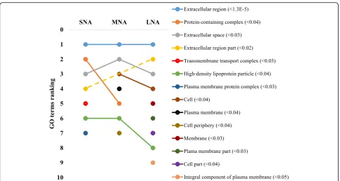
Gene Ontology (GO) and enrichment analyses
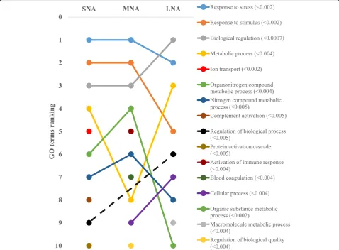
The STRING software allowed respective conversions of 1054, 1089, 1139 proteins in SNA, MNA, and LNA datasets, corresponding to ~ 65% of total proteins (see Additional file 2: Table S2). The highly significantly enriched GO terms in CC, MF, and BP were ranked according to significance levels in Figs. 2, 3, and 4, respectively. Dynamic ranking (increase, stable, or decrease) of GO terms were observed across SNA, MNA, and LNA.
In Fig. 2, proteins associated with “extracellular re-gion” GO term were the highest and significantly enriched (FDR < 1.3 × 10⁻⁵) in all follicle category. Mean-while, fluctuation and decrease significances in protein enriched were observed within the “protein-containing complex”, “extracellular space”, “cell”, and “high density lipoprotein particle” GO terms throughout follicle growth. In contrast, the significant enrichment of pro-teins in other GO terms (i.e., “extracellular region part”, membrane”, “plasma membrane part”), made them pro-imminent GO terms in LNA samples. Unlike in CC, the GO terms’patterns in MF showed high ranking fluctuations Fig. 1Total protein detection (a) and annotations (b). Venn Diagram of porcine follicular fluid was constructed with tools available at the
Table 1Functional classification of porcine follicular fluid proteomes during follicle development-Cellular component category
GO terms ~ GO names Number of annotations per follicle developmental stage, % Shared proteins
SNA MNA LNA n, %
GO:0005575 ~ Cellular component 435, 23.2 462, 23.6 479, 22.9 63, 21.0
GO:0005623 ~ Cell 345, 18.4 364, 18.6 379, 18.1 49, 16.3
GO:0005622 ~ Intracellular 286, 15.3 304, 15.5 336, 16.1 34, 11.3
GO:0016020 ~ Membrane 236, 12.6 242, 12.4 248, 11.8 29, 9.7
GO:0005737 ~ Cytoplasm 224, 11.9 233, 11.9 266, 12.7 24, 8.0
GO:0005634 ~ Nucleus 157, 8.4 155, 7.9 182, 8.7 21, 7.0
GO:0005576 ~ Extracellular region 76, 4.1 80, 4.1 82, 3.9 34, 11.3
GO:0005615 ~ Extracellular space 62, 3.3 62, 3.2 62, 3.0 35, 11.7
GO:0005694 ~ Chromosome 29, 1.5 30, 1.5 31, 1.5 3, 1.0
GO:0009986 ~ Cell surface 14, 0.7 13, 0.7 14, 0.7 4, 1.3
GO:0005578 ~ Extracellular matrix 11, 0.6 11, 0.6 14, 0.7 4, 1.3
Total annotations 1875, 100 1956, 100 2093, 100 300, 100
SNASmall non-atretic follicle (< 4 mm),MNAMedium non-atretic follicle (4–6 mm),LNALarge non-atretic follicle (> 6–12 mm)
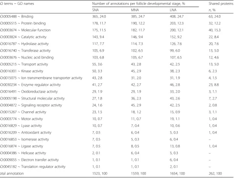
Table 2Functional classification of porcine follicular fluid proteomes during follicle development–Molecular function category
GO terms ~ GO names Number of annotations per follicle developmental stage, % Shared proteins
SNA MNA LNA n, %
GO:0005488 ~ Binding 365, 24.0 385, 24.7 408, 24.7 63, 24.0
GO:0005515 ~ Protein binding 178, 11.7 190, 12.2 203, 12.3 32, 12.2
GO:0003674 ~ Molecular function 175, 11.5 182, 11.7 200, 12.1 40, 15.3
GO:0003824 ~ Catalytic activity 143, 9.4 146, 9.4 152, 9.2 22, 8.4
GO:0016787 ~ Hydrolase activity 117, 7.7 114, 7.3 126, 7.6 20, 7.6
GO:0016740 ~ Transferase activity 105, 6.9 102, 6.5 99, 6.0 13, 5.0
GO:0003676 ~ Nucleic acid binding 103, 6.8 105, 6.7 107, 6.5 12, 4.6
GO:0005215 ~ Transport activity 55, 3.6 43, 2.8 42, 2.5 13, 5.0
GO:0016301 ~ Kinase activity 50, 3.3 45, 2.9 38, 2.3 6, 2.3
GO:0015075 ~ Ion transmembrane transporter activity 43, 2.8 31, 2.0 31, 1.9 4, 1.5
GO:0030234 ~ Enzyme regulator activity 41, 2.7 42, 2.7 46, 2.8 23, 8.8
GO:0016491 ~ Oxidoreductase activity 29, 1.9 29, 1.9 33, 2.0 3, 1.1
GO:0005198 ~ Structural molecular activity 27, 1.8 36, 2.3 43, 2.6 7, 2.7
GO:0004872 ~ Signaling receptor activity 24, 1.6 45, 2.9 42, 2.5 2, 0.8
GO:0015267 ~ Channel activity 23, 1.5 18, 1.2 15, 0.9 3, 1.1
GO:0003774 ~ Motor activity 10, 0.7 11, 0.7 19, 1.1 1, 0.4
GO:0016829 ~ Lyase activity 10, 0.7 7, 0.4 10, 0.6 1, 0.4
GO:0016209 ~ Antioxidant activity 7, 0.5 6, 0.4 5, 0.3 1, 0.4
GO:0016853 ~ Isomerase activity 7, 0.5 5, 0.3 6, 0.4 –
GO:0016874 ~ Ligase activity 7, 0.5 8, 0.5 13, 0.8 1, 0.4
GO:0004386 ~ Helicase activity 2, 0.1 6, 0.4 5, 0.3 –
GO:0009055 ~ Electron transfer activity 1, 0.1 1, 0.1 6, 0.4 –
GO:0045182 ~ Translation regulator activity 1, 0.1 1, 0.1 2, 0.1 –
Total annotation 1523, 100 1559, 100 1654, 100 262, 100
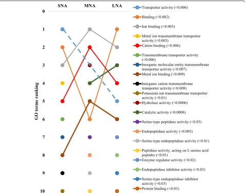
(Fig.3). Terms such as“transporter activity”,“binding”,“ cat-ion binding”, and metal ion binding”highly fluctuated across samples, to become the most enriched GO terms in LNA, together with“catalytic activity”. The patterns in the BP cat-egory (Fig.4) revealed similar fluctuations, mainly observed between MNA and LNA samples with decreased ranking of “response to stress”,“response to stimulus”,“organonitrogen compound metabolic process”, and “nitrogen compound metabolic process”. “Biological regulation”, metabolic process”, regulation of biological process”,“cellular process” GO terms were highly ranked in LNA, while“ion transport”, “complement activation”, and “protein activation cascade” were not significantly ranked as compared to LNA. Interest-ingly, proteins such as serine protease inhibitor, clade E (SERPINE); plasminogen activator, urokinase (PLAU); plasminogen activator, urokinase receptor (PLAUR) were detected only in SNA, MNA and LNA, respectively.
Protein-to-protein interactions (PPI) and pathway analyses
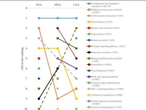
Data were generated through significantly enrichment (P< 10−16) and highest detection confidence (0.9).
Kmeans clustering networks showed variable patterns of PPI in SNA, MNA, and LNA, and“complement and co-agulation cascades” appeared as the major KEGG path-way in all samples (Fig. 5; FDR < 5.36 × 10⁻¹⁰). Other pathways such as “focal adhesion”, “protein digestion and adsorption”, “PI3K-Akt signaling pathway”, “ ECM-receptor interaction”, and ovarian steroidogenesis” were highly ranked in LNA samples.
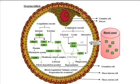
The Fig.6schematizes the“complement and coagulation cascades” pathway with contributing proteins of our data-sets, such as F12 (Coagulation factor XII); F2 (Thrombin); C5 (Complement 5a anaphylatoxin); C2 (Complement component 2); C3 (Complement 3); C7 (Complement com-ponent 7); CLU (Clusterin); A2M (Alpha-2-macroglobulin), F5 (Coagulation factor V); C4 (Uncharacterized protein; complement C4); PLG (Plasminogen); SERPIN (Serine pep-tidase inhibitor) that were found in all follicle categories.
Discussion
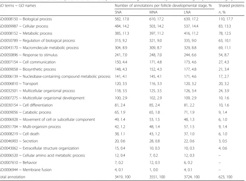
Ovarian follicular fluid is rich in various biocompounds (i.e., fatty acids, metabolites, proteins, steroids) that Table 3Functional classification of porcine follicular fluid proteomes during follicle development–Biological process category
GO terms ~ GO names Number of annotations per follicle developmental stage, % Shared proteins
SNA MNA LNA n, %
GO:0008150 ~ Biological process 582, 17.0 610, 17.2 639, 17.2 110, 17.7
GO:0009987 ~ Cellular process 484, 14.2 503, 14.2 537, 14.4 83, 13.3
GO:0008152 ~ Metabolic process 385, 11.3 397, 11.2 416, 11.2 78, 12.5
GO:0050789 ~ Regulation of biological process 313, 9.2 321, 9.0 335, 9.0 63, 10.1
GO:0043170 ~ Macromolecule metabolic process 304, 8.9 309, 8.7 329, 8.8 69, 11.1
GO:0050896 ~ Response to stimulus 241, 7.0 248, 7.0 244, 6.6 54, 8.7
GO:0007154 ~ Cell communication 150, 4.4 171, 4.8 173, 4.6 27, 4.3
GO:0009058 ~ Biosynthetic process 148, 4.3 152, 4.3 177, 4.8 21, 3.4
GO:0006139 ~ Nucleobase-containing compound metabolic process 141, 4.1 145, 4.1 171, 4.6 17, 2.7
GO:0006810 ~ Transport 120, 3.5 116, 3.3 120, 3.2 20, 3.2
GO:0032501 ~ Multicellular organismal process 118, 3.5 125, 3.5 126, 3.4 24, 3.9
GO:0007275 ~ Multicellular organismal development 100, 2.9 102, 2.9 109, 2.9 10, 1.6
GO:0030154 ~ Cell differentiation 81, 2.4 85, 2.4 81, 2.2 10, 1.6
GO:0009056 ~ Catabolic process 65, 1.9 65, 1.8 71, 1.9 9, 1.4
GO:0006928 ~ Movement of cell or subcellular component 49, 1.4 53, 1.5 48, 1.3 6, 1.0
GO:0051704 ~ Multi-organism process 42, 1.2 48, 1.4 57, 1.5 9, 1.4
GO:0008219 ~ Cell death 38, 1.1 43, 1.2 37, 1.0 6, 1.0
GO:0046903 ~ Secretion 20, 0.6 28, 0.8 22, 0.6 3, 0.5
GO:0043062 ~ Extracellular structure organization 15, 0.4 10, 0.3 10, 0.3 4, 0.6
GO:0006520 ~ Cellular amino acid metabolic process 12, 0.4 7, 0.2 12, 0.3 –
GO:0007610 ~ Behavior 7, 0.2 12, 0.3 6, 0.2 –
GO:0006944 ~ Membrane fusion 4, 0.1 1, 0.0 4, 0.1 –
Total annotation 3419, 100 3551, 100 3724, 100 623, 100
diversely and time-mannerly regulate oogenesis and folli-culogenesis [26–32]. Previous studies have demonstrated differential developmental competence of oocytes [17–19] derived from small (SNA: < 4 mm), medium (MNA: 4–7 mm) and large (LNA: > 6–12 mm) follicles [20, 21, 38], while others revealed the dynamic content of the follicular fluid playing critical roles during folliculogenesis [20, 21]. The present study characterizes the proteomes of specific developmental stage ovarian follicular fluids in pigs. It provides sets of predicted protein functions and networks that will contribute to the identification of key regulators of follicle and oocyte growth and potential biomarkers for oocyte quality assessment in assisted reproduction.
In the present study, colorless follicles with homogenous texture were selected and classified as small, medium, and large. Because of their reduced volumes, follicular fluids (FF) of different small or medium follicles of the same ovary were mixed during collection. A further reduction of individual various among ovaries and pigs was achieved by pooling FF of comparable higher or lower estradiol levels within the same follicle size; high estradiol levels in FF have been reported as features of healthy and non-atretic follicles [43, 44]. The follicle size-dependent in-crease of estradiol was in agreement with previous studies primate human [45] and non-human [46], rat [47] and livestock [48, 49], reporting association between estradiol synthesis and follicular cell viability. In contrast, intrafolli-cular protein concentrations were comparable regardless
of their origins, and were consistent with previous findings in pigs [50], cows [51], buffaloes [52], and goats [13].
The benefits of proteomic tools in the study of ovarian follicular fluids have been highlighted in various species, despite its known limitations (e.g., dynamic range of samples [53]. In the present work, proteome analyses re-vealed follicle size-dependent protein contents of follicu-lar fluids with 1627, 1699 and 1756 proteins in SNA, MNA and LNA, respectively. This increase total protein numbers contrasted with the unchanged protein concen-trations across samples, but therefore confirmed the in-creasing FF volume throughout follicle growth. A similar increase pattern of total protein numbers across follicle sizes has been reported in bovine FF [35]. It is specu-lated that protein increased numbers may result from higher 1) secretions of granulosa and thecal cells and 2) permeability of the ovarian-blood-follicle barrier allow-ing more protein flux into the FF to support follicle growth and oocyte maturation.
species, including human [54], buffalo [12], goat [13], and horse [37,55].
Focusing on the 50 most abundant proteins with NCBI annotations, 92% (46) of proteins was found across all follicle developmental stages. As expected, proteins such as serum albumin, fibrinogen beta chain, serotransferrin, ceruloplasmin, immunoglobulin G, serpin A3–8, alpha-2-HS-glycoprotein, and inhibitor of carbonic anhydrase were among the highly abundant proteins across samples. Within the abundant protein lists, fibulin-1 and (complement factor) properdin were not detected in the LNA samples. Fibulin-1 is a calcium-binding glycopro-tein found in the blood that participates to the extracel-lular matrix organization and plays a role in thrombosis leading to blood clot [56]. In the present study, the ab-sence of fibulin-1 in the LNA following its lower detec-tion in MNA are indicative of the follicle’s preparation
for the ovulation process that could be impeded by blood clotting. Furthermore, properdin is a member of the complement family found in the blood plasma and stabilizing the alternative pathway of the complement system (C3 and C5 convertases) by extending the half-life of the C3 and C5 converting enzymes [57]. The expression of properdin seems associated with early apoptotic cells and its absence in LNA and comparable detection levels in SNA and MNA samples coincide with the incidence of apoptosis during folliculogenesis.
proportion (88%) of protein annotations for fundamental predictions of protein functions through gene ontologies.
Gene ontology (GO) analyses revealed higher proportions (> 41%) of both“cellular component”and“cell”GO terms in cellular component category, while over 35% of general “binding” and “protein binding” GO terms in molecular function category and 42% of“biological process”,“cellular process”, and “metabolic process” GO terms in biological process category were the most prominent in all generated proteome datasets. These distributions were consistent with previous reports in bovine [35] and caprine FF [13]. The high proportions (> 15%) of intracellular proteins found in all proteome datasets were in agreement with a report in buffalo [12], and may reflect the perpetual remodeling (apoptosis and/or mitogenic turnover rate) of granulosa cells to support osmotic force of the growing liquid volume in the antrum cavity [58,59]. In all proteome datasets, pro-teins associated with“extracellular region”were the highly
enriched, while the FDR-based ranking of GO terms re-vealed the high significance of “high-density lipoprotein particle”GO term correlating with high level of lipid in fol-licular fluid [5]. Also, high enrichments of “binding”, “ion binding”, “metal ion binding”, and “cation binding” GO terms coincide with reported high levels of albumin and immunoglobulin in follicular fluids [12,13,37]. The FDR-based ranking revealing great fluctuations of significantly enriched GO terms (per functional category) across prote-ome datasets indicates likely changes of protein functions throughout folliculogenesis that can affect female fertility. Further investigations of critically enriched GO terms could permit the identification of potential protein candidates for follicle growth and/or oocyte maturation.
and“amoebiasis”exhibited higher significances in SNA and MNA, while LNA samples were characterized by pathways such as“protein digestion and absorption”,“focal adhesion”, and“PI3K-Akt signaling pathway”. In agreement with pre-vious reports in women [60], goats [13] and mares [55], the “complement and coagulation cascades”was the highly sig-nificant pathway across all samples. Both cascades shared protein inhibitors and activators of serine endopeptidase that are associated with inflammatory response [61], a required process for ovulation [62]. Numerous proteins that contribute to blood coagulation and fibrin degradation through plasminogen (PLG) or proteolysis by thrombin activation [61] were detected. For example, alpha-2-macroglobulin (A2M), a protease inhibitor that binds small or large proteinases such as the plasmin that regulates proteolytic activity [63], influences cumulus cells ex-pansion by inhibiting zinc-dependent metalloproteases [64]. In this way, the excess of A2M lead a reduction in porcine cumulus expansion [64, 65]. Clusterin
(CLU), another protease inhibitor, is an important regulator of the complement [66], preventing apoptosis of follicular cells [67].
The current study reveals the presence of serine protease inhibitor clade E (SERPINE) and plasmino-gen activator urokinase (PLAU) in SNA and MNA follicular fluids, respectively. Both proteins have shown potential roles on cumulus cells expansion in a previous study [68]. On the other hand, the detec-tion of PLAU receptors (PLAUR) in large follicles suggests a possible role of the PLAU/PLAUR complex at the later stage of folliculogenesis to favor oocyte maturation. We speculate that the establishment of this complex allows cell-surface plasminogen activa-tion through its conversion into plasmin inducing localized degradation of the extracellular matrix, which in turn influence the expansion of cumulus cells surrounding the maturing oocyte and prepare the ovarian tissues for ovulation.
Conclusion
Our study provides the proteome profiles of pFF during follicle growth for comparative analyses. Functional analyses of proteome datasets revealed protein clusters and networks that constitute the “core signature” of folliculogenesis, from which any deviations may serve for developmental biomarkers search. Hierarchical GO term arrangements were in-dicative of (qualitative and/or quantitative) protein variations during follicle growth. Several detected proteins and functions were already known, and the current study highlights follicle developmental-stage specific proteins that regulates oocyte maturation in small (SERPINE), medium (PLAU), and large (PLAUR) follicle sizes. Meantime, properdin and fibulin-1 were found as proteins of interest for follicle growth. The proposed datasets provide a useful basis for future studies to better comprehend ovarian folliculogenesis.
Supplementary information
Supplementary informationaccompanies this paper athttps://doi.org/10. 1186/s40104-019-0400-3.
Additional file 1:Table S1.Proteome datasets.
Additional file 2:Table S2.Functional analyses.
Abbreviations
A2M:Alpha-2-macroglobulin; BP: Biological process; C2: Complement component 2; C3: Complement 3; C4: Uncharacterized protein, complement C4; C5: Complement 5a anaphylatoxin; C7: Complement component 7; CC: Cellular component; CLU: Clusterin; E2: Estradiol; F12: Coagulation factor XII; F2: Thrombin; F5: Coagulation factor V; FF: Follicular fluid; GO: Gene ontology; LNA: Large non-atretic; MF: Molecular function; MNA: Medium non-atretic; PLAU: Plasminogen activator, urokinase; PLAUR: Plasminogen activator, urokinase receptor; PLG: Plasminogen; PPI: Protein-to-protein interactions; SERPIN: Serine peptidase inhibitor; SERPINE: Serine protease inhibitor, clade E; SNA: Small non-atretic
Acknowledgements
Not applicable.
Authors’contributions
VMP and JMF performed the experiments, analyzed data, and wrote the first draft to the manuscript. SFL participated in data interpretation and development of the first draft of the manuscript. JRF, STW, and PLR contributed to the experimental design and manuscript revision. All authors read and approved the final manuscript.
Funding
This study was supported by the USDA-ARS Biophotonics (Grant # 58–6402– 3-018) and the Coordenação de Aperfeiçoamento de Pessoal de Nível Superior–Brasil (CAPES)–Finance Code 001. VM Paes is the recipient of a PhD scholarship from CAPES.
Availability of data and materials
All data generated or analyzed during this study are included in this published article [and its supplementary information files].
Ethics approval and consent to participate
Not applicable.
Consent for publication
Not applicable.
Competing interests
The authors declare that they have no competing interests.
Author details
1Department of Animal and Dairy Sciences, Mississippi State University, 4025
Wise Center, PO Box 9815, Starkville, Mississippi State MS 39762, USA.
2Laboratory of Manipulation of Oocyte and Preantral follicles, State University
of Ceará, Fortaleza, CE, Brazil.
Received: 30 May 2019 Accepted: 18 October 2019
References
1. Armstrong DT. Effects of maternal age on oocyte developmental competence. Theriogenology. 2001;55:1303–22.
2. Shah JS, Sabouni R, Cayton Vaught KC, Owen CM, Albertini DF, Segars JH. Biomechanics and mechanical signaling in the ovary: a systematic review. J Assist Reprod Genet. 2018;35:1135–48.
3. Van Den Hurk R, Zhao J. Formation of mammalian oocytes and their growth, differentiation and maturation within ovarian follicles. Theriogenology. 2005;63:1717–51.
4. Gosden RG, Hunter RH, Telfer E, Torrance C, Brown N. Physiological factors underlying the formation of ovarian follicular fluid. J Reprod Fertil. 1988;82:813–25.
5. Grupen CG. The evolution of porcine embryo in vitro production. Theriogenology. 2014;81:24–37.
6. Ginther OJ. The theory of follicle selection in cattle. Domest Anim Endocrinol. 2016;57:85–99.
7. Chou CH, Chen MJ. The effect of steroid hormones on ovarian follicle development. Vitam Horm. 2018;107:155–75.
8. Savion N, Gospodarowicz D. Patterns of cellular peptide synthesis by cultured bovine granulosa cells. Endocrinology. 1980;107:1798–807. 9. Fortune JE. Ovarian follicular growth and development in mammals. Biol
Reprod. 1994;50:225–32.
10. O’callaghan D, Yaakub H, Hyttel P, Spicer LJ, Boland MP. Effect of nutrition and superovulation on oocyte morphology, follicular fluid composition and systemic hormone concentrations in ewes. J Reprod Fertil. 2000;118:303–13. 11. Nandi S, Kumar VG, Manjunatha BM, Grupta PS. Biochemical
composition of ovine follicular fluid in relation to follicle size. Develop Growth Differ. 2007;49:61–6.
12. Fu Q, Huang Y, Wang Z, Chen F, Huang D, Lu Y, et al. Proteome profile and quantitative proteomic analysis of buffalo (Bubalusbubalis) follicular fluid during follicle development. Int J Mol Sci. 2016;17:618.
13. Paula Junior AR, van Tilburg MF, Lobo MDP, Monteiro-Moreira ACO, Moreira RA, Melo CHS, et al. Proteomic analysis of follicular fluid from tropically-adapted goats. Anim Reprod Sci. 2018;188:35–44.
14. Fair T, Hyttel P, Greve T. Bovine oocyte diameter in relation to maturational competence and transcriptional activity. Mol Reprod Dev. 1995;42:437–42. 15. Crozet N, Ahmed-Ali M, Dubos MP. Developmental competence of goat
oocytes from follicles of different size categories following maturation, fertilization and culture in vitro. J Reprod Fertil. 1995;103:293–8. 16. Marchal R, Vigneron C, Perreau C, Bali-Papp A, Mermillod P. Effect of
follicular fluid size on meiotic and developmental competence of porcine oocytes. Theriogenology. 2002;57:1523–32.
17. Bagg MA, Nottle MB, Armstrong DT, Grupen CG. Relationship between follicle size and oocyte developmental competence in prepubertal and adult pigs. Reprod Fertil Dev. 2007;19:797–803.
18. Crozet N, Dahirel M, Gall L. Meiotic competence of in vitro grown goat oocytes. J Reprod Fertil. 2000;118:367–73.
19. Otoi T, Yamamoto K, Koyama N, Tachikawa S, Suzuki T. Bovine oocyte diameter in relation to developmental competence. Theriogenology. 1997;48:769–74. 20. Vatzias G, Hagen DR. Effects of porcine follicular fluid and
oviduct-conditioned media on maturation and fertilization of porcine oocytes in vitro. Biol Reprod. 1999;60:42–8.
21. Algriany O, Bevers M, Schoevers E, Colenbrander B, Dieleman S. Follicle size-dependent effects of sow follicular fluid on in vitro cumulus expansion, nuclear maturation and blastocyst formation of sow cumulus oocyte complexes. Theriogenology. 2004;62:1483–97.
22. Dumesic DA, Meldrum DR, Katz-Jaffe MG, Krisher RL, Schoolcraft WB. Oocyte environment: follicular fluid and cumulus cells are critical for oocyte health. Fertil Steril. 2015;103:303–16.
23. Revelli A, Delle Piane L, Casano S, Molinari E, Massobrio M, Rinaudo P. Follicular fluid content and oocyte quality: from single biochemical markers to metabolomics. Reprod Biol Endocrinol. 2009;7:40.
24. Leroy JL, Vanholder T, Delanghe JR, Opsomer G, Van Soom A, Bols PE, et al. Metabolite and ionic composition of follicular fluid from different-sized follicles and their relationship to serum concentrations in dairy cows. Anim Reprod Sci. 2004;80:201–11.
25. Paramio MT, Izquierdo D. Recent advances in in vitro embryo production in small ruminants. Theriogenology. 2016;86:152–9.
26. Younis AI, Brackett BG, Fayrer-Hosken RA. Influence of serum and hormones on bovine oocyte maturation and fertilization in vitro. Gamete Res. 1989;23:189–201.
27. Ding J, Foxcroft GR. Epidermal growth factor enhances oocyte maturation in pigs. Mol Reprod Dev. 1994;39:30–40.
28. Xia P, Tekpetey FR, Armstrong DT. Effect of IGF-I on pig oocyte maturation, fertilization, and early embryonic development in vitro, and on granulosa and cumulus cell biosynthetic activity. Mol Reprod Dev. 1994;38:373–9. 29. Grupen CG, Nagashima H, Nottle MB. Role of epidermal growth factor and
insulin-like growth factor-I on porcine oocyte maturation and embryonic development in vitro. Reprod Fertil Dev. 1997;9:571–5.
30. Akaki Y, Yoshioka K, Noguchi M, Hoshi H, Funahashi H. Successful piglet production in a chemically defined system for in-vitro production of porcine embryos: dibutyryl cyclic amp and epidermal growth factor-family peptides support in-vitro maturation of oocytes in the absence of gonadotropins. J Reprod Dev. 2009;55:446–53.
31. Arunakumari G, Shanmugasundaram N, Rao VH. Development of morulae from the oocytes of cultured sheep preantral follicles. Theriogenology. 2010; 74:884–94.
32. Magalhães DM, Duarte AB, Araújo VR, Brito IR, Soares TG, Lima IM, et al. In vitro production of a caprine embryo from a preantral follicle cultured in media supplemented with growth hormone. Theriogenology. 2011;75:182–8. 33. Angelucci S, Ciavardelli D, Di Giuseppe F, Eleuterio E, Sulpizio M, Tiboni GM,
et al. Proteome analysis of human follicular fluid. Biochim Biophys Acta. 1764;2006:1775–85.
34. Twigt J, Steegers-Theunissen RP, Bezstarosti K, Demmers JA. Proteomic analysis of the microenvironment of developing oocytes. Proteomics. 2012;12:1463–71.
35. Ferrazza RA, Garcia HDM, Schmidt EMDS, Mihm Carmichael M, Souza FF, Burchmore R, et al. Quantitative proteomic profiling of bovine follicular fluid during follicle development. Biol Reprod. 2017;97:835–49.
36. Kamalludin MH, Garcia-Guerra A, Wiltbank MC, Kirkpatrick BW. Proteomic analysis of follicular fluid in carriers and non-carriers of the trio allele for high ovulation rate in cattle. Reprod Fertil Dev. 2018;30:1643–50. 37. Fahiminiya S, Labas V, Roche S, Dacheux JL, Gérard N. Proteomic analysis of
mare follicular fluid during late follicle development. Proteome Sci. 2011;9:54. 38. Sun YL, Ping ZG, Li CJ, Sun YF, Yi KL, Chen L, et al. Comparative proteomic analysis of follicular fluids from normal and cystic follicles in sows. Reprod Domest Anim. 2011;46:889–95.
39. Feugang JM, Liao SF, Willard ST, Ryan PL. In-depth proteomic analysis of boar spermatozoa through shotgun and gel-based methods. BMC Genomics. 2018;19:62.
40. Van Den Berg BH, Harris T, McCarthy FM, Lamont SJ, Burgess SC. Non-electrophoretic differential detergent fractionation proteomics using frozen whole organs. Rapid Commun Mass Spectrom. 2007;21:3905–9.
41. Sokale A, Peebles ED, Zhai W, Pendarvis K, Burgess S, Pechan T. Proteome profile of the pipping muscle in broiler embryos. Proteomics. 2011;11:4262–5. 42. Yates JR, Eng JK, McCormack AL, Schieltz D. Method to correlate tandem
mass spectra of modified peptides to amino acid sequences in the protein database. Anal Chem. 1995;67:1426–36.
43. Matsuda F, Inoue N, Manabe N, Ohkura S. Follicular growth and atresia in mammalian ovaries: regulation by survival and death of granulosa cells. J Reprod Dev. 2012;58:44–50.
44. Ireland JJ, Roche JF. Development of antral follicles in cattle after prostaglandin-induced luteolysis: changes in serum hormones, steroids in follicular fluid, and gonadotropin receptors. Endocrinology. 1982;111:2077–86. 45. Guzel Y, Bildik G, Oktem O. Sphingosine-1-phosphate protects human
46. Xu J, Lawson MS, Xu F, Du Y, Tkachenko OY, Bishop CV, et al. Vitamin D3 regulates follicular development and intrafollicular vitamin D biosynthesis and signaling in the primate ovary. Front Physiol. 2018;9:1600. 47. Wu J, Tu D, Yuan LY, Yi JE, Tian Y. T-2 toxin regulates steroid hormone
secretion of rat ovarian granulosa cells through cAMP-PKA pathway. Toxicol Lett. 2015;232:573–9.
48. Murdoch WJ. Inhibition by oestradiol of oxidative stress-induced apoptosis in pig ovarian tissues. J Reprod Fertil. 1998;114:127–30.
49. Cheewasopit W, Laird M, Glister C, Knight PG. Myostatin is expressed in bovine ovarian follicles and modulates granulosal and thecal steroidogenesis. Reproduction. 2018;156:375–86.
50. Schuetz AW, Anisowicz A. Cation and protein composition of ovarian follicular fluid of the pig: relation to follicle size. Biol Reprod. 1974;11:64–72. 51. Andersen MM, Kroll J, Byskov AG, Faber M. Protein composition in the fluid
of individual bovine follicles. J Reprod Fertil. 1976;48:109–18.
52. Shiny J, Deshmukh BT, Dhaware SD, Borkar SD. Follicular fluid protein profile in buffalo. Vet Sci Res J. 2015;6:71–9.
53. Lippolis JD, Reinhardt TA. Utility, limitations, and promise of proteomics in animal science. Vet Immunol Immunopathol. 2010;138:241–51.
54. Bayasula IA, Kobayashi H, Goto M, Nakahara T, Nakamura T, et al. A proteomic analysis of human follicular fluid: comparison between fertilized oocytes and non-fertilized oocytes in the same patient. J Assist Reprod Genet. 2013;30:1231–8.
55. Dutra GA, Ishak GM, Pechanova O, Pechan T, Peterson DG, Jacob JCF, et al. Seasonal variation in equine follicular fluid proteome. Reprod Biol Endocrinol. 2019;17:29.
56. Tran H, Tanaka A, Litvinovich SV, Medved LV, Haudenschild CC, Argraves WS. The interaction of fibulin-1 with fibrinogen. A potential role in hemostasis and thrombosis. J Biol Chem. 1995;270:19458–64.
57. Kemper C, Hourcade DE. Properdin: new roles in pattern recognition and target clearance. Mol Immunol. 2008;45:4048–56.
58. Rodgers RJ, Irving-Rodgers HF. Formation of the ovarian follicular antrum and follicular fluid. Biol Reprod. 2010;82:1021–9.
59. Regan SLP, Knight PG, Yovich JL, Leung Y, Arfuso F, Dharmarajan A. Granulosa cell apoptosis in the ovarian follicle-a changing view. Front Endocrinol. 2018;9:61.
60. Zamah AM, Hassis ME, Albertolle ME, Williams KE. Proteomic analysis of human follicular fluid from fertile women. Clin Proteomics. 2015;12:5. 61. Oikonomopoulou K, Ricklin D, Ward PA, Lambris JD. Interactions between
coagulation and complement—their role in inflammation. Semin Immunopathol. 2012;34:151–65.
62. Jesam C, Salvatierra AM, Schwartz JL, Croxatto HB. Suppression of follicular rupture with meloxicam, a cyclooxygenase-2 inhibitor: potential for emergency contraception. Hum Reprod. 2010;25:368–73.
63. Rehman AA, Ahsan H, Khan FH. Alpha-2-macroglobulin: a physiological guardian. J Cell Physiol. 2013;228:1665–75.
64. Appeltant R, Beek J, Maes D, Bijttebier J, Van Steendam K, Nauwynck H, et al. Hampered cumulus expansion of porcine cumulus-oocyte complexes by excessive presence of alpha2-macroglobulin is likely mediated via inhibition of zinc-dependent metalloproteases. Anim Sci J. 2017;88:1279–90. 65. Bijttebier J, Tilleman K, Dhaenens M, Deforce D, Van Soom A, Maes D.
Comparative proteome analysis of porcine follicular fluid and serum reveals that excessiveα2-macroglobulin in serum hampers successful expansion of cumulus-oocyte complexes. Proteomics. 2009;9:4554–65.
66. Humphreys DT, Carver JA, Easterbrook-Smith SB, Wilson MR. Clusterin has chaperone-like activity similar to that of small heat shock proteins. J Biol Chem. 1999;274:6875–81.
67. Zwain IH, Amato P. cAMP-induced apoptosis in granulosa cells is associated with up-regulation of P53 and bax and down-regulation of clusterin. Endocr Res. 2001;27:233–49.