O R I G I N A L A R T I C L E
Molecular characterization and bioactivity profile of the tropical
sponge-associated bacterium
Shewanella algae
VCDB
R. S. Rachanamol•A. P. Lipton• V. Thankamani• A. R. Sarika•J. Selvin
Received: 12 August 2013 / Revised: 1 January 2014 / Accepted: 11 February 2014 / Published online: 5 March 2014
ÓSpringer-Verlag Berlin Heidelberg and AWI 2014
Abstract The pigmented, rod-shaped, Gram-negative, motile bacteria isolated from marine spongeCallyspongia diffusaexhibiting bioactivity was characterized as Shewa-nella algae (GenBank: KC623651). The 16S rRNA gene sequence-based phylogenetic analysis showed its similarity with the member of Shewanella and placed in a separate cluster with the recognized bacteria S. algae (PSB-05 FJ86678) with which it showed 99.0 % sequence similar-ity. Growth of the strain was optimum at temperature 30°C, pH 8.0 in the presence of 2.0–4.0 % of NaCl. High antibiotic activity against microbes such as Escherichia coli(MTCC 40),S.typhii(MTCC 98),P.vulgaris(MTCC 426),V.fluvialis,V.anguillarum,E.cloacae, andL.lactis was recorded. The growth of fungal pathogens such as Aspergillus niger, Aspergillus fumigatus, Saccharomyces cerevisiae, andColletotrichum gloeosporioideswas effec-tively controlled.
Keywords Sponge bacteria Callyspongia diffusa
Shewanella algaeMolecular characterization
Antimicrobial activity
Introduction
In marine sponges, increased evidences indicated the involvement of associated microorganisms for the pro-duction of secondary metabolite (Proksch et al. 2002; Chelossi et al. 2007; Flemer et al.2012). The functional interaction between associated microbes and host sponge is considered as essential in the bioprospecting sphere (Selvin et al. 2010). Among the microbes, the bacterial community in the sponge differs significantly from sea-water and sediments in density and diversity. The sponge-associated bacteria could be viewed as a highly potential source for the production of antibiotic compounds (Waters et al. 2010; Graca et al. 2013). The recovery of culturable bacterial strains with different bioactivity pro-files shows that the sponge tissues could be rich sources for isolating new strains of bacteria with potential capa-bilities of producing novel bioactive secondary metabo-lites (Xiong et al. 2013). Molecular and metagenomic approaches opened up new scientific possibilities, which also revealed the metabolic pathways involved in the production of natural compound by the diverse uncultur-able symbiotic bacteria. The marine spongeCallyspongisa diffusa has been investigated for isolating novel bacterial strains. Bacteria such as Pseudomonas aeruginosa, Escherichia coli, Vibrio parahaemolyticus, and Vibrio cholera have been reported to be associated with C. dif-fusa(Boobathy et al.2009). An in-depth knowledge about the C. diffusa-associated bacteria and their bioactivity profile is necessary to promote their bioprospecting ave-nues. Considering this, the marine sponge C.diffusa was chosen to retrieve maximum number of bacteria for exploring their bioactivity potential in a sponge-free environment. The results of isolation and characterization of a potential strain of bacteria viz., Shewanella algae, Communicated by G. Gerdts.
R. S. Rachanamol (&)V. Thankamani
Department of Biotechnology, University of Kerala, Kariyavattom, Trivandrum 695581, Kerala, India e-mail: [email protected]
A. P. LiptonA. R. Sarika
Marine Biotechnology Laboratory, Vizhinjam Research Centre of CMFRI, Vizhinjam 695521, Kerala, India
J. Selvin
Centre for Microbiology, Pondicherry University, Puducherry 605014, India
and its characteristics together with bioactive potential are presented in this paper.
Materials and methods
Collection and preparation of extract fromCallyspongia diffusa
Specimens ofC.diffusawere collected by SCUBA during September 2011 from southwest coast of India at depth ranging from 6 to 7 m off Vizhinjam (8°22045°N: 76°59029°E), at a distance of about 1.5 km from the shore. The sponge specimens were found attached to submerged rocks. Immediately upon collection, the sponge specimens were transferred to new polythene covers in situ to mini-mize the external contaminants and transient bacteria. The aqueous extract of the sponge was prepared by squeezing the sand-free specimen in sterile seawater. The resultant solution was filtered using Whatman filter paper (No. 1) and stored at 4.0°C in refrigerator for further experiments. Isolation of sponge-associated bacteria
The surface particles and microbes of freshly collected C. diffusa were removed by rinsing with sterile seawater and immediately transferred to sterile container. From the surface-sterilized sponge, required quantity of sponge tis-sue was excised using sterile scalpel and then homoge-nized. The homogenate was diluted in sterile saline, and appropriate dilutions were spread on Zobell Marine Agar (ZMA-HiMedia) and plates were incubated at 28.0±2.0°C. The colonies of bacteria developed on the agar plates were enumerated to determine the CFU/g. Seawater sample from the site of collection was also examined to enumerate the ambient bacterial load. Biochemical characterization of the isolate
The morphology and biochemical characteristics of the sponge isolates viz., Gram staining, motility, utilization of carbon sources, H2S production, and MR–VP test, were
carried out as per Smibert and Krieg (1994). Screening of bacterial isolates from sponge tissue for antimicrobial activity
The bacterial isolates obtained from the sponge were grown individually in Zobell Marine Broth (ZMB) for 48 h. The cell-free culture media of each isolate was pre-pared by centrifuging the growth medium and filtering of the supernatant using 0.2-lm filters. Sterile discs (6 mm) loaded with 15ll cell-free culture extracts of the
individual bacterial isolates associated withC.diffusawere placed on Muller-Hinton agar plates seeded with human and fish pathogenic bacteria. The presence or absence of inhibitory activity against the indicator organisms was determined after incubating the agar plates for 24 h at 37°C and measuring the zone of inhibition.
The antifungal activity of bacterial strains isolated from sponge tissue against a few food-borne fungi such as Aspergillus niger, Aspergillus fumigatus, Saccharomyces cerevisiae,and plant pathogensSclerotia rolfsii, Phytoph-thora palmivora, Phytophthora colocasiae, and Colleto-trichum gloeosporioides was evaluated. The activity was determined by dual-well agar diffusion technique. For testing antifungal activity, malt extract agar (MEA) med-ium was used as given by Mushtaq et al. (2010). After solidification of the media, 6.0-mm wells were cut on the agar surface and each well in a plate was loaded with 30ll of 24-h sponge-isolated bacterial broth culture; similarly, the cell-free supernatant (30 ll) of the same sponge bac-terial strains was also added. ZMB served as the control. A circular piece of 4.0 mm diameter from 7-day-old fungal cultures were cut and removed by cork borer, and was stabbed on agar previously loaded with bacterial culture and its cell-free supernatant. The observations with respect to the inhibition of fungal growth were observed after 5 days of incubation.
Determination of growth and antibacterial activity pattern at different conditions
Considering the preliminary results of bioactivity, the potential isolate VCDB was grown in ZMB and response with respect to maximum growth and metabolite produc-tion was recorded at varied temperature and pH. For determining the optimum temperature, the inoculated broth was incubated at varying temperatures from 20 to 50°C at 5 °C intervals. The pH optima was determined by varying the initial pH of the ZMB from 5.0 to 10.0 and inoculated with the isolate VCDB. The samples were removed after every 12 h to determine the growth OD (660 nm) and antibacterial activity.
Molecular characterization and phylogenetic analysis of the isolate VCDB
The similarity and homology of the 16S rRNA gene sequence was compared with existing sequences available in the data bank of NCBI using BLAST search. The DNA sequence was aligned, and phylogenetic tree was con-structed by Mega 5.05 using neighbor-joining method, and the sequence was deposited in GenBank.
Results
Identification ofCallyspongia diffusa and characteristics of bacterial isolates
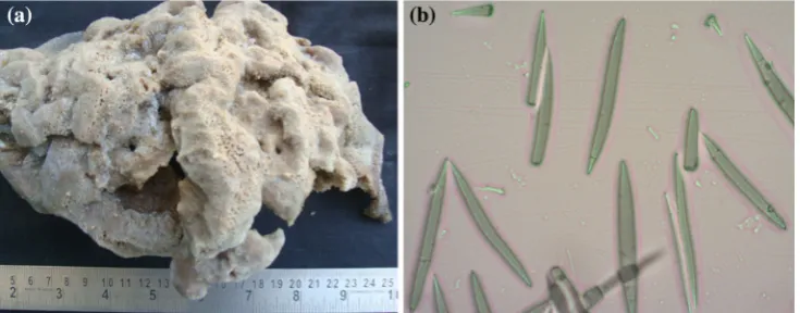
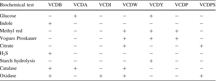
The sponge was identified asC.diffusabased on spicules morphology and further confirmed by Dr. P. A. Thomas, Sponge Taxonomist (Fig.1a, b). From the randomly chosen sponge tissue, seven morphologically and cultur-ally distinct bacterial strains were isolated and desig-nated—VCDB, VCDA, VCDW, VCDI, VCDY, VCDP, and VCDPS; the cultural and biochemical characteristics of the isolates are given in Tables1 and 2. The total number of bacterial colonies from the sponge interior was
recorded to be 50.0 % higher than the bacterial load from the ambient seawater. The black-pigmented VCDB isolate that proved highly potential by further observations was not recorded from the seawater, and the CFU of VCDB constituted 12.5 % of total microbial load of 169104CFU/g.
Screening of bacterial isolates from sponge tissue for antimicrobial activity
The results of the cell-free supernatant of associated bac-teria showed that all the bacbac-terial isolates except VCDI showed a significant antibacterial activity against human and fish pathogens (Table3). The VCDB isolate showed mild activity againstV.harveyi,V.anguillarum, L.lactis, P.vulgarisMTCC 426, andE.coliMTCC 40 with zone of inhibition of 10 mm. Significant activity was exhibited against S.typhi MTCC 92 (11 mm),V.fluvialis(14 mm), and E. cloacae (11 mm). The isolate VCDA exhibited inhibitory activity againstE.cloacae(12 mm)V.fluvialis, and V. anguillarum (11 mm) and mild activity of 7 mm against V.harveyiand 9 mm againstP.vulgaris.
Fig. 1 Morphological characters ofCallyspongia diffusa(a) and shape of spicules (9400) (b)
Table 1 Cultural characteristics of sponge-associated bacteria from
Callyspongia diffusa
Isolates Cultural characters of sponge-associated bacteria
Colony shape
Color Form Gram staining
and shape
Motility
VCDB Round Creamy white with black pigmentation
Convex, mucoid with black pigmentation
-rod ?
VCDA Spreading Creamy white Rhizoid ?rod ?
VCDW Round White Mucoid ?rod –
VCDI Round White Flattened ?cocci –
VCDY Round Yellow Raised, pin-headed -cocci –
VCDP Round Pink Small concentric ring ?rod –
Antifungal potential of isolates from sponge tissue The experimental results on antifungal activity of bacterial strains fromC.diffusaand its aqueous extract showed that only VCDB and its cell-free supernatant possessed effec-tive biocontrol potential against A. niger, A. fumigatus, S.cerevisiae,andC.gloeosporioides.
Determination of growth and metabolite production at different conditions
The growth (OD 660 nm) of the strain VCDB increased drastically between 12 and 48 h after incu-bation, and a maximum growth was recorded at
exponential phase after 48 h of incubation (Fig.2a, b). The sponge isolate VCDB showed its pigmentation at the late log phase of 24 h and maximized at 48 h. The antibacterial activity increased after 48 h and noted maximum when incubated at 30.0 °C with an initial media pH of 8.0.
Molecular characterization of the VCDB isolate
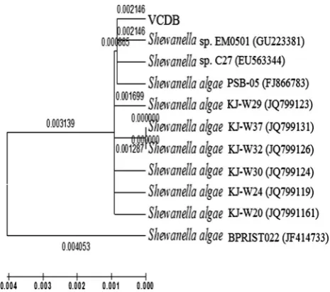
The BLAST search of the 16S rRNA gene sequence of the isolate VCDB showed a lineage withS.algaewith 99.0 % similarity to representative strains of genus Shewanella (Fig.3). The gene sequence was deposited in GenBank with accession number KC623651.
Table 2 Biochemical characteristics of the sponge isolates
Biochemical test VCDB VCDA VCDI VCDW VCDY VCDP VCDPS
Glucose - ? - - ? -
-Indole ? - - -
-Methyl red - - - ? ? ?
-Vogues Proskauer - - - ? ? ?
-Citrate - - - ? - - ?
H2S ? - - -
-Starch hydrolysis - - - - ? -
-Catalase ? ? - ? - -
-Oxidase ? - ? ? - - ?
Table 3 Antimicrobial activity ofCallyspongia diffusa -associated bacterial isolates
Test pathogens VCDB VCDA VCDW VCDI VCDY VCDP VCDPS
E.coli 10 10 9 0 9 9 9
S.typhi 11 0 0 0 0 0 0
P.vulgaris 10 9 0 0 0 0 0
V.harveyi 10 7 0 0 0 0 7
V.vulnificus 0 11 0 0 0 0 8
V.fluvialis 14 11 7 0 0 0 0
V.anguillarum 10 11 0 0 7 8 0
E.cloacae 11 12 13 0 0 0 0
L.lactis 10 0 0 0 0 7 0
Discussion
Several studies have indicated that sponge–microbe inter-actions are important to produce potential bioactive metabolites in sponge (Selvin et al. 2009; Prabha et al.
2010; Thomas et al. 2010). The results of the present investigation revealed that the tropical sponge C. diffusa yielded the bacterial isolate S. algae VCDB KC623651 with potential antibacterial activity. Earlier, Hentschel et al. (2001) gave strong evidences about the presence of taxonomically diverse bacteria within mesohyl of the sponge Aplysina, which created an environment favoring the production of defense compounds and antimicrobials. Penesyan et al. (2011) identified an antibacterial compound from sponge-associated bacteriaPseudovibrioD323.
The bacterial isolateS.algaeobtained from the tropical spongeC.diffusawas characterized as Gram-negative, rod-shaped, motile, aerobic, catalase, and oxidase-positive strain-producing H2S with black pigmentation at the late
exponential phase. Similar observation for differentiating S. algae by the production of Pyomelanin at the late exponential phase was reported by Turick et al. (2008). Optimal growth of VCDB strain occurred at temperature 30°C, pH 8.0 in the presence of 2.0–4.0 % of NaCl. The strain also showed a high antimicrobial activity in these optimized conditions. Yang et al. (2006) described a sim-ilar psychrophilic Shewanella spongiae from a marine sponge living at 20 m water depth of East Sea (Sea of Japan) with optimum growth at temperature 40°C in pH level of 6.0–6.5 in the presence of 2.5 % of NaCl. The phylogenetic analysis revealed their type strain HJ037
shared phyletic line with S. algae and Shewanella ama-zonensis. Strains of Shewanella isolated from aquatic environments and sediments were described by Bowman et al. (1997), Bozal et al. (2002), and Satomi et al. (2006). The bacteria of this genus have attracted great attention due to their diverse respiratory capacities, illustrated by their ability to utilize a wide range of terminal electron accep-tors, including oxygen, nitrate, metals, and sulfur com-pounds (Kostka et al.2002), and to degrade pollutants such as chlorinated solvents (Petrovskis et al. 1994), petroleum (Semple and Westlake 1987), and RDX (1,3,5-trinitroper-hydro-1,3,5-triazine) (Zhao et al. 2004). Lee et al. (2006) isolatedShewanella iricinae sp. nov. from marine sponge Ircinia dendroides associated with Posidonia sea grass from Mediterranean Sea that showed 95.0 % sequence similarity with the recognized bacterial strain S. algae (UST040317). Chelossi et al. (2007) isolated 461 bacterial strains from sponge Chondrilla nucula, among them 60 strains were reported to be potential. The study also inferred CHC2 strain isolated from C. nucula and was sharing 89.0 % similarity to the Gram-negativeS.algaeas well as to uncultured bacterial clone with antibacterial activities towardS.aureus,P. atlantica,and P.elongata. The VCDB strain KC 623651 isolated from C. diffusa showed 99.0 % similarity with S. algae on molecular analysis exhibited high antimicrobial activity against human and fish pathogens,E.coli(MTCC 40)E.cloacae, P.vulgaris(MTCC 426),S.typhii(MTCC 98),andVibribo sps. The vibriostatic efficacy of S. algae isolated from Penaeus monodon against V. parahaemolyticus and V. alginolyticuswas statistically proved by Shakibazadeh et al. (2008). A highly potential bioactivity by VCDB against fish pathogens likeE.cloaceaceand vibrios such as V. fluvialis and V. anguillarum mostly isolated as patho-genic strains from marine ornamental fishes as well as shrimp aquaculture was observed in this study.
Sponge associates with antifungal activity was reported by Li Zheng et al. (2005) in his study on marine sponge Hymeniacidon perleve-associated bacterium NJ6-31, which inhibited the growth of S. cerevisia ACCC 2.1882. The antifungal property exerted byS.algae KC623651 and its exocellular products against plant fungal pathogen Col-lectotrichum gloeosporioides and food-borne fungus A.niger, A.fumigatus,andS.cerevisiaeindicate the pos-sibility of using S.algae and its exocellular products as a biocontrol agents against fungal pathogens. Wang (2006) explored the relationship between microbial diversity and host specificity of marine sponge–bacteria associations in Cymbastela concentrica, Callyspongia,andStylinosp. by 16S rDNA sequencing of excised DGGE bands. Three distinct types of bacteria with specific characteristics like ‘‘specialists’’ found on only host species, ‘‘sponge associ-ates’’ found on multiple hosts but not in seawater, and Fig. 3 Phylogenetic tree of strain VCDB drawn in Mega 5.05 using
‘‘generalists’’ found from multiple hosts and seawater. The molecular study on VCDB strain fromC. diffusashowed 99.0 % similarity withS.algae isolated from mangroves. Thus, the presentS.algaeKC623651 could be considered as a true ‘‘sponge associates’’ as the same strain could not be isolated from seawater collected along with the sponge sample from same vicinity of the Vizhinjam coast. Inter-estingly, this species constituted about 12.5 % of the total retrievable bacterial species recorded.
Microbial associates of sponges gained significance only when a remarkable similarity was found between the compounds isolated predominantly from sponges and those found in terrestrial of entirely different taxa (Perry et al.
1998). The rapid development of pharmaceutical markets and technological development has increased the demand for the production of novel products from sponges toward human health concerns (Yung et al. 2011). At the same time, due considerations are to given to limit and regulate the mass collection of sponges from their natural habitat as suggested by Sipkema et al. (2011). In this context, the sponge-associated bacteria gain much relevance and importance making the least dependence from natural collection and consequent destructions. The findings could encourage the development of antimicrobial metabolites fromS.algaeKC623651 alone, originally isolated from the sponge C. diffusa and least further dependence on host collection from marine habitat.
References
Boobathy S, Soundarapandian P, Subasri V, Vembu N, Gunasundari V (2009) Bioactivities of protein isolated from marine sponge,
Sigmadocia fibulatus. Current Res J Biol Sci1(3):160–162 Bowman J, McCammon S, Nichols D, Skerratt J, Rea S, Nichols P,
McMeekin T (1997) Shewanella gelidimarina sp. nov. and
Shewanella frigidimarinasp. nov. novel Antarctic species with the ability to produce eicosapentaenoic acid (20:5v3) and grow anaerobically by dissimilatory Fe(III) reduction. Int J Syst Bacteriol 47:1040–1047
Bozal N, Montes MJ, Tudela E, Jimenez F, Guinea J (2002)
Shewanella frigidimarina and Shewanella livingstonensis sp. nov. isolated from Antarctic coastal areas. Int J Syst Evol Microbiol 52:195–205
Chelossi E, Pantile P, Pronzato R, Milanese M, Hentschel U (2007) Bacteria with antimicrobial properties isolated from the Medi-terranean sponges Chondrilla nucula and Petrosia ficiformis. Aquat Microb Ecol 49:157–163
Flemer B, Kennedy J, Margassery JP, O’Gara F, Dobson ADW (2012) Diversity nad antimicrobial activities of microbe from two Irish marine sponge,Suberites carnosusand Leucosolenia
sp. J Appl Microbiol 112:289–301
Graca AP, Bondoso J, Gaspar H, Xavier JR, Monteiro MC, Cruz M, Oves-Costales D, Vicente F, Lage OM (2013) Antimicrobial activity of heterotrophic bacterial communities from the marine sponge Erylus discophorus (Astrophorida, Geodiidae). PLoS One 8(11):e78992
Hentschel U, Schmid M, Wager M, Fiseseler L, Genert C, Hacker J (2001) Isolation and phylogenetic analysis of bacteria with antimicrobial activities from the Mediterranean spongeAplysina cavernicola. FEMS Microbiol Ecol 35:305–312
Kamke J, Michael MW, Schmitt S (2010) Activity profiles for marine sponge-associated bacteria obtained by 16S rRNA vs 16S rRNA gene comparisons. ISME J 4:498–508
Kostka JE, Dalton DD, Skelton H, Dollhopf S, Stucki JW (2002) Growth of iron(III)-reducing bacteria on clay minerals as the sole electron acceptor and comparison of growth yields on a variety of oxidized iron forms. Appl Environ Microbiol 68(12):6256–6262
Lee OO, Lau SCK, Tsoi MMY et al (2006)Shewanella irciniaesp. nov. a novel member of the familyShewanellaceaeisolated from the marine spongeIrcinia dendroidesin the Bay of Villefranche Mediterranean Sea. Int J Syst Evol Microbiol 56:2871–2877 Penesyan A, Tebben J, Lee M, Thomas T, Harder SKT, Egan S
(2011) Identification of the antibacterial compound produced by the marine epiphytic bacterium Pseudovibrio sp. D323 and related sponge-associated bacteria. Mar Drugs 9:1391–1400 Perry NB, Blunt JW, Munro MHG, Mycalamide A (1998) An
antiviral compound from a New Zealand sponge of the genus
Mycale. J Am Chem Soc 110:4850–4851
Petrovskis EA, Vogel TM, Adriaens P (1994) Effects of electron acceptors and donors on transformation of tetrachloromethane by
Shewanella putrefaciens MR-1. FEMS Microbiol Lett 121:357–363
Prabha D, Wahidullah S, Rodrigues C, D’Souza L (2010) The sponge-associated bacterium Bacillus licheniformis SAB1: a source of antimicrobial compounds. Mar Drugs 8(4):1203–1212 Proksch P, Edrada RA, Ebel R (2002) Drugs from the seas—current status and microbiological implications. Appl Microbiol Bio-technol 5:125–134
Satomi M, Vogel BF, Gram L, Venkateswaran K (2006)Shewanella hafniensissp. nov. and Shewanella morhuae sp. nov. isolated from marine fish of the Baltic Sea. Int J Syst Evol Microbiol 56:243–249
Selvin J, Shanmughapriya S, Gandhimathi R, Kiran GS, Ravji TR, Natarajaseenivasan K, Hema TA (2009) Optimization and production of novel antimicrobial agents from sponge associated actinomycetesNocardiopsis dassonvilleiMAD08. Appl Micro-biol Biotechnol 83:435–445
Selvin J, Ninawe AS, Kiran GS, Lipton AP (2010) Sponge–microbial interactions: ecological implications and bioprospecting ave-nues. Crit Rev Microbiol 36:82–90
Semple KM, Westlake DWS (1987) Characterization of iron reducing
Alteromonas putrefaciens strains from oil-field fluids. Can J Microbiol 33:366–371
Shakibazadeh S, Saad CR, Christianus A, Kamarudin MS, Sijam K, Shamsudin MN, Neela VK (2008) Evaluation of in vitro
vibriostatic activity ofShewanella algaeisolated from healthy
Penaeus monodon. African J Biotechnol 7(21):3952–3961 Sipkema D, Schippers K, Maalcke WJ, Yang Yu, Salim S, Blanch
HW (2011) Multiple approaches to enhance the cultivability of bacteria associated with the marine spongeHaliclona(gellius) sp. Appl Environ Microbiol 77(6):2130–2140
Smibert RM, Krieg NR (1994) Phenotypic characterization. In: Gerhardt P, Murray RGE, Wood WA, Krieg NR (eds) Methods for general and molecular bacteriology. ASM Press, Washington DC, pp 607–654
Mushtaq S, Ali A, Khokhar I, Mukhtar I (2010) Antagonistic potential of soil bacteria against food borne fungi. World J Appl Sci 11(8):966–969
Turick CE, Caccavo F Jr, Tisa LS (2008) Pyomelanin is produced by
Shewanella algae BrY and affected by exogenous iron. Can J Microbiol 54(4):334–339
Wang G (2006) Diversity and biotechnological potential of the sponge associated microbial consortia. J Ind Microbiol Biotech-nol 33(7):545–551
Waters AL, Hill RT, Place AR, Hamann MT (2010) The expanding role of marine microbes in pharmaceutical development. Curr Opin Biotechnol 23:539–543
Xiong Z-Q, Wang J-F, Hao Y-Y, Wang Y (2013) Recent trends in the discovery and development of marine microbial natural pro-ducts. Mar Drugs 11:700–717
Yang SH, Know KK, Lee HS, Kim SJ (2006)Shewanella spongiae
sp. nov. isolated from a marine sponge. Int J Syst Evol Microbiol 56:2879–2882
Yung PY, Burke C, Lewis M, Kjelleberg S, Thomas T (2011) Novel antibacterial proteins from the microbial communities associated with the spongeCymbastela concentricaand the green algaUlva australis. Appl Environ Microbiol 77(4):1512–1515
Zhao JS, Greer CW, Thiboutot S, Ampleman G, Hawari J (2004) Biodegradation of the nitramine explosives hexahydro-1,3,5-trinitro-1,3,5-triazine and octahydro-1,3,5,7-tetranitro-1,3,5,7-tetrazocine in cold marine sediment under anaerobic and oligotrophic conditions. Can J Microbiol 50:91–96