D I S C O V E R Y N O T E S
Open Access
Evolutionary patterns of phosphorylated serines
Yerbol Z Kurmangaliyev
1,2, Alexander Goland
1, Mikhail S Gelfand
1,3*Abstract:Posttranslationally modified amino acids are chemically distinct types of amino acids and in terms of evolution they might behave differently from their non-modified counterparts. In order to check this possibility, we reconstructed the evolutionary history of phosphorylated serines in several groups of organisms. Comparisons of substitution vectors have revealed some significant differences in the evolution of modified and corresponding non-modified amino acids. In particular, phosphoserines are more frequently substituted to aspartate and glutamate, compared to non-phosphorylated serines.
Reviewers:This article was reviewed by Arcady Mushegian and Sandor Pongor.
Findings
Post-translational modifications play an important role in diversifying protein structure and function [1,2]. Pro-tein phosphorylation is one of the most important and widely distributed types of post-translational modifica-tions. In eukaryotes, reversible protein phosphorylation plays a key role in the signal transduction and other processes [3,4]. Recent advances in mass spectrometry allowed for large-scale identification of phosphorylation events [5]. Analyses of these data have already revealed some specific structural and evolutionary features of phosphoserines. Phosphoserines tend to occur in intrin-sically disordered regions [6-8] and regions correspond-ing to alternatively spliced gene segments [9]. Phosphorylated amino acids are more conserved than their non-phosphorylated counterparts [7,10-12]. Some very old phosphorylation events potentially can be com-mon to organisms fromArchaeato human [10].
Here we investigated another evolutionary aspect of protein modification sites. Since modified amino acids chemically are a distinct type of amino acids, in terms of evolution they might behave differently from their non-modified counterparts (on the top of the different level of conservation). To analyse differences in the evolution of standard amino acids and their modified counterparts, we reconstructed the evolution of phosphorylated amino acids in three groups of organisms. Particularly, we stu-died phosphorylation of serine in the human, fruit fly and yeast proteomes.
Phosphorylation sites were downloaded from the PHOSIDA [7] and PhosphoPEP [13] databases. For yeast and fruit fly we studied phosphoserines obtained in two high-throughput experiment each, by different groups of researchers [13-16]. For human we used data-sets obtained in four different high-through experiments [17-20]. Phosphorylation is highly dynamic process, and the overlap of phosphorylation events identified in dif-ferent experiments from various cell lines and tissues is relatively small. Sites observed to be phosphorylated in more than one high-througput experiment likely are modified in a more constitutive manner, or at least represent a more reliable dataset of phosphoserines.
We analysed the evolution of modification sites and their non-modified counterparts separately among eight vertebrates (humanHomo sapiens;chimpanzeePan tro-golodytes; mouse Mus musculus; rat Rattus norvegicus; cow Bos taurus; dog Canis lupus familiaris; chicken Gallus gallus; and zebrafish Danio rerio), eleven fruit flies (Drosophila melanogaster; D. yakuba;D. erecta; D. sechecellia; D. ananassae; D.pseudoobscura; D. persimi-lis; D. wilistoni; D. mojavensis; D. viripersimi-lis; D. grimshawi) and fifteen fungi (Saccharomyces cerevisiae; S. para-doxus; S. mikatae; S. bayanus; Candida glabrata; S. cas-telli; Kluyveromyces waltii; K. lactis; Ashbya gossypii; Debaryomyces hansenii; C. albicans; Yarrowia lipolytica; Aspergillus nidulans; Neurospora crassa; Schizosaccharo-myces pombe). Orthologs of modified H. sapiens pro-teins were obtained from HomoloGene [21]; for D. melanogaster, from FlyBase [22]; and forS. cerevisiae, from FungalOrthogroups [23]. Only orthologs with the highest identity to the modified protein were selected * Correspondence: [email protected]
1
Institute for Information Transmission Problems (the Kharkevich Institute) RAS, Bolshoi Karetny pereulok 19, Moscow, 127994, Russia
Full list of author information is available at the end of the article
from each species. Multiple alignments were constructed using ClustalW [24].
As mentioned above, the evolutionary features and frequencies of phosphoserines may depend on structural context. Especially, phosphoserines tend to occur within intrinsically disordered regions of proteins [6-8]. To take this into account, we analysed serines from disordered regions and ordered regions of phosphoproteins sepa-rately. Intrinsically disordered regions were predicted by PONDR VSL2 [25].
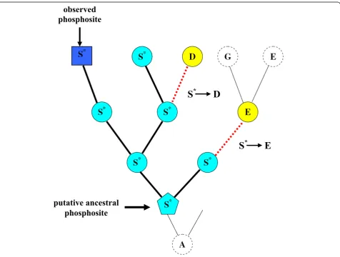
For each phosphorylated serine, we have reconstructed the evolution of this site in the corresponding taxonomi-cal group using a fast modification of the maximum likelihood algorithm (A. Goland, in preparation). Since we cannot reconstruct the moment in evolution when a residue had become modified, we assumed that it coin-cides with the oldest residue of the given type in a given tree (Figure 1). Then we calculated the number of sub-stitutions of ancestral putative modification sites to
other amino acids, and calculated the vectors of substi-tution frequencies.
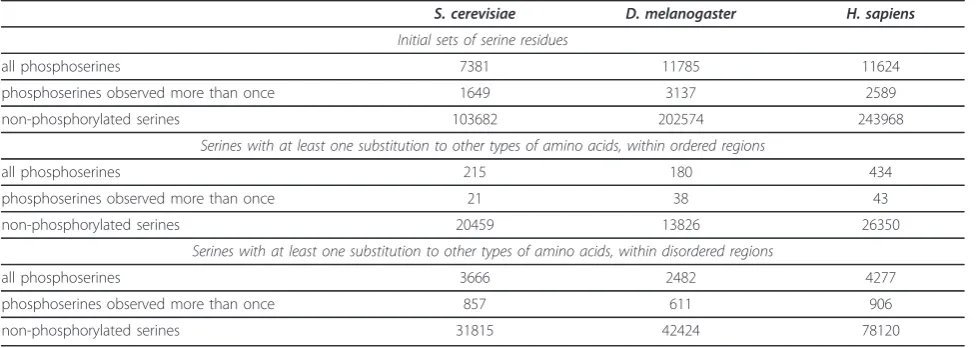
Only a fraction of phosphoserines from the initial datasets were aligned to other types of amino acids in our data, and very small number of them occurred in ordered regions. Thus further analyses were performed only for serines from regions predicted to be intrinsi-cally disordered. The final datasets of phosphorylated and non-phosphorylated serines included only sites that experienced at least one substituition to other types of amino acids and originated from disordered regions of phosphoproteins (Table 1). Some phosphorylation events were observed in more than one experiment, and this subset was also analyzed separately.
The control sets consisted of non-modified serine resi-dues from disordered regions of the same proteins. To measure the statistical significance of the difference between substitution vectors of modified and non-modi-fied serines we performed bootstraping of control sets.
putative ancestral
phosphosite
observed
phosphosite
S
*D
S
*E
To do that that, we generated 10000 random control sets of non-phosphorylated serines. Each control set was of the same size as the corresponding phosphorylated set (generic sets and subsets of reliable phophosites).
Structural features of phosphoserines may not be lim-ited to disorder of surrounding protein regions, and may include other specific properties such as secondary structures, solvent availability etc. Therefore, to maxi-mally eliminate the confounding effects, we created additional control sets containing non-modified serines located at the same protein regions as modification sites. Non-modified serines, was collected at the maxi-mal distance of 10, 11 and 9 amino acid residues from phosphoserines, for yeast, fruit fly and human respec-tively. Again, the size of the control sets was the same as the size of the respective phosphoserine sets.
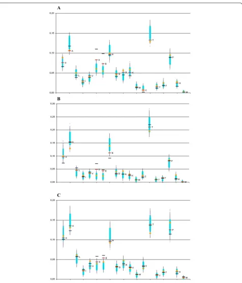
Differences in the substitution vectors between phos-phorylated and non-phosphos-phorylated serines from disor-dered regions varied among different groups of organism, but some trends were stable and significant (Figure 2). Rather unexpectedly, we did not observe any preference for substitution of phosphoserines to other aminoacids that may be phosphorylated, that is as threo-nine and tyrosine. At the same time, phosphorylation converts serine into a negatively charged amino acid, and, as one can see in Figure 2 in all three datasets phosphoserines are more frequently substituted to aspartate and glutamate than non-phosphorylated ser-ines. In both cases the substitution rates of phosphoser-ines are much higher than in all bootstraps of control sets (P-value << 10-4). In the case of the more reliable subsets of phosphoserines observed in several experi-ments, the subtitution rate to aspartate and glutamate is even higher, and also lies outside the interval of boot-straps that in this case is wider, as the sample size is
smaller. At that, artificial substitution of serine to aspar-tate and glutamate, called phosphomimetic mutation, is widely used to confirm phosphorylation of serine [26,27].
There are considerable other shifts of substitution rates common to all three taxa. Particularly, phosphoser-ines are relatively rarely substituted to alanphosphoser-ines and cysteines (Figure 2). However, in these cases, the con-trol-set substitution vectors of non-phosphorylated ser-ines located in the same regions as phosphoserser-ines were also shifted in the same direction as phosphoserines (as compared to all non-phosphorylated serines). Hence, these shifts are likely related not to modifications, but to specific features of these regions.
The rates of substitutions to aspartate and glutamate in the additional control sets of nearest non-phosphory-lated serines also are not shifted, with the exception of vertebrates where they are also shifted toward higher values (but still to a much weaker extent than in case of phosphoserines). Note that these control sets may be contaminated by phosphoserines. Indeed, phosphoser-ines tends to co-occur, forming clusters [28]. Therefore the sets of nearest non-phosphorylated serines likely contain phosphoserines which were not detected yet. Removing these phosphoserines would increase the sig-nificance of our observations.
The comparison with nearest non-phosphorylated ser-ines takes into account the fact that phosphoserser-ines tend to occur in intrinsically disordered regions. Meth-ods used in large-scale phosphoproteomic experiments are based on selection of negatively charged peptides which results in a bias towards enrichment of phospho-peptides with acidic residues [29,30]. This fact, coupled with the fact that phosphoserines may shift positions within rapidly evolving disordered regions [31] and
Table 1 Datasets of phosphorylated and non-phosphorylated serines
S. cerevisiae D. melanogaster H. sapiens
Initial sets of serine residues
all phosphoserines 7381 11785 11624
phosphoserines observed more than once 1649 3137 2589
non-phosphorylated serines 103682 202574 243968
Serines with at least one substitution to other types of amino acids, within ordered regions
all phosphoserines 215 180 434
phosphoserines observed more than once 21 38 43
non-phosphorylated serines 20459 13826 26350
Serines with at least one substitution to other types of amino acids, within disordered regions
all phosphoserines 3666 2482 4277
phosphoserines observed more than once 857 611 906
non-phosphorylated serines 31815 42424 78120
$
%
&
general problems of alignments of such regions could distort our analysis. But this would have the same influ-ence on our control sets of non-modified serines from the same regions of proteins. Hence the observed differ-ences between these controls and phosphoserines can-not be explained by such artifacts.
In addition to serine phosphorylation, we analysed the evolution of another abundant type of protein modifica-tion, lysine acetylation. Recently two large datasets of human acetylation sites became available [32,33]. We observed some differences between substitution vectors of acetylated and non-acetylated lysines, but the results obtained for these two sets of acetyllysines were discor-dant (data not shown). As noted in one of these papers [32], the spectrum of acetylated proteins is different between these two datasets obtained from different tis-sues. We observed that less than 2% of sites are com-mon for both datasets. It is seems that the available acetyllysine data are not sufficient for meaningful analysis.
It should be taken into account that our substitution vectors are probably enriched with false-positive phos-phosites. This results from of our over-simplified assumption that a site is modified from the first appear-ance of the corresponding residue in the evolutionary record. Additionally, phosphoserines from large-scale experiments may be false-positive sites. There is evi-dence that many phosphorylation sites could be non-functional or non-specific, as sometimes non-functional tar-gets of phosphorylation are not particular sites, but entire protein regions [31,34,35]. On the other hand, the control sets could contain not yet detected phosphoser-ines. These false positives and false negatives should blur the differences between the substitution vectors of modified and non-modified residues. Most likely, the real level of differences is higher than the one observed here.
Reviewers’comments
Reviewer’s Report 1
Reviewer 1: Arcady Mushegian - Stowers Institute, Kan-sas City, USA
Reviewer’s comment
The idea of comparing of evolutionary substitution pat-terns of modified and non-modified residues in proteins is good, and the approach proposed by the authors, i.e., to reconstruct, using an ML model, the point at which the target of modification first emerged and then to see what it mutates to, is probably the only computational approach plausible at the moment.
I trust the authors that their implementation of this approach is technically sound, but, unfortunately, this is hard to ascertain from the submitted version of the manuscript, which reads as a preliminary draft devoid of
the quantitative details. This has to change - please pro-vide at least the following:
1. The collection of phosphorylated and acetylated sites: how many sites of each type in each organism are there?
Author’s Response A table with a description of the final datasets used for the construction of substitution vectors has been added to the revised version (Table 1).
Reviewer’s comment
2. The phosphorylation sites at least (also acetylated sites?) are said to occur more often in the intrinsically disorded regions. Taking the non-globular regions in the proteins (which can be identified, e.g., using Wootton and Federhen’s SEG program) as a proxy for “intrinsic disorder”, can it be said that the actual sample of modi-fied residues that the authors were working with is indeed more commonly occurring in such regions? And how does this sit with the ability to align the proteins in these regions?
Author’s ResponseWe predicted intrinsically disordered regions and recalculated substitution vectors separately for serine residues from disordered and ordered regions. Most of phosphoserines from the initial datasets came from protein regions predicted to be disordered (Table 1). Problems with alignments of such region are dis-cussed in the revised version. Additional controls of non-modified serines from same regions of proteins were introduced to address this problem.
Reviewer’s comment
3. The “control sets” of non-modified serines (more accurately, not-observed-to-be-modified serines): are these found in the disordered/non-globular regions to the same extent as the modified ones? If not, the con-trols may be biased with regard to amino acid composi-tion and to the regions of the protein molecules (e.g., buried vs exposed) - test this directly please.
Author’s ResponseIndeed, the amino acid composition of disordered regions and regions with a regular struc-ture differs strongly. As described in response to com-ment #2, in the revised version we considered both phosphosrylated and non-phosphorylated serines from disordered and regular regions separately. Moreover, as discussed in the revised text, sets including only closest non-modified serines provide an even better control for artifacts that could be caused by specifics of regions sur-rounding modification sites.
Reviewer’s comment
4. The trends that the authors discuss are interesting but weak - to what extent this may be explained by the small sample sizes? What was the statistical test for which the P-values are reported?
evolutionary reconstruction. The final datasets are described in Table 1.
To measure the statistical significance, we used boot-straps of control sets of non-modified serines. For all phosphoserines and, separately, for the subset of phos-phoserines observed in more than one experiment, we generated 10000 random sets of non-modified serines of appropriate size. For additional controls using neigh-bouring sites, we compiled sets of nearest serines of the same size as the corresponding sets of phosphorylated serines.
Reviewer’s comment
5. In vertebrates, the“neighboring”serines from control set 2 seem to be faithfully following the trend towards change into D or E, with some separation from the con-trol set 1. If this trend withstands the possible correc-tion proposed in #2, perhaps this means that, in a “disordered” region that has several serines, any or all of them may targets of phosphorylation. Perhaps then it would be interesting to sum the substitution vectors over the region that has several serines, at least one of which is phosphorylated (i.e., how likely is it that at least one serine in this region is substituted by amino acid X?)
Author’s Response The phosphoserines tends to cluster in the sequence [28]. Thus, as discussed in the revised version, the control set consisting of nearest non-phos-phorylated serines could be contaminated by false-nega-tive phosphoserines, not yet detected in experiments. On the other hand, as the trend in the control set of nearest serines is weaker, averaging of the substitution vectors would simply dilute the observation.
Reviewer’s Report 2
Reviewer 2: Sandor Pongor - International Centre for Genetic Engineering and Biotechnology, Trieste, Italy
Reviewer’s comment
There is mounting evidence in recent years that the study of post-translational modifications has important lessons for understanding diverse aspects of protein evo-lution. It has been noted among others that phosphory-lated sites tend to occur in those segments of the proteins that are intrinsically disordered and/or corre-spond to alternative splice sites. Currently there are insufficient data on the conservation of modified sites. Kurmangalyev and associates address this problem using carefully selected datasets and well-designed statistical analyses.
The authors conclude that there are significant differ-ences in the evolution of modified and corresponding non-modified amino acids. In particular, phosphoserines are more frequently substituted to aspartate and gluta-mate, compared to non-phosphorylated serines. Similarly, acetyllysines are more rarely substituted to isoleucine and
valine. These findings underline the importance of post-translational modifications when discussing the variation of residue conservations within sequence regions. The methodology is straightforward and sound and will be a useful template for future studies. The authors may want to add a few examples for situation where this approach can or can not be used.
Author’s ResponseAs discussed in the revised text, the analysis of a newly available dataset of human acetyla-tion sites [33] did not confirm our initial observaacetyla-tions. This is likely due to low reproducibility of currently available datasets of avetyllysines (the overlap between two datasets is extremely small). This suggests that con-clusions based on such analyses should be done care-fully, on data obtained from different sources and for a variety of organisms. We have encountered a similar problem with phosphothreonines and phosphotyrosines, where the datasets were simply too small for reliable conclusions.
Acknowledgements
We are grateful to Dmitry Malko, Ekaterina Ermakova and Anna Lyubetskaya who shared their programs and data, and to Stefka Tyanova and Jürgen Cox for useful discussions. This study was partially supported by the state contract 2.740.11.0101, Russian Foundation of Basic Research (09-04-92745), and program“Molecular and Cellular Biology”of the Russian Academy of Sciences.
Author details
1Institute for Information Transmission Problems (the Kharkevich Institute)
RAS, Bolshoi Karetny pereulok 19, Moscow, 127994, Russia.2National Center for Biotechnology of the Republic of Kazakhstan, Valikhanov str., 13/1, Astana, 010000, Republic of Kazakhstan.3Faculty of Bioengineering and Bioinformatics, Moscow State University, Vorobievy Gory 1-73, Moscow, 119991, Russia.
Authors’contributions
YK and MG conceived the study. YK compiled the data. AG developed algorithms. YK and MG performed calculations. YK and MG analyzed the results and wrote the paper. All authors have approved the final version.
Competing interests
The authors declare that they have no competing interests.
Received: 28 September 2010 Accepted: 9 February 2011 Published: 9 February 2011
References
1. Mann M, Jensen ON:Proteomic analysis of post-translational modifications.Nat Biotechnol2003,21:255-261.
2. Seo J, Lee KJ:Post-translational Modifications and Their Biological Function: Proteomic Analysis and Systematic Approaches.Journal of Biochemistry and Molecular Biology2004,37:35-44.
3. Hunter T:Signaling-2000 and beyond.Cell2000,100:113-127. 4. Cohen P:The origins of protein phosphorylation.Nat Cell Biol2002,4:
E127-E130.
5. Ptacek J, Snyder M:Charging it up: global analysis of protein phosphorylation.Trends Genet2006,22:545-554.
6. Iakoucheva LM, Radivojac P, Brown CJ, O’Connor TR, Sikes JG, Obradovic Z, Dunker AK:The importance of intrinsic disorder for protein
phosphorylation.Nucleic Acids Res2004,32:1037-1049.
7. Gnad F, Ren S, Cox J, Olsen JV, Macek B, Oroshi M, Mann M:PHOSIDA (phosphorylation site database): management, structural and evolutionary investigation, and prediction of phosphosites.Genome Biol
8. Collins MO, Yu L, Campuzano I, Grant SG, Choudhary JS:
Phosphoproteomic analysis of the mouse brain cytosol reveals a predominance of protein phosphorylation in regions of intrinsic sequence disorder.Mol Cell Proteomics2008,7:1331-1348.
9. Kurmangaliev EZh, Gel’fand MS:[Alternative splicing tends to involve phosphorylation sites].Mol Biol (Mosk)2009,43:572-574.
10. Macek B, Gnad F, Soufi B, Kumar C, Olsen JV, Mijakovic I, Mann M:
Phosphoproteome analysis of E. coli reveals evolutionary conservation of bacterial Ser/Thr/Tyr phosphorylation.Mol Cell Proteomics2008,
7:299-307.
11. Malik R, Nigg EA, Körner R:Comparative conservation analysis of the human mitotic phosphoproteome.Bioinformatics2008,24:1426-1432. 12. Boekhorst J, van Breukelen B, Heck A Jr, Snel B:Comparative
phosphoproteomics reveals evolutionary and functional conservation of phosphorylation across eukaryotes.Genome Biol2008,9:R144.
13. Bodenmiller B, Campbell D, Gerrits B, Lam H, Jovanovic M, Picotti P, Schlapbach R, Aebersold R:PhosphoPep - a database of protein phosphorylation sites in model organisms.Nat Biotechnol2008,
26:1339-1340.
14. Bodenmiller B, Malmstrom J, Gerrits B, Campbell D, Lam H, Schmidt A, Rinner O, Mueller LN, Shannon PT, Pedrioli PG, Panse C, Lee HK, Schlapbach R, Aebersold R:PhosphoPep - a phosphoproteome resource for systems biology research in Drosophila Kc167 cells.Mol Syst Biol
2007,3:139.
15. Hilger M, Bonaldi T, Gnad F, Mann M:Systems-wide analysis of a phosphataseknock-down by quantitative proteomics and phosphoproteomics.Mol Cell Proteomics2009,8:1908-1920.
16. Gnad F, de Godoy LM, Cox J, Neuhauser N, Ren S, Olsen JV, Mann M: High-accuracy identification and bioinformatic analysis of in vivo protein phosphorylation sites in yeast.Proteomics2009,9:4642-4652. 17. Olsen JV, Blagoev B, Gnad F, Macek B, Kumar C, Mortensen P, Mann M:
Global, in vivo, and site-specific phosphorylation dynamics in signaling networks.Cell2006,127:635-648.
18. Daub H, Olsen JV, Bairlein M, Gnad F, Oppermann FS, Körner R, Greff Z, Kéri G, Stemmann O, Mann M:Kinase-selective enrichment enables quantitative phosphoproteomics of the kinome across the cell cycle.Mol Cell2008,31:438-448.
19. Oppermann FS, Gnad F, Olsen JV, Hornberger R, Greff Z, Kéri G, Mann M, Daub H:Large-scale proteomics analysis of the human kinome.Mol Cell Proteomics2009,8:1751-1764.
20. Olsen JV, Vermeulen M, Santamaria A, Kumar C, Miller ML, Jensen LJ, Gnad F, Cox J, Jensen TS, Nigg EA, Brunak S, Mann M:Quantitative phosphoproteomics reveals widespread full phosphorylation site occupancy during mitosis.Sci Signal2010,3:ra3.
21. Wheeler Geer LY, Marchler-Bauer A, Geer RC, Han L, He J, He S, Liu C, Shi W, Bryant SH:The NCBI BioSystems database.Nucleic Acids Res2010,
38:D492-D496.
22. Tweedie S, Ashburner M, Falls K, Leyland P, McQuilton P, Marygold S, Millburn G, Osumi-Sutherland D, Schroeder A, Seal R, Zhang H, The FlyBase Consortium:FlyBase: enhancing Drosophila Gene Ontology annotations. Nucleic Acids Res2009,37:D555-D559.
23. Wapinski I, Pfeffer A, Friedman N, Regev A:Natural history and evolutionary principles of gene duplication in fungi.Nature2007,
449:54-61.
24. Larkin MA, Blackshields G, Brown NP, Chenna R, McGettigan PA, McWilliam H, Valentin F, Wallace IM, Wilm A, Lopez R, Thompson JD, Gibson TJ, Higgins DG:Clustal W and Clustal X version 2.0.Bioinformatics
2007,23:2947-2948.
25. Peng K, Radivojac P, Vucetic S, Dunker AK, Obradovic Z:Length-dependent prediction of protein intrinsic disorder.BMC Bioinformatics2006,7:208. 26. Tarrant MK, Cole PA:The chemical biology of protein phosphorylation.
Annu Rev Biochem2009,78:797-825.
27. Song Q, Pallikkuth S, Bossuyt J, Bers DM, Robia SL:Phosphomimetic mutations enhance phospholemman oligomerization and modulate its interaction with the NA/K-ATPase.J Biol Chem2011.
28. Schweiger R, Linial M:Cooperativity within proximal phosphorylation sites is revealed from large-scale proteomics data.Biol Direct2010,5:6. 29. Ficarro SB, McCleland ML, Stukenberg PT, Burke DJ, Ross MM,
Shabanowitz J, Hunt DF, White FM:Phosphoproteome analysis by mass spectrometry and its application to Saccharomyces cerevisiae.Nat Biotechnol2002,20:301-305.
30. Mann M, Ong SE, Grønborg M, Steen H, Jensen ON, Pandey A:Analysis of protein phosphorylation using mass spectrometry: deciphering the phosphoproteome.Trends Biotechnol2002,20:261-268.
31. Holt LJ, Tuch BB, Villén J, Johnson AD, Gygi SP, Morgan DO:Global analysis of Cdk1 substrate phosphorylation sites provides insights into evolution. Science2009,325:1682-1686.
32. Zhao S, Xu W, Jiang W, Yu W, Lin Y, Zhang T, Yao J, Zhou L, Zeng Y, Li H, Li Y, Shi J, An W, Hancock SM, He F, Qin L, Chin J, Yang P, Chen X, Lei Q, Xiong Y, Guan K:Regulation of Cellular Metabolism by Protein Lysine Acetylation.Science2010,327:1000-1004.
33. Choudhary C, Kumar C, Gnad F, Nielsen ML, Rehman M, Walther TC, Olsen JV, Mann M:Lysine acetylation targets protein complexes and co-regulates major cellular functions.Science2009,325:834-840.
34. Landry CR, Levy ED, Michnick SW:Weak functional constraints on phosphoproteomes.Trends Genet2009,25:193-197.
35. Tan CS, Jørgensen C, Linding R:Roles of“junk phosphorylation”in modulating biomolecular association of phosphorylated proteins?Cell Cycle2010,9:1276-1280.
doi:10.1186/1745-6150-6-8
Cite this article as:Kurmangaliyevet al.:Evolutionary patterns of phosphorylated serines.Biology Direct20116:8.
Submit your next manuscript to BioMed Central and take full advantage of:
• Convenient online submission
• Thorough peer review
• No space constraints or color figure charges
• Immediate publication on acceptance
• Inclusion in PubMed, CAS, Scopus and Google Scholar
• Research which is freely available for redistribution