T E C H N I C A L A D V A N C E
Open Access
Sparse reconstruction of compressive
sensing MRI using cross-domain stochastically
fully connected conditional random fields
Edward Li
1, Farzad Khalvati
2, Mohammad Javad Shafiee
1, Masoom A. Haider
2and Alexander Wong
1*Abstract
Background: Magnetic Resonance Imaging (MRI) is a crucial medical imaging technology for the screening and diagnosis of frequently occurring cancers. However, image quality may suffer from long acquisition times for MRIs due to patient motion, which also leads to patient discomfort. Reducing MRI acquisition times can reduce patient
discomfort leading to reduced motion artifacts from the acquisition process. Compressive sensing strategies applied to MRI have been demonstrated to be effective in decreasing acquisition times significantly by sparsely sampling the k-space during the acquisition process. However, such a strategy requires advanced reconstruction algorithms to produce high quality and reliable images from compressive sensing MRI.
Methods: This paper proposes a new reconstruction approach based on cross-domain stochastically fully connected conditional random fields (CD-SFCRF) for compressive sensing MRI. The CD-SFCRF introduces constraints in both k-space and spatial domains within a stochastically fully connected graphical model to produce improved MRI reconstruction.
Results: Experimental results using T2-weighted (T2w) imaging and diffusion-weighted imaging (DWI) of the prostate show strong performance in preserving fine details and tissue structures in the reconstructed images when compared to other tested methods even at low sampling rates.
Conclusions: The ability to better utilize a limited amount of information to reconstruct T2w and DWI images in a short amount of time while preserving the important details in the images demonstrates the potential of the proposed CD-SFCRF framework as a viable reconstruction algorithm for compressive sensing MRI.
Keywords: Compressive sensing, Conditional random fields, Magnetic resonance imaging
Abbreviations: MRI, Magnetic resonance imaging; CRF, Conditional random field; CD-SFCRF, Cross-domain stochastically fully connected conditional random fields; T2w, T2-weighted; DWI, Diffusion-weighted imaging; MP-MRI, Multi-parametric MRI; CDI, Correlated diffusion imaging; TV, Total variation; ADC, Apparent diffusion coefficient; PSNR, Peak signal-to-noise ratio; stdev, Standard deviation; CAD, Computer-aided diagnosis; CD-MRI, Computational diffusion MRI
*Correspondence: [email protected]
1Department of Systems Design Engineering, University of Waterloo, Waterloo, Ontario, Canada
Full list of author information is available at the end of the article
Introduction
Magnetic Resonance Imaging (MRI) is a medical imaging technology that is currently used for diagnostic imag-ing of a wide range of diseases. In particular, since MRI does not use ionizing radiation, it has become a crucial imaging modality for screening frequently occurring can-cers such as prostate cancer in men, breast cancer in women, as well as lung and colorectal cancer for both men and women. In 2015, 196,900 new cases of cancer (excluding non-melanoma skin cancers) were expected, with 51 % of these belonging to the four aforemen-tioned types of cancer in Canada [1]. As such, cancer screening methods with accurate and reliable informa-tion such as MRI is highly desired. Of particular interest for cancer screening is multi-parametric MRI (MP-MRI) since more information can be acquired through different modalities. MP-MRI contains different techniques such as diffusion weighted imaging (DWI), correlated diffu-sion imaging (CDI) [2–4], dynamic contrast enhancement (DCE), T2-weighted (T2w) imaging, and T1-weighted (T1w) imaging [5]. Although this approach provides a more complete information, acquisition times are signif-icantly longer which causes more patient discomfort and motion artifacts that decrease image quality. As a result, new methods to improve MRI acquisition times are highly desired to facilitate for reliable MP-MRI data acquisition.
Compressive sensing has been demonstrated to be an effective strategy for reducing MRI acquisition times by acquiring significantly fewer samples ink-space. A com-plete signal can then be fully reconstructed through sparse, yet sufficient number of samples [6–8]. In MRI, compressive sampling strategies have been demonstrated to be highly effective at reducing acquisition time while maintaining image quality as different types of tissue structure have been shown to be sparse in certain domains [9]. Furthermore, different techniques have been proposed to improve the imaging process [10] as well as the reconstruction process [11–23] in compressive sens-ing. Due to the limited amount of data available through compressive sensing, advanced reconstruction algorithms are required to produce high quality and reliable images.
Different methods have been proposed for sparse recon-struction of compressive sensing MRI [11–23]. As a notable example, Block [14] proposed an iterative image reconstruction technique using a modified total variation (TV) constraint [20, 21] for sparse reconstruction of com-pressive sensing brain MRI. Trzasko [15] introduced a homotopicl0minimization method for the sparse recon-struction of compressive sensing spinal MRI. Wong [12] extended upon this idea and proposed a regional sparsi-fied domain for the sparse reconstruction of breast MRI. A similar technique was also demonstrated by Qu using combined sparsifying transforms and smoothedl0norm minimization [13], where they showed that the use of
combined transforms can improve image quality com-prised of the reconstructed images from compressive sensing MRI when compared to methods using a single sparsifying transform. However, the downside of the l0 norm minimization is the fact that its performance sig-nificantly depends on the tuning parameters where these tuning parameters can greatly affect the convergence rate of the algorithm. Otherlnoptimization techniques such as the standardl2(least squares) minimization can have high error rates as reported in [24].
An area that is little explored but can reap significant potential benefits is the application of random field mod-eling for improved sparse reconstruction of compressive sensing MRI. Random field modeling such as Markov ran-dom fields (MRF) [25, 26] and conditional ranran-dom fields (CRF) [27] have long been shown to be powerful tools for incorporating spatial context within a probabilistic graphical modeling framework, which can have significant benefits for reconstructing images from sparse measure-ments. Despite powerful modeling capabilities and poten-tial benefit to sparse reconstruction, one of the biggest hurdles in leveraging random field models for compres-sive sensing MRI is the fact that all MRI measurements are made in k-space, whereas the images are reconstructed in spatial domain. As the majority of random field mod-els are typically modeled in a single domain, such modmod-els cannot be used directly for the purpose of sparse recon-struction of compressive sensing MRI. This is further complicated by the fact that the MRI measurements ink -space are sparse and incomplete, which make it difficult to leverage existing random field models for this problem. Therefore, a probabilistic graphical modeling framework that can consolidate the fact that partial measurements are made in a domain different than the desired states of the reconstruction images is needed to truly leverage the power of random field modeling for sparse reconstruction of compressed sensing MRI.
data as well as prostate MRI data captured using T2w and DWI imaging modalities, which also yields appar-ent diffusion coefficiappar-ent (ADC) map images, were used to illustrate the efficacy of the proposed CD-SFCRF frame-work for sparse reconstruction of compressive sensing MRI. To the best of the authors’ knowledge, this is the first time that constraints in bothk-space and spatial domains are used in conjunction within a stochastically fully con-nected graphical model for the sparse reconstruction of compressive sensing MRI, which is the main contribution of this paper.
The paper is formatted as follows. The method-ology behind the proposed CD-SFCRF framework is described in Section “Methodology”. The experimen-tal setup is described in Section “Experimenexperimen-tal setup”. Results and discussions are presented and discussed in Sections “Results” and “Discussion”, respectively. Finally, the conclusion is presented in Section “Conclusions”.
Methodology
In MRI, measurements are made in the k-space [30], with the lower frequency coefficients in thek-space con-taining coarse-grained contrast information while higher frequency coefficients contain fine-grained image detail information. The MRI measurements from the k-space are transformed into the spatial domain to form the recon-structed MRI image. Most compressive sensing strate-gies [6, 15] sparsely sample thek-space to reduce image acquisition time significantly. Therefore, to fully utilize available information in the reconstruction process, data-driven constraints in the k-space domain and data and spatial driven constraints in the spatial domain would be highly beneficial in improving image reconstruction quality from compressive sensing MRI.
Motivated by this, the proposed cross-domain stochas-tically fully connected conditional random field (CD-SFCRF) introduced here for the purpose of sparse reconstruction of compressive sensing MRI, extends upon
the seminal work on stochastically fully connected con-ditional random fields (SFCRF) first proposed in [29] to facilitate for this cross-domain optimization. SFCRFs are fully-connected conditional random fields with stochas-tically defined cliques. Unlike traditional conditional random fields (CRF) where nodal interactions are deter-ministic and restricted to local neighborhoods, each node in the graph representing a SFCRF is connected to every other node in the graph, with the cliques for each node is stochastically determined based on a distribution prob-ability. Therefore, the number of pairwise cliques might not be the same as the number of neighborhood pairs as in the traditional CRF models. By leveraging long-range nodal interactions in a stochastic manner, SFCRFs facil-itate for improved detail preservation while maintaining similar computational complexity as CRFs, which makes SFCRFs particularly enticing for the purpose of improved sparse reconstruction of compressive sensing MRI. How-ever, here the problem is to reconstruct an MRI image in the spatial domain while the available measurements are made ink-space domain. Similar to most CRF mod-els, SFCRFs cannot be leveraged directly for this purpose. Motivated by the significant potential benefits of using SFCRFs in improving reconstruction quality of compres-sive sensing MRI, we extend the SFCRF model into a cross-domain stochastically fully connected conditional random field (CD-SFCRF) model that incorporates cross-domain information and constraints from k-space and spatial domains to reconstruct the desirable MRI image from sparse observations ink-space.
The theory pertaining to sparse reconstruction via a cross-domain stochastically fully connected conditional random field model is detailed in Appendix 1.
Implementation
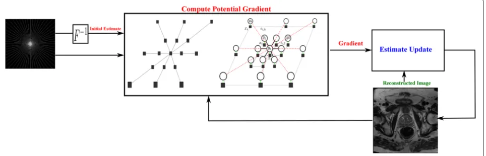
An implementation of the proposed CD-SFCRF framework for the purpose of sparse reconstruction from compressive sensing MRI is illustrated in Fig. 1. Here,
an iterative gradient descent optimization approach is employed, and can be described as follows. First, the original compressive sensing MRI data in k-space is transformed to the spatial domain to provide an initial estimate of the reconstructed image. Second, the gradient of the unary and pairwise energy potentials is computed, where the unary data driven consistencies with respect to the original observations are enforced in thek-space, and spatial and data driven consistencies are enforced in the spatial domain. Third, the estimate of the reconstructed image is updated based on the previous estimate and the computed gradient. The second and third steps of this process are repeated until convergence.
Experimental setup
To study the efficacy of the proposed CD-SFCRF method for the purpose of sparse reconstruction of compressive sensing MRI, experiments were performed including: i) MRI data acquired of a MRI training phantom, and ii) prostate MP-MRI data of 20 patient cases. A detailed description of the phantom data, patient data, and MRI image acquisition procedure to facilitate for the various experiments are described below.
Phantom data
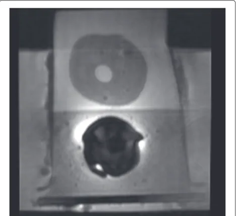
The MRI training phantom used in the experiments, shown in Fig. 2, was a multi-modality prostate training phantom from Computerized Imaging Reference Systems Inc (CIRCS MODEL 053). The phantom is composed of a clear acrylic container with dimensions 11.5×7.0×9.5cm with a front probe opening of 3.2cmdiameter and a rear
Fig. 2Example slice of the prostate training phantom from Computerized Imaging Reference Systems Inc (CIRCS MODEL 053) used for evaluation purposes
probe opening of 2.6 cmdiameter. The prostate is com-posed of high-scattering Blue Zerdine with dimensions 5×4.5×4.0cmand is placed in a background gel similar to water with little backscatter attenuation(≤0.07dB/cm− MHz). Within the prostate, there are 3 randomly placed lesions of sizes between 0.5−1.0cmplaced hypoechoic to the prostate. The urethra and rectal wall are made of low scattering Zerdine with diameter of 0.7 cm with dimensions 6×11×0.5cm, respectively. This phantom was imaged with an inflatable Medrad eCoil ERC using DWI. The DWI MRI was acquired by a 3T GE Discovery MR750. DWI was collected atb = 0mm2/sat 3-NEX2. For the DWI data, the echo time (TE) was 71.70msand repetition time (TR) was 10, 000.00ms.
Patient data experiments
To test the efficacy of the proposed CD-SFCRF framework within a real clinical scenario, MRI data of 20 patients (17 with cancer and 3 without cancer) were acquired using a Philips Achieva 3.0T machine at Sunnybrook Health Sciences Centre, Toronto, Ontario, Canada. All data was obtained retrospectively under the local insti-tutional research ethics board (Research Ethics Board of Sunnybrook Health Sciences Centre). For each patient, the following MP-MRI modalities were obtained (Table 1): T2w and DWI. The patients’ age ranged from 53 to 83. Table 1 summarizes the information about the 20 patients’ datasets used in this study, which includes displayed field of view (DFOV), resolution, echo time (TE), and repeti-tion time (TR).
Compressed sensing configuration
In order to evaluate the efficacy of the proposed CD-SFCRF framework at different sample rates, we first acquired MRI measurements at all k-space coefficients. Based on this fully-sampled set ofk-space measurements, sparse sampling was then conducted using radial sampling patterns with different numbers of radial sampling lines to achieve a desired sampling rate. For example, Fig. 3 shows a radial sampling pattern which corresponds to a sampling rate of 32 % of thek-space. Different sampling rates were tested and evaluated in this study.
Results
In order to evaluate the efficacy of the proposed CD-SFCRF framework for sparse reconstruction of com-pressive MRI sensing, a comparative evaluation analysis
Table 1Description of the prostate T2w and DWI images Modality DFOV (cm2) Resolution (mm3) TE (ms) TR (ms)
T2w 22×22 0.49×0.49×3 110 4,687
Fig. 3Radialk-space sampling pattern at 32 % sampling rate
was performed alongside a baselinel2minimization (L2) reconstruction method, and a state-of-the-art homo-topicl0minimization (HL0) [15] reconstruction method. The tested methods were compared quantitatively through peak signal-to-noise ratio (PSNR) analysis, and
qualitatively via visual assessment. All tested methods were implemented based on the original literature, with optimal parameters used in this study. All tested methods were run until convergence.
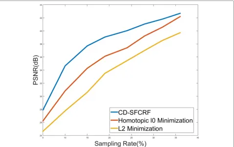
Figure 4 shows the PSNR versus sampling rate plots for the tested methods for the phantom MRI data.
Tables 2, 3, and 4 show the PSNR results for the three reconstructed methods for the T2w, DWI, as well as ADC map images for the patient data experiments at different sampling rates.
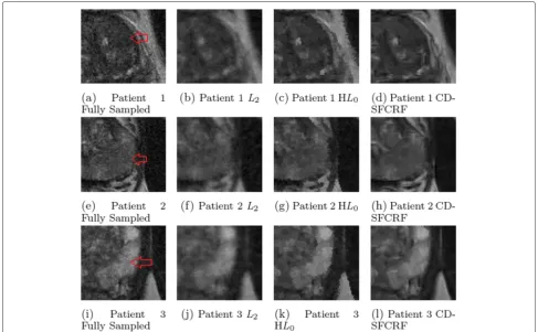
Figures 5 and 6 shows the visual comparison between the reconstructed images produced using the pro-posed CD-SFCRF framework compared with that produced using the L2 and homotopicl0 minimization reconstruction methods for three cases for T2w images.
Figures 7 and 8 shows the visual comparison between the reconstructed images produced using the proposed CD-SFCRF framework compared with that produced using theL2and HL0methods for three patient cases for DWI (b=100s/mm2) and ADC maps.
Discussion
As it can be observed from Fig. 4, the proposed CD-SFCRF framework achieved noticeable PSNR improve-ments over the other tested methods at all tested sampling
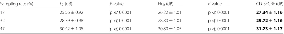
Table 2PSNR±standard deviation (stdev) for T2w images for the patient data experiments (24 images/patient for 20 patients) across different methods withP-values comparing L2and HL0methods with CD-SFCRF, respectively
Sampling rate (%) L2(dB) P-value HL0(dB) P-value CD-SFCRF (dB)
17 25.56±0.92 p0.0001 26.22±1.01 p0.0001 27.34±1.16
32 28.39±0.98 p0.0001 28.80±1.01 p0.0001 29.72±1.16
47 30.42±1.05 p0.0001 30.80±1.05 p0.0001 31.23±1.17
Bold face indicates the method with the highest performance metric
rates. The CD-SFCRF produced improvements of up to 4 dB over HL0 and 7dB over L2 in low sampling con-ditions. It can also be observed that as sampling rates increase, the performance differences decrease. This is due to the fact that as the sampling rate increases, the amount of available measurements increases, and as such the level of reconstruction quality improvements that can be achieved will naturally decrease given the amount of available information becomes increasingly sufficient for high quality reconstruction. The ability of the CD-SFCRF framework to produce high quality reconstruction at very low sampling rates can be demonstrated visually as well.
From additional quantitative analysis of patients MRI data presented in Tables 2, 3, and 4, it can be observed that the proposed CD-SFCRF framework achieved the greatest PSNR improvements for the lowest sampling rate (i.e., 17 %) where for T2w, CD-SFCRF improved PSNR by 1.78dBand 1.12dBover theL2and HL0methods, respec-tively. For DWI, CD-SFCRF improved PSNR by 1.85dB and 0.28 B over the L2 and HL0 methods, respectively. Interestingly for ADC maps, the best improvements in PSNR were achieved for the highest sampling rate (47 %) where CD-SFCRF improved PSNR by 4.44dBand 0.21B over theL2and HL0methods, respectively.
Tables 2, 3 and 4 also show theP-values calculated by comparing the proposed CD-SFCRF method withL2and HL0 methods, respectively. As it can be seen, P-values show significant difference between CD-SFCRF and the other two methods for T2w and DWI images. For ADC maps, the proposed CD-SFCRF was significantly differ-ent than L2 method as well. The only comparison that did not show significantly different results was CD-SFCRF compared to HL0 method for ADC maps. This shows that the PSNR improvement for the proposed CD-SFCRF
framework was meaningful for the majority of cases when compared to other tested methods.
Comparing the results for phantom MRI data (Fig. 4) and patients MRI data shown in Tables 2, 3 and 4 shows that the proposed CD-SFCRF framework yields higher performance improvement at 10 - 20 % sampling range for phantom MRI data compared to patients MRI data. The reason for this difference on PSNR improvement is the fact that the morphological and textural properties of the phantom is significantly less complex than that of real patients’ prostates, and thus the reconstruction problem is a simpler one for the phantom and as a result, greater PSNR gains were achieved using the proposed method.
Qualitative observations from Figs. 5 and 6 show that the L2 method resulted in blurry T2w images as well as noticeable radial artifacts at low sampling rates as expected due to the least squares reconstruction being prone to errors. The HL0 approach performed better than the L2 minimization and was able to noticeably reduce artifacts and provide a higher quality reconstruc-tion. However, in comparison, the CD-SFCRF was able to better restore details and fine tissue structure in the reconstructed image when compared to HL0. This is to be expected as the CD-SFCRF takes advantage of more complete data and spatial driven consistencies in a fully connected nature, thus better modeling the underlying tissue detail and structures.
Furthermore, as it can be seen in Figs. 7 and 8, theL2 method resulted in blurry DWI and ADC map images again with noticeable radial artifacts. Although the HL0 approach performed better than the L2 method, it can be observed once again that the proposed CD-SFCRF approach was able to preserve more fine tissue structure and detail in the reconstructed image when compared to the HL0 method. Nevertheless, an inherent trade-off
Table 3PSNR±stdev for DWI images for the patient data experiments (24 images/b-value for 4 b-values/patient for 20 patients) across different methods withP-values comparing L2and HL0methods with CD-SFCRF, respectively
Sampling rate (%) L2(dB) P-value HL0(dB) P-value CD-SFCRF (dB)
17 26.90±1.86 p0.0001 28.46±2.50 p0.0001 28.75±2.22
32 31.92±2.32 p0.0001 33.39±3.03 p0.0001 33.61±2.17
47 36.45±2.67 p0.0001 37.85±2.48 p0.0001 37.99±2.11
Table 4PSNR±stdev for ADC images for the patient data experiments (24 images/patient for 20 patient) across different methods withP-values comparing L2and HL0methods with CD-SFCRF, respectively
Sampling rate (%) L2(dB) P-value HL0(dB) P-value CD-SFCRF (dB)
17 17.20±0.61 p0.0001 19.35±0.58 0.88 19.50±0.57
32 18.05±0.48 p0.0001 21.66±0.54 0.89 21.72±0.55
47 18.72±0.32 p0.0001 22.94±0.39 0.89 23.16±0.37
Bold face indicates the method with the highest performance metric
exists between preserving fine textural granularity and reducing artifacts due to compressed sensing which can be well utilized in the proposed CD-SFCRF framework to achieve a balance between the two competing constraints. In Figs. 5, 6, 7 and 8, the tumourous regions marked by a radiologist and confirmed by pathology report (biopsy results) are shown by red arrow or white boundary. It can be seen that the proposed CD-SFCRF method pre-serves the separability of the cancerous and healthy tissue in all cases, which is an important measure for usability of the proposed method in practice. As it can be seen, the tumourous regions are blurred in theL2 method, which may make it difficult to detect for radiologists.
Both quantitative and qualitative analysis demonstrate the potential of the proposed CD-SFCRF framework as a reliable reconstruction approach for compressive sens-ing in MRI. It demonstrates the ability to produce edge and tissue details at very low sampling rates. The CD-SFCRF framework better utilize available information to
produce high quality reconstructed images given very lim-ited available information. Preservation of tissue structure and detail enhancement, and noise and artifact mitiga-tion are very important for MRI as the diagnostic quality is directly related to the image quality. This demonstrates that the CD-SFCRF framework can be a viable clini-cal technique as the reduction in acquisition can lead to faster acquisitions and lower patient wait times. With a lower acquisition time and hence lower patient wait time, patients can have access to the necessary treatments in a timely manner, significantly improving the patient outcome and survival rates.
The compressive sensing method used to recon-struct MR images can influence the performance of the computer-aided diagnosis (CAD) tools. For exam-ple, several radiomics-based CAD algorithms have been proposed for automatic prostate cancer detection which use T2w and DWI to extract texture and morphologi-cal features fed into a classifier [31–36]. These algorithms
Fig. 6Sample T2w results (zoomed in) for 3 patient images produced using CD-SFCRF,L2, and HL0at 32 % sampling ratio. Compared to other methods, CD-SFCRF preserves tissue details and contrast especially in the tumourous regions. The arrow shows tumourous region in the fully sampled image (a, e, i)
heavily rely on the quality of regions of interests in similar cases in DWI and therefore, it is expected that a recon-structed MRI with better quality will improve the perfor-mance. As future work, we will investigate the effect of the proposed compressive sensing method on the detection accuracies of these radiomics-based CAD algorithms with respect to the L2and HL0 methods. Moreover, recently, computational diffusion MRI (CD-MRI) has been intro-duced which utilizes the wealth of information in DW-MRI to computationally construct new sequences of DW-MRI that potentially will help radiologists with more accurate and consistent diagnosis [2, 3]. The proposed CD-SFCRF framework will be integrated into CD-MRI algorithms [2, 3] to investigate whether CD-SFCRF improves the separability of cancerous and healthy tissues in prostate for these computationally generated MR sequences with respect to theL2and HL0methods.
The limitations of the proposed CD-SFCRF method that will be addressed in the future direction of this work include the limited sample size. A larger and more diverse dataset will be used to address this limitation. Moreover, in this work, the proposed method was applied only to the prostate. Future work also includes applications of the proposed method to the MRI acquisitions of other organs
such as breast or moving organs such as heart. In addition, although the CD-SFCRF can significantly decrease MRI acquisition times, because of the fully-connected nature of this method, the algorithm may require a considerable processing time to complete (although not comparable to original MRI acquisition time). As future work, we will modify the proposed method to improve processing time and the efficiency of the algorithm.
Conclusions
Fig. 7Sample DWI results (b=100s/mm2) for three patient cases produced using CD-SFCRF,L
2, and HL0at 32 % sampling ratio. Compared to other methods, CD-SFCRF preserves tissue details and contrast especially in the tumourous regions. The tumourous region in the fully sampled image is marked (a, e, i)
especially at low sampling rates. The ability to better utilize available information given very limited informa-tion demonstrates the potential of the proposed CD-SFCRF framework as a viable reconstruction algorithm for compressive sensing MRI. The proposed CD-SFCRF can significantly reduce MRI acquisition times with-out sacrificing quality and potential reduction in the accuracy of diagnosis. Reducing MRI acquisition time would reduce related cost significantly and lead to less patient discomfort during the MRI acquisition and more importantly, it would reduce the patient wait times con-siderably. A fast access to MRI would directly trans-late to better care given to patients who need it the most.
Appendix 1
Sparse reconstruction via cross-domain stochastically fully connected conditional random field
The main goal here is to reconstruct imageY given orig-inal sparsely sampledk-space observationsX. We model the conditional probabilityP(Y|X)of the full state setYin spatial domain given the set of sparse measurementsXin k-space, which can be written as:
P(Y|X)= 1
Z(X)exp(−ψ(Y|X)) (1)
where Z(X) is the normalization function and ψ(.) is a combination of unary and pairwise potential functions:
ψ(Y|X)= n
i=1
ψu(yi,X)+
ϕ∈C
ψp(yϕ,X) (2)
Hereyi∈Yis a single state in the setY = {yi}ni=1,yϕ ∈Y encodes a clique structure in the setC, andX = {xj}nj=1 is the observations (radially sub-sampled frequency coef-ficients) in the frequency domain (k-space). The unary potentialψuis enforced in thek-space while the pairwise potentialψpis applied in the spatial domain. The unary potential enforces original observations to preserve data fidelity. Since the available observations are captured ink -space in MRI, the model must be formulated in a way to be consistent in bothk-space and spatial domain.
Fig. 8Sample ADC map results for three patient cases produced using CD-SFCRF,L2, and HL0at 32 % sampling ratio. Compared to other methods, CD-SFCRF preserves tissue details and contrast especially in the tumourous regions. The tumourous region in the fully sampled image is marked (a, e, i)
fully utilize available data within this random field model is to formulate the unary potential in thek-space and the pairwise potential in the spatial domain.
One of main differences between the proposed CD-SFCRF framework from conventional CRF models is to incorporate long-range information in the model and pre-serve boundaries and image structural properties more effectively which is important here due to sparse avail-able observation. To capture long-range information, CD-SFCRF assumes fully connected neighboring structure for the underlying graph which each node i has a set of neighbors
N(i)=j|j=1 :n,j=1 (3)
where |N(i)| = n− 1 and includes all other nodes in the graph as neighbors of nodei. Here the pairwise clique structures are utilized such that:
C=Cp(i)ni=1 (4)
Cp(i)=
(i,j)|j∈N(i), 1S{i,j} =1. (5)
The active cliques in the inference procedure are deter-mined by the stochastic indicator function 1S{i,j} =1. The indicator function decides whether or not nodes can con-struct a clique,Cp(i)for nodei. This stochastic indicator function combines spatial and data driven information to model the probability distribution of informative cliques which informative cliques have higher probability to par-ticipate in the inference. The set of active cliques are obtained to extract pairwise potentials in Eq. 2.
As mentioned before, ψ(·) in Eq. 2 is the combina-tion of two potential funccombina-tionsψu(.), the unary potential and ψp(.), the pairwise potential. These potential func-tions are formulated with their corresponding weightsλ, respectively as:
ψu(Y,X)= K
j=1
λu
jFj(Y,X) (6)
ψp(yϕ,X)= K
{yi,yj}∈yϕ,k=1
λp
kfk(yi,yj,X) (7)
training stages. Although it is possible to provide several arbitrary feature functions to model the conditional prob-abilityP(Y|X), here two feature functions are provided to formulate the image reconstruction for the purpose of sparse reconstruction from compressive sensing MRI. The conditional distribution of Y givenX is trained to pro-mote/suppress different features in both the unary and pairwise potentials. Higherλuj values promotes a higher reinforcement of original observations while highλpk val-ues promotes higher consideration of spatial and data driven neighborhood constraints. In Eq. 6,Frefers to the frequency domain potential function. The unary poten-tial is calculated in thek-space while the pairwise remains in the spatial domain. This is the novelty of the CD-SFCRF whihc facilitates for better preservation of fine tissue details and contrast in the reconstructed image. The unary potential functionFj(yi,X)can be formulated as:
Fj(Y,X)=
π
2
ω=−π2
F(Y,ω)−xω (8)
whereF(·,·) is the Fourier operator and returns thek -space coefficient corresponding to frequencyω. Based on this formulation, the unary potential is enforced in the k-space and in the inferencing step, the model tries to estimate imageY to be consistent to the originalk-space observationX= {xω}
π
2
ω=−π2.
The pairwise functionfk(yi,yj,X)can be formulated as:
fk(yi,yj,X)=exp
−(yi−yj)2·(xi−xj)2 3σ2
(9)
whereσ is a control variable for the amount of weighting node pairs in the cliqueϕ = {i,j}. Contrary to the unary potential, the pairwise potential is enforced in the spatial domain.
Graph representation
GraphG(V,E)(Fig. 9) is the realization of the CD-SFCRF where V is the set of nodes of the graph representing states Y = {yi}ni=1, E is the set of edges in the graph. Observationsxi∈Xare made in thek-space domain. Our final state estimationsYare in the spatial domain (image). Figure 9 shows the graphical representation of how the spatial and k-space domain are incorporated to model the conditional probabilityP(Y|X).xicomes from sparse measurements in thek-space. In the inference procedure, thek-space observations are transformed into the spatial domain using the Fourier transform to compute the pair-wise potentials. Pairpair-wise potentials are calculated in the spatial domain and transformed into thek-space to com-bine with the unary potential and perform data fidelity. For different types of MRI data, different sparse sampling
Fig. 9Realization of CD-SFCRF graph.Xirepresents original
observations made in thek-space,xirepresents spatial domain
representation of thek-space measurements andyirepresent states.
Fdenotes the Fourier operator used in transformingk-space observations into the spatial domain. Connectivity is determined based on probability distributions. Nodes with higher connectivity have solid black edges while lower probable connections are represented as dashed red lines
patterns can be used. Furthermore, pairwise connectiv-ity can be trained for specific types of details and tissue structure.
Funding
This research has been supported by the Canada Research Chairs programs, Natural Sciences and Engineering Research Council of Canada (NSERC), the Ministry of Research and Innovation of Ontario, and Ontario Institute of Cancer Research (OICR).
Availability of data and materials
The datasets during and/or analysed during the current study available from the corresponding author on reasonable request pending the approval of the institute.
Authors’ contributions
EL, FK, MJS, and AW contributed to the design and implementation of the concept. EL, FK, MJS, and AW contributed to the design and implementation of the experiments, and performing statistical analysis. FK and MAH were involved in collecting and reviewing the data. All authors contributed to the writing and reviewing of the paper. All authors read and approved the final manuscript.
Competing interests
The authors declare that they have no competing interests.
Consent for publication Not applicable.
Ethics approval and consent to participate
The institutional research ethics board (Sunnybrook Health Sciences Centre Research Ethics Board) approved this retrospective single institution study and waived the requirement for informed consent.
Author details
1Department of Systems Design Engineering, University of Waterloo, Waterloo, Ontario, Canada.2Department of Medical Imaging, University of Toronto and Sunnybrook Research Institute, Toronto, Ontario, Canada.
Received: 5 April 2016 Accepted: 15 August 2016
References
1. Canadian Cancer Statistics Special topic : Predictions of the future burden of cancer in Canada. Technical Report 2015 Canadian Cancer Society www.cancer.ca/statistics.
2. Wong A, Glaister J, Cameron A, Haider MA. Correlated diffusion imaging. BMC Med Imaging. 2013;13(1):26. doi:10.1186/1471-2342-13-26. 3. Wong A, Khalvati F, Haider MA. Dual-Stage Correlated Diffusion Imaging.
In: IEEE International Symposium on Biomedical Imaging (ISBI). Brooklyn; 2015. p. 75–8.
4. Khalvati F, Wong A, Haider MA. Enhanced Dual-Stage Correlated Diffusion Imaging. In: International Conference of the IEEE Engineering in Medicine and Biology Society (EMBC). Orlando; 2016. p. 5537–40. 5. Esen T, Turkbey B, Patel A, Futterer J. Multiparametric mri in prostate
cancer. BioMed Research International. 2014;2014:296810. doi:10.1155/2014/296810.
6. Donoho DL. Compressed sensing. IEEE Trans Inform Theory. 2006;52(4): 1289–306. doi:10.1109/TIT.2006.871582.
7. Candès EJ. Compressive sampling. Int Congr Math. 2006;3:1433–52. 8. Baraniuk R. Compressive Sensing. IEEE Signal Process Mag.
2007;24:1–9.
9. Lustig M, Donoho D, Pauly JM. Magn Reson Med. 2007;58(6):1182–95. doi:10.1002/mrm.21391.
10. Liang D, Liu B, Wang J, Ying L. Accelerating SENSE using compressed sensing. Magn Reson Med. 2009;62(6):1574–84. doi:10.1002/mrm.22161. 11. Ye JC, Tak S, Han Y, Park HW. Projection reconstruction MR imaging using
FOCUSS,. Magn Reson Med. 2007;57(4):764–5. doi:10.1002/mrm.21202. 12. Wong A, Mishra A, Fieguth P, Clausi DA. Sparse reconstruction of breast
MRI using homotopic L0 minimization in a regional sparsified domain. IEEE Trans Bio-med Eng. 2013;60(3):743–52. doi:10.1109/TBME.2010.2089456. 13. Qu X, Cao X, Guo D, Hu C, Chen Z. Compressed Sensing MRI with
Combined Sparsifying transforms and Smooted L0 Norm Minimization.
In: IEEE International Conference on Acoustics, Speech and Signal Processing (ICASSP). Dallas; 2010. p. 626–9.
14. Block KT, Uecker M, Frahm J. Magn Reson Med. 2007;57(6):1086–98. doi:10.1002/mrm.21236.
15. Trzasko J, Manduca A. Highly undersampled magnetic resonance image reconstruction via homotopic-minimization. Med Imaging IEEE Trans. 2009;28(1):106–21.
16. Chartrand R. Fast algorithms for nonconvex compressive sensing: Mri reconstruction from very few data. In: Biomedical Imaging: From Nano to Macro, 2009. ISBI’09. IEEE International Symposium On. Boston: IEEE; 2009. p. 262–5.
17. Yin W, Osher S, Goldfarb D, Darbon J. Bregman iterative algorithms for
\ell_1-minimization with applications to compressed sensing. SIAM J Imaging Sci. 2008;1(1):143–68.
18. Figueiredo MA, Nowak RD, Wright SJ. Gradient projection for sparse reconstruction: Application to compressed sensing and other inverse problems. Selected Topics Signal Process IEEE J. 2007;1(4):586–97. 19. Goldstein T, Osher S. The split bregman method for l1-regularized
problems. SIAM J Imaging Sci. 2009;2(2):323–43.
20. Wang Y, Yang J, Yin W, Zhang Y. A new alternating minimization algorithm for total variation image reconstruction. SIAM J Imaging Sci. 2008;1(3):248–72.
21. Osher S, Burger M, Goldfarb D, Xu J, Yin W. An iterative regularization method for total variation-based image restoration. Multiscale Model Simul. 2005;4(2):460–89.
22. Qu X. Compressed sensing MRI based on nonsubsampled contourlet transform. 2008 IEEE International Symposium on IT in Medicine and Education. 2008693–696. doi:10.1109/ITME.2008.4743955.
23. Yu Y, Hong M, Liu F, Wang H, Crozier S. Compressed sensing MRI using Singular Value Decomposition based sparsity basis. Conf Proc Ann Int Conf IEEE Eng Med Biol Soc. 2011;2011(1):5734–7.
doi:10.1109/IEMBS.2011.6091419.
24. Aster R, Borchers B, Thurber C. Parameter estimation and inverse problems. Int Geophys. Series, 90: Elsevier; 2005.
25. Held K, Kops ER, Krause BJ, Wells III WM, Kikinis R, Muller-Gartner HW. Markov random field segmentation of brain mr images. Med Imaging, IEEE Trans. 1997;16(6):878–6.
26. Blake A, Kohli P, Rother C. Markov Random Fields for Vision and Image Processing. Cambridge: MIT Press; 2011.
27. Lafferty J, McCallum A, Pereira FCN. Conditional random fields: Probabilistic models for segmenting and labeling sequence data. In: ICML (International Conference on Machine Learning); 2001. p. 282–9. 28. Nyquist H. Certain topics in telegraph transmission theory. Am Inst
Electrical Eng Trans. 1928;47(2):617–44.
29. Shafiee MJ, Wong A, Siva P, Fieguth P. IEEE International Conference on Image Processing (ICIP). Paris; 2014, pp. 4289–93.
30. Ljunggren S. A simple graphical representation of fourier-based imaging methods. J Magn Reson (1969). 1983;54(2):338–43.
31. Khalvati F, Wong A, Haider M. Automated Prostate Cancer Detection via Comprehensive Multi-Parametric Magnetic Resonance Imaging Texture Feature Models. Biomed Central Med Imaging. 201515–27.
doi:10.1186/s12880-015-0069-9.
32. Khalvati F, Modhafar A, Cameron A, Wong A, Haider M. A
multi-parametric diffusion magnetic resonance imaging texture feature model for prostate cancer analysis. In: MICCAI 2014 Workshop on Computational Diffusion MRI. Boston; 2014. p. 79–88.
33. Cameron A, Khalvati F, Haider M, Wong A. MAPS: A Quantitative Radiomics Approach for Prostate Cancer Detection. IEEE Trans Biomed Eng. 2016;63(6):1145–56.
34. Cameron A, Modhafar A, Khalvati F, Lui D, Shafiee M, Wong A, Haider MA. Multiparametric MRI Prostate Cancer Analysis via a Hybrid Morphological-Textural Model. In: International Conference of the IEEE Engineering in Medicine and Biology Society (EMBC). Chicago; 2014. p. 3357–60.
35. Chung A, Scharfenberger C, Khalvati F, Wong A, Haider M. Textural Distinctiveness in Multi-Parametric Prostate MRI for Suspicious Region Detection. In: International Conference on Image Analysis and Recognition (ICIAR). Ontario; 2015. p. 368–76.