Open Access
Research
Effect of Carnitine and herbal mixture extract on obesity induced by
high fat diet in rats
Kamal A Amin*
1and Mohamed A Nagy
2Address: 1Biochemistry Department Faculty of Veterinary Medicine, Beni-Suef University, Beni-Suef, Egypt and 2Chemistry Department Faculty of Science Beni-Suef, Beni-Suef University, Beni-Suef, Egypt
Email: Kamal A Amin* - kaamin10@yahoo.com; Mohamed A Nagy - nagy_bio@yahoo.com * Corresponding author
Abstract
Background: Obesity-associated type 2 diabetes is rapidly increasing throughout the world. It is generally recognized that natural products with a long history of safety can modulate obesity.
Aim: To investigate the development of obesity in response to a high fat diet (HFD) and to estimate the effect of L-carnitine and an Egyptian Herbal mixture formulation (HMF) (consisting of T. chebula, Senae, rhubarb, black cumin, aniseed, fennel and licorice) on bodyweight, food intake, lipid profiles, renal, hepatic, cardiac function markers, lipid Peroxidation, and the glucose and insulin levels in blood and liver tissue in rats.
Method: White male albino rats weighing 80-90 gm, 60 days old. 10 rats were fed a normal basal diet (Cr), 30 rats fed a high-fat diet (HFD) for 14 weeks during the entire study. Rats of the HFD group were equally divided into 3 subgroups each one include 10 rats. The first group received HFD with no supplement (HFD), the 2nd group HFD+L-carnitine and the third group
received HFD+HMF. Carnitine and HMF were administered at 10th week (start time for treatments) for 4 weeks.
Body weight, lipid profile & renal function (urea, uric acid creatinine) ALT & AST activities, cardiac markers, (LDH, C.K-NAC and MB) the oxidative stress marker reduced glutathione (GSH), and Malondialdehyde (MDA) catalase activity, in addition to glucose, insulin, and insulin resistance in serum & tissues were analyzed.
Results: Data showed that feeding HFD diet significantly increased final body weight, triglycerides (TG), total cholesterol, & LDL concentration compared with controls, while significantly decreasing HDL; meanwhile treatment with L-carnitine, or HMF significantly normalized the lipid profile.
Serum ALT, urea, uric acid, creatinine, LDH, CK-NAC, CK-MB were significantly higher in the high fat group compared with normal controls; and administration of L-carnitine or herbal extract significantly lessened the effect of the HFD. Hyperglycemia, hyperinsulinemia, and high insulin resistance (IR) significantly increased in HFD in comparison with the control group. The treatment with L-carnitine or HMF improved the condition. HFD elevated hepatic MDA and lipid peroxidation associated with reduction in hepatic GSH and catalase activity; whereas administration of L-carnitine or herbal extract significantly ameliorated these hepatic alterations.
Conclusion: HFD induced obesity associated with a disturbed lipid profile, defective antioxidant stability, and high values of IR parameters; this may have implications for the progress of obesity related problems. Treatment with L-carnitine, or HMF extract improved obesity and its associated metabolic problems in different degrees. Also HMF has antioxidant, hypolipidaemic insulin sensitizing effects. Moreover HMF might be a safe combination on the organs whose functions were examined, as a way to surmount the obesity state; and it has a distinct anti-obesity effect.
Published: 16 October 2009
Diabetology & Metabolic Syndrome 2009, 1:17 doi:10.1186/1758-5996-1-17
Received: 20 July 2009 Accepted: 16 October 2009
This article is available from: http://www.dmsjournal.com/content/1/1/17
© 2009 Amin and Nagy; licensee BioMed Central Ltd.
Introduction
The global prevalence of obesity is increasing rapidly among adults as well as among children and adolescents in places where high dietary fat intake is a major risk factor for the development of obesity [1]. Obesity is reaching epidemic proportions worldwide; it is correlated with var-ious comorbidities, among which the most relevant are dyslipidemia [2], diabetes mellitus T2DM [3], fatty liver (which can later progress to nonalcoholic fatty liver dis-ease [4]), cardiovascular (CV) disdis-eases such as heart fail-ure (HF) and coronary heart disease (CHD) [5].
L-carnitine is essential for the transfer of long-chain fatty acids from the cytosol to mitochondria for subsequent beta oxidation. Its lack impairs the ability to use fat as fuel. This can result in an acute metabolic decompensation, most often early in life, with hepatic encephalopathy and hypoketotic hypoglycemia [6].
Carnitine (L-beta-hydroxy-gamma-N,N,Ntrimethyl-ami-nobutyric acid) is one of the "nutraceuticals" that has plei-otropic biologic effects. L-carnitine administration to carnitine-deficient rats led to normalization in myocardial function including indices of contractility relaxation, systolic diastolic blood pressure [7].
The liver is a central organ for carnitine metabolism; therefore it is not surprising that carnitine metabolism is impaired in patients and experimental animals with cer-tain types of chronic liver disease. L-carnitine can have a therapeutic role in some of these conditions [8].
Nowadays there is an increased demand for using plants in therapy "back to nature" instead of using synthetic drugs which may have adverse effects. Traditional medic-inal plants are often cheaper, locally available, and easily consumable (raw or as simple medicinal preparations). These simple medicinal preparations often mediate bene-ficial responses due to their active chemical constituents.
The reason for using herbs in combination is that herbs have chemicals components which can bring strong effects on body. When herbs are used in combination it helps the body better manage potentially undesirable effects of any one and each herb in the combination/for-mulation plays a curative or pacifying role. It is therefore preferable to use herbal combinations instead of depend-ing on sdepend-ingle herbs [9].
The HMF contains antioxidants, stimulant laxatives that are present in T. chebula, senna, licorice and rhubarb. The herbal supplement also includes carminatives which are represented by black cumin, aniseed, fennel, and licorice [10]. These carminative herbs are utilized to improve digestion or to treat dyspepsia or irritable bowel
symp-toms of ulcerative colitis; and to treat maladies of specific organs in the digestive system, such as the pancreas, liver, stomach or large and small intestines.
Demulcents and carminatives are often used to soften and physiologically balance the harsh effects of stimulant lax-atives [11]. Both licorice and aniseed are considered to be flavors that by altering the characteristics of the solute, causing sweet and sour tanginess.
The ethanolic extract of roots of rhubarb led to the isola-tion of anthraquinones: chrysophanol, physcion, emo-din, glucopyranoside, stilbenes: desoxyrhaponticin, rhaponticin, resveratrol, rhapontigenin, glucopyranoside, ampelopsin B and rhaponticin [12].
The mechanism of rhubarb action is twofold: 1) stimula-tion of colonic motility, which augments propulsion and accelerates colonic transit which in turn reduces fluid absorption from the fecal mass; 2) an increase in the para-cellular permeability across the colonic mucosa, probably owing to an inhibition of Na+/K+ exchanging ATPase or to
an inhibition of chloride channels. This results in an increase in the water content in the large intestine. Purga-tion is followed by an astringent effect owing to the tan-nins presence [13].
Licorice root is the dried peeled or unpeeled root of Gly-cyrrhiza glabra known as Spanish licorice; or of other vari-eties of Glycyrrhiza glabra [14].
Constituents with antioxidant capacity were isolated from
Glycyrrhiza glabra. The isolated compounds were
identi-fied as the isoflavans, Hispaglabridin A, Hispaglabridin B, Glabridin; the two chalcones, isoprenyl chalcone deriva-tive isoliquiritigenin; theisoflavone, formononetin. Among these compounds, Glabridin constituted the major component in the crude extract and the most potent antioxidant toward LDL oxidation [15].
Glycyrrhizin inhibited histamine release from rat mast cells prevented carbon Tetrachloride-induced liver lesions and macrophage-mediated cytotoxicity. Glycyrrhizin pro-tected the liver apparently through its membrane stabili-zation effects [16]. Glycyrrhizic acid its glucoside, glycyrrhizin impart the unique licorice taste. Glycyrrhizin is 50 times sweeter than sucrose. Licorice sweetness has a slower onset than sugar so it is important for the palata-bility of the HMF.
Aniseed is the dried ripe fruits of the Pimpenella anisum
Fennel is the ripe fruit, or seed, of Foeniculum vulgare. It has a strongly aromatic odor somewhat bitter sweet pun-gent taste. The British Herbal Pharmacopoeia reported its action as a carminative is considered to be one of the best additions to purgative medicines, and it is often com-pounded with T. chebula, senna and rhubarb, in infusions or mixture [18]. Fennel seed extracts may possess a radical scavenging antioxidant activity due to the occurrence of some phenol compounds in fennel being responsible for such an activity [19].
Black cumin is the dried ripe seeds of Nigella sativa (NS), strongly aromatic when crushed, reminiscent of anise or nutmeg, also slightly bitter tasting at first, then spicy and somewhat pungent. It contains fixed, volatile oils which contain thymoquinone and several monoterpenes, including p-cymene a-pinene [20]. Many therapeutic effects of NS extracts have been documented, including immunomodulative, inflammatory, antitumor, anti-diabetic and antiulcerogenic [21] effects in both clinical and experimental studies.
Recently, the use of powerful drugs has become a popular means to overcome excess weight. Adverse effects may limit their overall usefulness. Accordingly, recent prelimi-nary reports suggesting that herbs with along history of use of other natural substances is less likely to produce toxicity and might be effective in reducing food intake, promoting significant weight loss, are encouraging. Pre-liminary findings suggest that, at least, one formulation of herbs has such promise.
However, the effect of HMF on lipid peroxidation and antioxidant enzymes activities in obesity has not been examined and so far, little is known on the medicinal val-ues of HMF. Thus, the present investigation was carried out in order to study the possible antiobesity, hypolipi-demic, hypoglycemic and antioxidant effect of HMF. The effect was compared with L-carnitine in a model of high fat diet-induced obesity. Moreover, the extract was also tested for its hepatic cardiac and renoprotective effects in rats.
Material and Methods
This study was approved by the Committee of Scientific Ethics at Beni Suef University
Materials
1- Diets
composition of the experimental diet (g/kg diet) was according to the formula of Kim et al. [22]. It included the normal diet for control rats (fat 5%, carbohydrates 65%, proteins, 20.3% fiber 5%, salt mixture and 3.7% vitamin mixture 1%). The high fat diet contained fat 46%, carbo-hydrates 24%, proteins, 20.3%, fiber 5%, salt mixture
3.7%, vitamin mixture 1%. Normal and HFD constituents were purchased from El-Gomhoria Company, Cairo, Egypt. HFD was preserved at 4°C until used.
2- Experimental animals
40 white male albino rats weighing 80-90 gm, 60 day old were used for this study. They were purchased from the National Research Center, Cairo, Egypt. All animals were housed in stainless steel cages contain barriers for each rat for individual housing and the cage contain 6 rats and each rat had a tag number. They kept under standard envi-ronmentally controlled, clean-air room with temperature 24 ± 5°C, illumination (12 h light/12 h dark cycles), a rel-ative humidity of 60 ± 4%, and water and rodent chow were available ad libitum throughout the period of the investigation. They were housed for two weeks after their arrival in the laboratory for accommodation.
Our work was carried out in accordance with the guide-lines of Faculty of Science at Beni Suef University for ani-mal use. These aniani-mals were used for induction of obesity.
Food consumption was calculated daily at the same time by subtracting the amount of food left over in each cage barrier for each rat from the measured amount of food provided at the previous day (gm/day/rat). The mean of food consumption per each rat was considered by divid-ing the amount of food eaten in a week by 7.
The average of food consumptions were represented in gm/day/rat and the body weight for each rat was deter-mined once a week (g).
3- Drug administration
L-carnitine (dietary supplement): 1 ml containing 250 mg carnitine was purchased from the Arab Company for Pharmaceuticals Medicinal Plants (MEPACO, Egypt (Enshas El Raml-Sharkeiya). Oral administration of a dose of 250 mg/kg daily was done according to the method described by Oka et al [23]. Handling of the ani-mal was the same for all groups and did not affect weight gain.
4- Plant material preparation of the herbal formulation
Herbs were purchased from local Mohey El-Attar Com-pany in El-Minia city. Identification and extractions of medicinal plants were completed in department of Phar-macognacy, faculty of pharmacy, El Minia University.
weight [26], Sweet fennel 300 mg/kg body weight [11], Aniseed 10 mg/kg body weight [27], licorice (Glycyrrhiza
galabra) 300 mg/kg body weight [15] Black cumin
(Nig-ella sativa) 300 mg/kg body weight [28].
The extraction was done by water extract for T. chebula, ethanol for senna and Nigella sativa, ethanol-water extract for rhubarb, licorice, Pimpenella anisum and Foeniculum vulgare (fennel). The extract of each plant was collected and the mixture administered orally as a suspension by stomach tube at a dose of 790 mg/kg body weight daily, the volume of the vehicle being kept constant at 1 ml/kg.
The identified compounds belonged to some specific structural types: flavenoids glabridin, and sennosides. After isolation by several column chromatographic steps from the extract and characterization by spectroscopic methods, the main compounds were identified as triterpe-noids, coumarin, gallic acid, chebulin and ellagic acid as well as other phenolic compounds from T. chebula extrac-tion.
Anthraquinones, rhein, and tannins were isolated from rhubarb extraction. Glabridin from Glycyrrhiza galabra
extraction, thymoquinone from Nigella sativa extraction. Phenolic content, d-limonene β-myrcene from Foeniculum
vulgare (fennel) extraction and anethole form Pimpenella
anisum extract.
Methods
1-Experimental design and animal grouping
A total 40 rats were randomly assigned into two groups, normal 10 rats and obese 30 rats. Obesity was induced in rats for 70 day by feeding the high fat diet. The rats were included in four groups after induction of obesity. In the experiment 10 rats were used in each group.
• Normal group, 10 rats fed on normal diet during the entire study (98 day).
• Obese group, 10 rats given high-fat diet during the entire study (98 day) and saline oral daily by using stom-ach tube at 10th week.
• HFD+carnitine group, 10 obese rats received HFD dur-ing the entire study and 250 mg/kg L-carnitine for 28 days as a single daily dose in the morning.
• HFD+ HMF group, 10 obese rats received HFD during the entire study and 790 mg/kg mixed herbal extract for 28 days as single oral daily by using stomach tube.
Our goal is to achieve obesity model in 10 weeks follow-ing by treatment period for 4 weeks. This model provided us reliable method and resembles the clinical cases of
obesity and its treatments; also this period of treatment is safe and recommended in previous research.
2- Sampling and tissue preparation Blood Sampling
By the end of the experimental periods, venous blood samples were collected from the orbital sinus of normal, obese control, obese treated rats via glass capillaries at fasting state. The blood samples were collected in dry glass centrifuge tubes, allowed to coagulate at room tempera-ture and centrifuged at 3500 rpm for 15 minutes at room temperature for separation of serum.
The clear, non-haemolysed supernatant sera were sepa-rated using clean dry disposable plastic syringes and stored at -20°C for subsequent biochemical measure-ments as follows: lipid profile, liver enzyme activities related to its function, kidney function, heart biomarkers, glucose, insulin concentration, and oxidative stress mark-ers.
Tissue samples
Rats were sacrificed by decapitation and an abdominal incision was immediately done for separation of hepatic, perirenal, and visceral adipose tissues. These were dried on filter paper and weighed (g.). The liver was immedi-ately excised and weighed (g.) and underwent homogeni-zation for GSH & MDA catalase measurements.
3- Biochemical analysis of Serum and tissue
Serum glucose was estimated according to the method of
Trinder [29] using Stanbio Laboratory USA Kits. Serum insulin was assayed in the Radioactive Isotopes Unit, Cen-tral Department of Scientific Analysis and Testing, National Research Center (Dokki, Giza) using radioim-munoassay kits (Diagnostic Products Corporation, Los Angeles, USA) [Coat-A-Count] according to the method of
Marschner et al. [30]. Insulin resistance was calculated using the Homeostasis Model Assessment [31]. ALT and AST activities were measured according to the method of
Reitman et al., [32] using kits purchased from Rox Com-pany, United Kingdom.
Serum urea and creatinine levels were measured colori-metrically [33]; serum uric acid was measured according to the method of Sanders et al.[34] using kits purchased from Diamond Diagnostic Egypt. Serum was analyzed for total cholesterol [35], triglycerides [36], VLDL [37], and HDL [38], by enzymatic colorimetric methods using kits.
according to the procedure of Beutler et al. [42] and
Cohen et al. [43] respectively. Statistical analysis
Statistical analysis was carried out using Graph Pad Instat software (version 3, ISS-Rome, Italy). Unless otherwise specified, groups of data were compared with an unpaired t-test one-way analysis of variance (ANOVA) followed by Tukey-Kramer (TK) multiple comparisons post-test. Val-ues of P < 0.05 were regarded as significant. Data were expressed in tables and figures as mean ± standard error (SEM).
Results
Body weight increased significantly in rats on the HFD compared with controls (Figure 1), while treatments with L-carnitine or HMF significantly reduced this gain during the treatment period (Table 1 and figure 2).
Food consumption increased significantly in the HFD group compared with controls (Figure 3), while treat-ments with L-carnitine or HMF significantly ameliorated the changes during the treatment period (Table 2 and fig-ure 4).
Serum TG, cholesterol, LDL/VLDL levels, and perirenal and mesenteric fat were significantly elevated in the HFD group, while HDL was significantly decreased compared
to the controls. Additional administration of L-carnitine or HMF significantly improved these changes (Table 3).
Serum LDH, CK, AST, ALT, urea, uric acid, and creatinine concentrations were significantly higher in the HFD group compared to the control one; these changes, were affected by supplementation with L-carnitine and HMF (Table 4).
Serum levels of glucose, insulin and hepatic MDA, were significantly raised, while hepatic GSH and catalase were significantly lowered in the HFD group compared to the controls. These alterations were ameliorated by
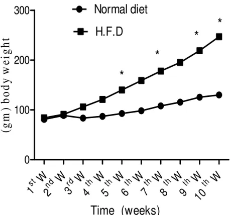
adminis-Effect of normal and HFD on whole body weights weekly during 10 weeks in rats
Figure 1
Effect of normal and HFD on whole body weights weekly during 10 weeks in rats. Body weight increased significantly in rats fed on the HFD during 5th, 7th, 9th and 10th
W. compared with controls. Values significantly different compared to normal *P < 0.05.
W
st
1
nd2
W
W
rd
3
th4
W
5
thW
W
th
6
W
th
7
W
th
8
W
th
9
W
th
10
0
100
200
300
Normal diet
H.F.D
*
*
*
*
Time (weeks)
(g
m
) body
w
e
ig
ht
Effect of HFD, L-carnitine and HMF on body weights during treatment period in rats
Figure 2
Effect of HFD, L-carnitine and HMF on body weights during treatment period in rats. Treatments with L-car-nitine or HMF significantly reduced the elevated body weight during the treatment period compared to HFD. (HFD vs. L carnitine or HMF) Means not sharing common letter are sig-nificantly different (p < 0.05).
W
th
10
W
th
11
W
th
12
W
th
13
W
th
14
200 220 240 260 280 300 320
H.F.D
L.carnitine
Herbal exract
a
a
a a
b
b
b b
b
b
b
b
Time (weeks)
(g
) b
o
d
y
w
e
ig
h
t
Table 1: Effect of HFD, L-Carnitine and HMF on whole body weights of rats.
Weeks HFD L. carnitine HMF
11th Week 255.0 ± 3.54a 231.2 ± 2.56b 222.8 ± 3.58b
12th Week 270.0 ± 2.60a 247.0 ± 3.36b 239.6 ± 4.19b
13th Week 282.6 ± 2.09a 261.0 ± 3.81b 256.4 ± 5.07b
14th Week 286.4 ± 3.97a 250.0 ± 3.41b 262.0 ± 6.04b
tration of L-carnitine HMF but there were no differences between L-carnitine and HMF (Table 5).
Discussion
Effect of diet treatments on body weight food consumption
Obesity is considered to be a disorder of energy balance, occurring when energy expenditure is no longer in equi-librium with daily energy intake, so as to ensure body weight homeostasis [44]. Although the etiology of obesity is complex, dietary factors, particularly the consumption of a HFD, is considered a risk factor for its development [45].
The current results showed that body weight increased sig-nificantly in the HFD group compared with the normal group (Figure 1), a result in accordance with that of Xu et al [46]; this is associated with increased food intake. Consumption of the HFD led to obesity because it facili-tates the development of a positive energy balance leading to an increase in visceral fat deposition; this led to abdom-inal obesity in particular. Moreover, Schrauwen-Hinder-ling et al [47] found that HFD feeding is accompanied by molecular adaptations that favor fat storage in muscle rather than oxidation.
In the current study, rats fed HFD consumed considerably more food than the control rats throughout the experi-ment (Table 2 and Figure 3). So their caloric intake was increased and they showed a large increase in perirenal visceral adipose tissue mass (Table 3), suggesting that the excess energy led to the build up of adiposity. This is the source of the increase in body weight. Rat consuming the high fat ration actually received about 27% more kilocal-ories, more weight, and had larger fat pads than rats fed only chow. Rats consuming a palatable dietary fat in addi-tion to a standard chow diet take in about 10% more cal-ories a day than rats fed only the chow diet and over time,
Effect of normal and HFD on food consumption during 10 weeks in gm/day/rat
Figure 3
Effect of normal and HFD on food consumption dur-ing 10 weeks in gm/day/rat. Food consumption increased significantly in the HFD group during 7th, 9th and 10th W.
compared with controls. Values significantly different com-pared to normal *P < 0.05.
W st
1 2nd W3rd W4th W5th W6th W W th
7 8th W9th W10th W
0 5 10
15 Normal diet
H.F.D
*
*
*
*
(Time) weeks
F
ood c
ons
um
pt
ion
(g
/d
a
y
/r
a
t)
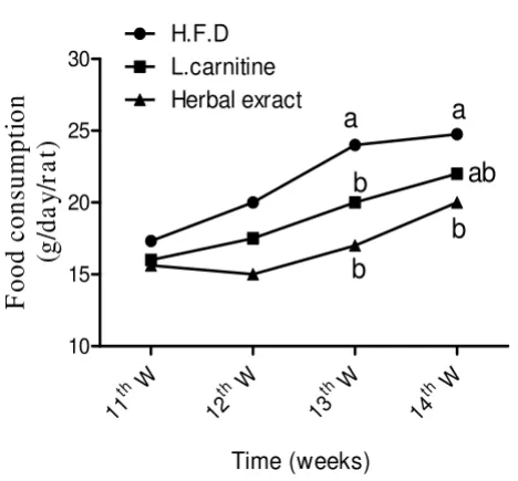
Effect of HFD, L-carnitine and HMF on food consumption during treatment period gm/day/rat
Figure 4
Effect of HFD, L-carnitine and HMF on food con-sumption during treatment period gm/day/rat. Treat-ments with L-carnitine or HMF significantly ameliorated the changes during 13th and 14th W. of the treatment period
(HFD vs. L carnitine or HMF). Means having different letter are significantly different (p < 0.05).
W
th
11
W
th
12
W
th
13
W
th
14
10 15 20 25 30
H.F.D
L.carnitine
Herbal exract
a
b
b
a
b
ab
Time (weeks)
F
ood consumpti
on
(g
/d
a
y
/r
a
t)
Table 2: Effect of HFD, L-Carnitine and HMF on food intake in rats
Weeks HFD L. carnitine HMF
11th Week 17.33 ± 1.05a 16 ± 1.58a 15.63 ± 0.62a
12 stWeek 20 ± 1.58a 17.50 ± 1.32a 15 ± 0.71a
13th Week 24 ± 2.27a 20 ± 1.63b 17 ± 1.23b
14th Week 24.75 ± 2.29a 22 ± 1.83a, b 20 ± 1.47b
become obese and exhibit a number of complications of obesity.
Our results showed a significant decrease in food intake, whole body weight, and adipose tissue accumulation from oral administration of L-carnitine (Tables 1, 2). Interest in the role of L-carnitine as a feed additive to improve whole body composition arose from the desire to partition nutrients away from lipid accretion, causing improvement of nitrogen balance; L-carnitine also attenu-ated visceral fat accumulation and accelerattenu-ated the normal-ization of food intake [48].
The current results due to the effect of HMF, showed a sig-nificant decrease in body weight and food consumption (Table 1, 2) in accordance with the findings of York et al
[49]. Several isoflavans constituents of licorice are unique phytoestrogens, which like estradiol, affect the serotoner-gic system, inhibiting serotonin re-uptake and thereby increasing the levels of serotonin in synaptic clefts. This enhances satiety and resembles the action of sibutramine, but naturally [50].
Dietary phytoestrogens may also activate AMPK leading to a reduction in food intake [51]. Our study is a novel trial for using a phytoestrogen in fennel, licorice and anise as a
Table 3: Effect of HFD, L-Carnitine and HMF on plasma lipid profile and adipose tissues weight in HFD rats
Normal diet HFD L-Carnitine HMF
TG (mg/dl) 63.94 ± 2.19 172.6 ± 1.73**a 118.5 ± 1.21 b 116 ± 1.83b
VLDL (mg/dl) 12.79 ± 0.44 34.52 ± 0.35**a 23.69 ± 0.24b 23.20 ± 0.37b
T.C (mg/dl) 95.29 ± 1.03 152.7 ± 2.56**a 109.6 ± 1.51b 113.1 ± 1.54b
HDL (mg/dl) 70.97 ± 1.89 53.34 ± 2.52**a 67.45 ± 1.4b 65.59 ± 1.36b
LDL (mg/dl) 12.48 ± 2.0 67.06 ± 1.65**a 17.29 ± 0. 85b 17.42 ± 2.08b
Visceral fat (gm) 3.54 ± 0.51 14.8 ± 1.07**a 10.10 ± 0.33b 10.40 ± 0.93b
Perirenal fat (gm) 3.54 ± 0.51 14.8 ± 1.07**a 10.10 ± 0.33 b 10.40 ± 0.93b
Values significantly different compared to normal P** < 0.01. Values are expressed as means ± SEM. Means not sharing common letter are significantly different (p < 0.05)
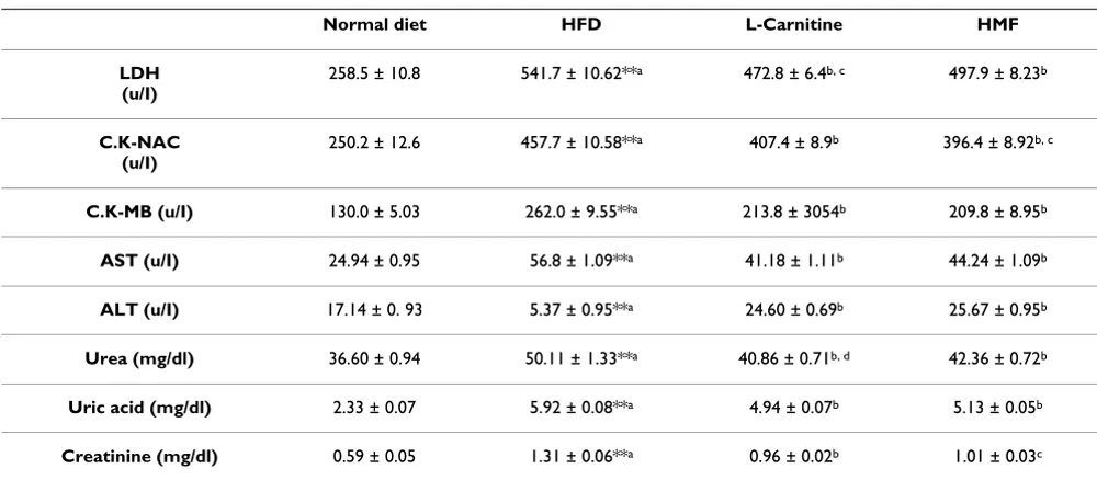
Table 4: Effect of HFD, L-Carnitine and HMF on cardiac biomarkers, liver enzyme activity and kidney function tests of HFD rats
Normal diet HFD L-Carnitine HMF
LDH (u/I)
258.5 ± 10.8 541.7 ± 10.62**a 472.8 ± 6.4b, c 497.9 ± 8.23b
C.K-NAC (u/I)
250.2 ± 12.6 457.7 ± 10.58**a 407.4 ± 8.9b 396.4 ± 8.92b, c
C.K-MB (u/I) 130.0 ± 5.03 262.0 ± 9.55**a 213.8 ± 3054b 209.8 ± 8.95b
AST (u/I) 24.94 ± 0.95 56.8 ± 1.09**a 41.18 ± 1.11b 44.24 ± 1.09b
ALT (u/I) 17.14 ± 0. 93 5.37 ± 0.95**a 24.60 ± 0.69b 25.67 ± 0.95b
Urea (mg/dl) 36.60 ± 0.94 50.11 ± 1.33**a 40.86 ± 0.71b, d 42.36 ± 0.72b
Uric acid (mg/dl) 2.33 ± 0.07 5.92 ± 0.08**a 4.94 ± 0.07b 5.13 ± 0.05b
Creatinine (mg/dl) 0.59 ± 0.05 1.31 ± 0.06**a 0.96 ± 0.02b 1.01 ± 0.03c
natural serotonin reuptake inhibitor instead of sibu-tramine.
Anetholein fennel as a carminative herb improves the digestion by stabilization of the gastrointestinal mucous membrane and causing the pancreas to increase its secre-tions [11]. Hydroxyl-anthracene glucosides, especially sennosides A B in senna and T. chebula of HMF improve gastrointestinal motility and influence colonic motility thereby reducing fluid absorption and facilitate weight loss.
Effect of diet treatments on serum lipid profile in rats In the present study, a high fat diet resulted in dyslipi-demic changes as illustrated by increasing triglycerides, VLDL, total cholesterol and low density lipoprotein LDL and a decrease in serum level of high density lipoprotein HDL (Table 3) a finding in accordance with that of Woo et al. [52].
L-carnitine supplementation produced significant decreases in serum TG, VLDL, and T-cholesterol, LDL-C while there were significant increases in HDL cholesterol in obese rats. These results are in agreement with those of
González-Ortiz et al. [53] and El-Metwally et al., [54] who reported that oral L-carnitine increases plasma free carnitine levels, improves dyslipidemia and decreases oxi-dative stress, with reduction of cardiac parameters.
L-carnitine administration to obese rats reduces signifi-cantly serum hypertriglyceridemia (Table 3) via decreased synthesis of triglycerides by the liver or by inhibition of triglyceride release from the liver. Also L-carnitine induced marked reductions in total serum cholesterol in skeletal muscles of obese rats [55].
L-carnitine is necessary for mitochondrial transport metabolism of long-chain fatty acids, thus for myocardial energetic metabolism. Fatty acids cross mitochondrial membranes as acyl-carnitine derivatives to enter pathways for oxidation, acylation, chain shortening or chain elon-gation-desaturation. Therefore, L-carnitine-dependent fatty acid transfer is central to lipid metabolism; dietary supplementation of L-carnitine improves the utilization of fat providing marked reduction in plasma levels of TG [56].
In our experimental rat model of obesity, treatment with HMF led to significant decrease in TG, VLDL, T-terol and LDL-C but a significant increase in HDL choles-terol (Table 3), a result in agreement with that of Murali et al [57].
T. chebula components of HMF have hypocholestero-lemic activity that might be mediated through increased cholesterol excretion in the feces. In addition anthraqui-none glycosides from rhubarb in the HMF have lipid-low-ering effects, resulting in depression of lipid accumulation. It consequently has anti-atherosclerotic properties [58].
Glycyrrhizin, the active constituent in licorice root, which is another plant in the mixture, exerts hypocholestero-lemic action by stimulating the conversion of cholesterol into bile acids without effect on cholesterol synthesis [59]. Another herbal supplement in the HMF, Nigella sativa, contains active constituents, phytosterols (mostly b-sito-sterol, stigmasterol campesterol) with the ability to reduce the intestinal absorption of diet biliary cholesterol [60].
Table 5: Effect of diet and treatments on serum glucose, insulin, insulin resistance lipid peroxidation in obese rats
Normal diet HFD L-Carnitine HMF
Glucose (mg/dl) 57.87 ± 1.96 145.1 ± 2.26**a 120.9 ± 1.96b, c 118.0 ± 3.23c, d
Insulin
(μIU/ml) 4.40 ± 0.15 6.69 ± 0.13**
a 4.5 ± 0.07b 4.41 ± 0.04b
Insulin resistance 1.17 ± 0.09 2.89 ± 0.07**a 2.46 ± 0.09b 2.40 ± 0.05b
M.D.A (n mol M.D.A/g/hr) 4.69 ± 0.06 9.72 ± 0.08**a 7.96 ± 0.08b, c 8.21 ± 0.07b
GSH (n mol/ 100 mg tissue)
72.65 ± 0.72 57.31 ± 0.72**a 68.77 ± 0.89b, c 68.45 ± 0.46b
Catalase (K×10-2) 52.60 ± 0.96 19.88 ± 0.63**a 36.97 ± 0.71b 32.22 ± 1.06c
Effect of diet treatments on cardiac biomarkers in rats The obese rats showed a significant increase in the activity of serum LDH, CK-NAC and CK-MB when compared to the normal rats (Table 4). Similar results were reported by
Diniz et al. [61]. Reduced muscle mitochondrial content function with increasing obesity would lower the total cel-lular ATP yield, which would result most notably in increased mitochondrial volume, and increased glycolytic enzymes necessitating increased activity of creatine kinase, as this enzyme is responsible for rapidly transfer-ring high-energy phosphate groups from the site of pro-duction to the site of use [62].
The increased blood levels of total cholesterol, LDL, VLDL as well as lowered levels of HDL in HFD rat have been identified in the development of hypercholestremia, which is one of the risk factors for CAD [63]. Administra-tion of L-carnitine produces a significant decrease in the activity of CK-NAC, CK-MB and LDH (Table 4). This is in agreement with Ferrari et al. [64], who reported a reduc-tion in cardiac markers, with L-carnitine having a good protective effect on myocardium.
In conclusion, L-carnitine stimulates the activity of pyru-vate dehydrogenase (PDH) which is an important enzyme catalyzing the rate-limiting step in lactate utilization. In type 2 diabetes this led to significant decrease in the activ-ity of LDH [65]. Moreover, L-carnitine in the β-oxidation of fatty acid as parallel source for energy acts synergisti-cally with the creatine/phosphocreatine/creatine kinase system to produce significantly decreased activity of crea-tine kinase.
This effect is mainly attributable to the vasodilatation property of L-carnitine, which both improves energetic metabolism of the hypoxic/damaged muscle and enhances wash-out of algogenic metabolites.
Our findings showed that obese rats treated with the HMF exhibited significant decreases in LDH, NAC and CK-MB activity (Table 4), in with the findings of Suchalatha et al. [66], who reported that T. chebula extract treatment ameliorates the effect of lipid peroxide formation that is related to the activities of diagnostic myocardial marker enzymes.
The rhubarb content in our mixture could prevent the development of atherosclerosis through regulating vascu-lar inflammatory processes in rats fed with an atherogenic diet [67]; and the thymoquinone content in N. sativa nor-malized cardiac histopathology, as it decreased lipid per-oxidation [20,21].
Effect of treatments on renal function tests of HFD in rat The obese rats showed a highly significant increase in the concentration of serum urea, uric acid creatinine, com-pared with the normal group (Table 4) which is in agree-ment with the results of Cindik et al. [68].
Abnormal renal function is mainly associated with dia-betic nephropathy. The pathophysiology involves glucose that binds irreversibly to proteins in the kidney circulation to form advanced glycosylation end products that can form complexes that contribute to renal damage by stim-ulation of fibrotic growth factors [69].
HFD induces alteration of renal lipid metabolism by an imbalance between lipogenesis and lipolysis in the kid-ney, as well as systemic metabolic abnormalities and sub-sequent renal lipid accumulation leading to renal injury [70]. There is an association between CKD and insulin resistance that contributes to increased VLDL production and decreased HDL levels, both of which are considered risk factors for the development of kidney dysfunction and markers for progression of CKD [71].
In addition HFD resulted in hyperinsulinemia, activation of the renin-angiotensin system, glomerular hyperfiltra-tion and structural changes in the kidney that may be the precursors of more severe glomerular injury associated with prolonged obesity [72].
The oral administration of L-carnitine (Table 4) shows that serum concentration of urea, uric acid and creatinine were highly significantly decreased. The effect of L-carni-tine on renal lipid metabolism could serve as a new ther-apeutic approach, as it counters the renal changes associated with metabolic syndrome [73]. Hence, L-carni-tine has beneficial effects on renal function.
Although there is still debate on the significance of uric acid as a risk factor for cardiovascular disease, many phy-sicians do consider elevated uric acid to be a component of the metabolic syndrome. There is little support for an independent causal role for serum uric acid in the devel-opment of CHD. However, uric acid may provide useful prognostic information in subjects with hypertensive vas-cular disease, suggesting that the influence of uric acid on CHD is explained by the secondary association of it with other risk factors such as dyslipidemia, hyperinsulinaemia and obesity [74].
the remnant kidney [75]. Rhein, another ingredient in rhubarb of HMF, improves cell metabolism through glu-cose transporter-1: it decreases cell hypertrophy, indicat-ing that there are multiple active indicat-ingredients even in a single herbal medicine involved in the multiple therapeu-tic effects of rhubarb in CKD, and suggesting that HMF might delay the progression of renal failure.
The herbal drugs containing tannins have a uremic-toxin-decreasing action, whereas rhubarb's tannins significantly improved BUN creatinine, glomerular filtration rate and renal blood flow [76]. In this respect rhubarb has proven effective as a diuretic in rabbit models, apparently by blocking sodium-potassium ATPase in the renal medulla [77].
The HMF therapy retains the balance between lipogenesis and lipolysis in the kidney to counteract the obesity-asso-ciated renal damage, in addition to maintaining cellular hydration due to laxative effect of senna, which is associ-ated with diuretic effect of rhubarb, collectively leading to improvement in renal blood flow and consequent improvement in kidney function.
Effect of treatments on glucose and insulin of HFD rats Our results showed that a high fat diet results in signifi-cant increase in serum glucose, insulin level and insulin resistance (Table 5), which parallels the results obtained from Zhang et al. [78]. Diminished hepatic and muscular uptake of glucose produced hyperlipidemia due to increased fat mobilization from adipose tissue and resist-ance to the antilipolytic actions of insulin. Impaired insu-lin action is associated with an oversupply of lipids. This increased availability led to either elevated lipid stored in insulin target tissues (e.g. muscle, liver adipose) or increased plasma FFA or triglyceride [79].
The weight loss in this study due to oral administration of L-carnitine is associated with hypoglycemia, as it pro-motes insulin sensitivity, thus lowering insulin resistance in obese rats, possibly by regulating the cell energy metab-olism or reducing free fatty acids as shown also by
González-Ortiz et al. [53] and [55].
Obesity is associated with endothelial dysfunction through direct mechanisms, as insulin resistance in asso-ciation with diabetes mellitus and dyslipidemia, indi-rectly, by the production of adipokines and pro-inflammatory cytokines, induces oxidative stress that affect the role of endothelium in modulating vascular function structure [80].
Our findings (Table 5) indicated a significant decrease of serum glucose, insulin and its resistance in obese rats treated with extracts of HMF. Some prenylflavonoids,
such as glycycoumarin, glycyrin, dehydroglyasperin C and dehydroglyasperin D, in licorice ethanolic extract, are effective in preventing and ameliorating diabetes, abdom-inal obesity and preventing hypertension and metabolic syndrome [81].
In addition, hypoglycemia induced by T. chebula is prob-ably mediated through enhanced secretion of insulin from the beta-cells of the pancreatic islets or through an extra pancreatic mechanism. Moreover, T. chebula may reduce the effect of inflammatory cytokine release during diabetes which may be one of the causative agents for the insulin resistance [69].
The hypoglycemic action of thymoquinone due to Nigella sativa in HMF could be partly due to preservation of β-cell integrity of the pancreatic islets causing a significant increase in insulin secretion. It also sensitized hepatocytes to the action of insulin. Furthermore, NS treatment exerts a therapeutic protective effect on diabetes by decreasing oxidative stress, preserving pancreatic beta-cell integrity [21].
Effect of treatments on hepatic enzymes and lipid peroxidation in HFD-rats
The current data showed a significant increase in the activ-ity of enzymes AST and ALT in the obese compared with normal rats (Table 4). Liver is bombarded by the free fatty acids (FFA) that pour out of the adipose tissue into the portal blood. This can directly cause inflammation within the liver cells, which then release further pro-inflamma-tory cytokines, leading to more hepatocyte injury and affecting the integrity of liver cells [82].
Administration of L-carnitine produces a significant low-ering effect in the activity of AST and ALT in obese rats, a result in agreement with Yapar et al. [83]. Improvement of hepatocyte integrity does not seem to be limited to its obligatory role in the transmembrane import of fatty acids for mitochondrial β-oxidation, but L-carnitine prevents lipotrope methyl group wastage and increased production of polyamines with known immunomodulatory proper-ties. Additionally L-carnitine can directly modify cytokine responses and reduce TNF-production. Moreover, lipid peroxidation is significantly blunted by oral administra-tion of L-carnitine [84].
The present results demonstrate that the mixture of HMF showed a significant decrease in the activity of both AST and ALT (Table 4) agreeing with the resulted obtained by
Celik and Isik [19].
by several enzymes that participate in the formation of the correct disulfide bonds of many proteins. Polypeptide hormones participate in the metabolism of xenobiotics.
β-myrcene elevates the levels of apoproteins CYP2B1 CYP2B2, which are subtypes of P450 enzyme system that catalyse the oxidative metabolism of a wide variety of exogenous chemicals including drugs, toxins, and endog-enous compounds such as fatty acids [85]. Nigella sativa and licorice root extract of HMF possesses hepatoprotec-tive effects in some models of liver toxicity [86].
HFD generates oxidative stress in obese rats as shown by a marked increase in the levels of MDA and a distinct dimi-nution in hepatic GSH, as well as activities of the antioxi-dant enzyme catalase. All showed reduced activity in hyperlipidemic rats (Table 5).
Hyperglycemia in the HFD group activates different path-ways leading to increased oxidative stress. Increased activ-ity of the polylol pathway inhibition of the pentose phosphate pathway as a result of hyperglycemia resulted in decreased intracellular levels of NADPH, which is required for regeneration of GSH from its oxidized form GSSG [87]. The net result was non-enzymatic disruption of H2O2 and increased levels of cellular superoxides, hydroperoxides, hydroxyl radicals as well as other radi-cals.
In addition oxidative stress may be increased in metabolic syndrome due to dyslipidemia resulting from increased levels of FFA and TGs that led to increased formation of foam cells, rendering LDL less dense and more vulnerable to oxidation and uptake by macrophages [88].
Our results indicated that L-carnitine produced a signifi-cant inhibition of MDA production and a signifisignifi-cant increase in GSH and activity of catalase (Table 5). L-carni-tine reduces significantly the content of thiobarbituric acid reactive substances (TBARS), and causes marked increase in activity of catalase in skeletal muscles of obese rats [55]. Moreover L-carnitine favorably modulates oxi-dative stress causing a reduction in oxidized LDL choles-terol levels [89].
L-carnitine effectively protects and improves mitochon-drial function in vivo: it acts as an antioxidant, so by inhibiting ROS it protects the vascular endothelial tissues against oxidative damage in hypertension [90]. Thus, L-carnitine treatment effectively protected the liver tissue against oxidative damage and showed marked improve-ment in its antioxidant status.
The current results demonstrated an antioxidative effect of HMF, as indicated by increased GSH level and hepatic cat-alase activity in comparison with obese rats (Table 5).
Administration of aqueous extract of T. chebula effectively modulated oxidative stress and enhanced antioxidant sta-tus in the liver [91], which may be due to the total polyphenol content, expressed as gallic acid. T. chebula extract has the potential to play a role in the prevention of oxidative damage in living systems which can be attrib-uted to its membrane stabilizing activities [92]. It has stronger antioxidant activity than α-tocopherol, due to the presence of hydroxybenzoic acid derivatives, flavonol aglycones and their glycosides.
Glabridin, a natural polyphenolic isoflavone antioxidant from licorice root extract in HMF possesses potent free radical scavenging activity that could promote a decrease in lipid peroxidation and could protect LDL from oxida-tion [93]. This would occur via a direct interacoxida-tion with the lipoprotein and/or an indirect effect through accumu-lation in arterial macrophages. Therefore licorice repre-sents a potent nutrient, which can attenuate the development of atherosclerosis, secondary to its antioxi-dant properties against lipids peroxidation in arterial cells [94].
Polyphenols, the main compound of N. sativa oil in HMF, have many biological properties. They possess powerful antioxidative components, which can inhibit membrane lipid peroxidation [28], and their administration exerts a therapeutic protective effect by decreasing oxidative stress.
Foeniculum vulgare extract, another constituent of the
HMF, has potent antioxidant activity, as has been docu-mented with various antioxidant assays. These activities include total antioxidant, free radical scavenging, super-oxide anion radical scavenging, and hydrogen persuper-oxide scavenging [95]. Antioxidant activity of the anthraqui-nones of rhubarb in the HMF may help protect against lipid peroxidation and free radical damage; its extracts will probably be useful for the development of safe food additives [12].
This study showed the antioxidant effect of dietary supple-ment represented by L-carnitine and free radical scavenger activities of herbal supplements represented by T. chebula, licorice, fennel, aniseed and Nigella sativa extract in the HMF.
Based on these broad observations, we suggest that high fat diet-induced obesity resulted in deleterious effects in kidney and liver tissues. HMF extract or carnitine supple-mentation counteracted the injuries, and ameliorated or normalized most the biochemical parameters.
for obesity, both L-carnitine and HMF have shown improvements in body weight, lipid profile, glucose lev-els, liver, and kidney and cardiac marker of function, as well as insulin resistance and oxidative stress markers.
It can be concluded that in this study HFD induced a model of obesity associated with high body weight, hyperlipaemia, hyperinsulinemia, insulin resistance, hepatic oxidative stress, associated hepatic, cardiac renal function marker disturbance in rats. The obesity and its associated problems in this study could be ameliorated in different degrees by using L-carnitine or HMF extract.
The study also showed that herbal extract mixture signifi-cantly improved body weight, cholesterol, LDL triglycer-ide concentrations, creatinine, urea, ALT, oxidative stress, resulting from the high-fat diet. So it also had hypolipi-demic, renoprotective, hapatoprotective, antioxidant and antiobesity effect.
It could be helpful to use HMF in conjunction with drugs; herbal extract might be a safer combination to surmount the overweight state. Positive results from such trials would confirm the medicinal usefulness of empirical combinations of traditional Egyptian medicines.
Competing interests
The authors declare that they have no competing interests.
Authors' contributions
KA and MN carried out experimental work; biochemical analysis, statistical analysis, interpretation and discussion of results related to their part of the work. KA, design and planning of the study; drafting and revision of the script. All authors read and approved the final manu-script.
Acknowledgements
This study was supported by Beni Suef University. We appreciate the assist-ance and advice of Prof. Dr Bastawy M., Vice Dean of College of Science, Beni Suef Univ. and Dr. Abdel Hameid H. for kind co-operation.
References
1. Canbakan B, Tahan V, Balci H, Hatemi I, Erer B, Ozbay G, Sut N, Haci-bekiroglu M, Imeryuz N, Senturk H: Leptin in nonalcoholic fatty liver disease. Ann Hepatol 2008, 7:249-54.
2. Fried M, Hainer V, Basdevant A, Buchwald H, Dietel M, Finer N, Greve JW, Horber F, Mathus-Vliegen E, Scopinaro N, Steffen R, Tsi-gos C, Weiner R, Widhalm K: Interdisciplinary European guide-lines on surgery for severe obesity. Rozhl Chir 2008, 87:468-76. 3. Pagotto U, Vanuzzo D, Vicennati V, Pasquali RG: Pharmacological therapy of obesity. G Ital Cardiol (Rome). 2008, 9(4 Suppl 1):83S-93S.
4. Marović D: Elevated body mass index fatty liver. Srp Arh Celok Lek 2008, 136:122-5.
5. Lavie CJ, Artham SM, Milani RV, Ventura HO: The obesity para-dox: impact of obesity on the prevalence prognosis of cardi-ovascular diseases. Postgrad Med 2008, 120:34-41.
6. Amat di San Filippo C, Taylor MR, Mestroni L, Botto LD, Longo N: Cardiomyopathy carnitine deficiency. Mol Genet Metab 2008, 94:162-6.
7. Kraemer WJ, Volek JS, Dunn-Lewis C: L-carnitine supplementa-tion: influence upon physiological function. Curr Sports Med Rep
2008, 7(4):218.
8. Krähenbühl S: Carnitine metabolism in chronic liver disease.
Life Sci 1996, 59(19):1579-99.
9. Moro CO, Basile G: Obesity and medicinal plants. Fitoterapia
2000.
10. Pitasawat B, Choochote W, Kanjanapothi D, Panthong A, Jitpakdi A, Chaithong U: Screening for larvicidal activity of ten carmina-tive plants. Southeast Asian J Trop Med Public Health 1998, 29:660-2. 11. Schöne F, Vetter A, Hartung H, Bergmann H, Biertümpfel A, Richter G, Müller S, Breitschuh G: Effects of essential oils from fennel (Foeniculi aetheroleum) caraway (Carvi aetheroleum) in pigs. J Anim Physiol Anim Nutr (Berl) 2006, 90:500-10.
12. Ngoc TM, Hung TM, Thuong PT, Na M, Kim H, Ha do T, Min BS, Minh PT, Bae K: Inhibition of human low density lipoprotein high density lipoprotein oxidation by oligostilbenes from rhu-barb. Biol Pharm Bull 2008, 31:1809-12.
13. Reynolds JEF, Martindale : The extra pharmacopoeia. 30th edi-tion. London, Pharmaceutical Press; 1993:903.
14. Ghazanfar SA: Hbook of Arabian medicinal plants. Boca Raton, FL, CRC Press; 1994:110-11.
15. Fuhrman B, Buch S, Vaya J, Belinky PA, Coleman R, Hayek T, Aviram M: Licorice extract its major polyphenol glabridin protect low-density lipoprotein against lipid peroxidation: in vitro ex vivo studies in humans in atherosclerotic apolipoprotein E-deficient mice. Am J Clin Nutr 1997, 66(2):267-75.
16. Tripathi M, Singh BK, Kakkar P: Glycyrrhizic acid modulates
t-BHP induced apoptosis in primary rat hepatocytes. Food
Chem Toxicol 2009, 47:339-47.
17. Al Mofleh IA, Alhaider AA, Mossa JS, Al-Soohaibani MO, Rafatullah S: Aqueous suspension of anise "Pimpinella anisum" protects rats against chemically induced gastric ulcers. World J Gastro-enterol 2007, 13(7):1112-1118.
18. Cos¸ge B, Kiralan M, Gürbüz B: Characteristics of fatty acids essential oil from sweet fennel (Foeniculum vulgare Mill. var. dulce) bitter fennel fruits (F. vulgare Mill. var. vulgare) grow-ing in Turkey. Nat Prod Res 2008, 22:1011-6.
19. Celik I, Isik I: Determination of chemopreventive role of Foe-niculum vulgare Salvia officinalis infusion on trichloroacetic acid-induced increased serum marker enzymes lipid peroxi-dation antioxidative defense systems in rats Nat. Prod Res
2008, 10, 22(1):66-75.
20. El-Tahir KEH, Al-Harbi MMS, Ashour MM: The cardiovascular actions of the volatile oil of the black seed (Nigella sativa) in rats: elucidation of the mechanism of action. Gen Pharmacol
1993, 24:1123-31.
21. El-Dakhakhny M, Mady N, Lembert N, Ammon HP: The hypoglyc-emic effect of Nigella sativa oil is mediated by extrapancre-atic actions. Planta Med 2002, 68:465-6.
22. Kim JH, Hahm DH, Yang DC, Kim JH, Lee HJ, Shim I: Effect of crude saponin of Korean red ginseng on high-fat diet-induced obes-ity in the rat. J Pharmacol Sci 2005, 97:124-31.
23. Oka T, Itoi T, Terada N, Nakanishi H, Taguchi R, Hamaoka K: A change in the membranous lipid composition accelerates lipid peroxidation in young rat hearts subjected to 2 weeks of hypoxia followed by hyperoxia. Circ J 2008, 72:1359-66. 24. Jin Kang, Tian You, Xin Jin, Inoue Masahisa, Setsu Kojun, Kan Rui Ryo
Tamamura : Hepatotoxicity Induced by Excessive Intake of rhubarb. J Hard Tissue Biology 2006, 15(1):16-19.
25. Mengs U, Mitchell J, McPherson S, Gregson R, Tigner J: A 13-week oral toxicity study of senna in the rat with an 8-week recov-ery period. Arch Toxicol 2004, 78(5):269-75.
26. Naik GH, Priyadarsini KI, Satav JG, Banavalikar MM, Sohoni DP, Biyani MK, Mohan H: Comparative antioxidant activity of individual herbal components used in Ayurvedic medicine. Phytochemis-try 2003, 63:97-104.
27. Özbek H, Özturk M, Özturk A, Ceylan E, Yener Z: Determination of Lethal Doses of Volatile Fixed Oils of Several Plants. East-ern Journal of medicine 2004, 9(1):4-6.
28. Musa D, Dilsiz N, Gumushan H, Ulakoglu G, Bitiren M: Antitumor activity of an ethanol extract of Nigella sativa seeds . Biologia Bratislava 2004, 59(6):735-740.
29. Trinder P: Determination of glucose in blood using glucose oxidase with alternative oxygen acceptor. Ann Clin Biochem
30. Marschner I, Bottermann P, Erhardt F, Linke R, Löffler G, Maier V, Schwt P, Vogt W, Scriba PC: "Group experiments on the radio-immunological insulin determination". Horm Metab Res 1974, l, 6(4):293-6.
31. Mattews DR, Hosker JP, Rudenski AS, Naylor BA, Treacher DF, Turner RC: "Homeostasis model assessment: insulin resist-ance beta cell function from fasting plasma insulin concen-trations in man". Diabetologia 1985, 28:412-19.
32. Reitman S, Frankel S: A colorimetric method for the determi-nation of serum glutamic oxaloacetic glutamic pyruvic transaminases. Am J Clin Path 1957, 28:56-65.
33. Thomas L: Clinical Laboratory Diagnostics. 1st edition. Frank-furt: TH-Books Verlagsgesellschaft; 1998:366-74.
34. Sanders GT, Pasman AJ, Hoek F: Determination of uric acid with uricase and peroxidase. Clin Chim Acta 1980, 101(2-3):299-303. 35. Deeg R, Ziegenohrm J: Kinetic enzymatic method for auto-mated determination of total cholesterol in serum. Clin Chem.
1983, 29(10):1798-1802.
36. Fossati P, Prencipe L: Serum triglycerides determined colour-metrically with an enzyme that produces hydrogen perox-ide. Clin Chem 1982, 28(1):2077-80.
37. Friede WT, Levy RT, Fredrickson DF: Estimation of the concen-tration of Low Density Lipoprotein Cholesterol in plasma without use of the prepation ultra centrifuge. Clin Chem 1972, 18:499-502.
38. Burestin M, Selvenick HR, Morfin R: Rapid method for the isola-tion of lipoproteins from human serum by precipitaisola-tion with polyanions. J Lipid Res 1970, 11:583-95.
39. Würzburg U, Hennrich N, Orth HD, Lang H, Prellwitz W, Neumeier D, Knedel M, Rick W: Quantitative determination of creatine kinase isoenzyme catalytic concentrations in serum using
immunological methods. J Clin Chem Clin Biochem 1977,
15(3):131-7.
40. Moss DW, Henderson AR: Clinical enzymology. In Titez Textbook of Clinical Chemistry 3rd edition. Edited by: Brutis CA, Ashwood ER. Philadelphia: W.B. Saunders Company; 1999:617-721.
41. Mihara M, Uchiyama M: Determination of malonaldehyde pre-cursor in tissue by thiobarbituric acid test. Anal Biochem 1978, 86(1):271-278.
42. Beutler E, Duron O, Kelly BM: Improved method for the deter-mination of blood glutathione. J Lab Clin Med 1963, 61:882-8. 43. Cohen G, Dembiec D, Marcus J: Measurement of catalase
activ-ity in tissue extracts. Analytical Biochem 1970, 34(1):30-38. 44. Van Herpen NA, Schrauwen-Hinderling VB: Lipid accumulation in
non-adipose tissue lipotoxicity. Physiol Behav 2008, 23:231-41. 45. Kim JY, Nolte LA, Hansen PA, Han DH, Ferguson K, Thompson PA,
Holloszy JO: High-fat diet-induced muscle insulin resistance: relationship to visceral fat mass. Am J Physiol Regul Integr Comp Physiol 2000, 279:R2057-65.
46. Xu RY, Wan YP, Tang QY, Wu J, Cai W: The effects of high fat on central appetite genes in Wistar rats: A microarray analysis.
Clin Chim Acta 2008, 397:96-100.
47. Schrauwen-Hinderling VB, Kooi ME, Hesselink MK, Moonen-Kornips E, Schaart G, Mustard KJ, Hardie DG, Saris WH, Nicolay K: Intramy-ocellular lipid content molecular adaptations in response to a 1-week high-fat diet. Obes Res 2005, 13:2088-94.
48. Kim YJ, Kim KY, Kim MS, Lee JH, Lee KP, Park T: A mixture of the aqueous extract of Garcinia cambogia, soy peptide L: -carni-tine reduces the accumulation of visceral fat mass in rats rendered obese by a high fat diet. Genes Nutr 2008, 2:353-8. 49. York DA, Thomas S, Greenway FL, Liu Z, Rood JC: Effect of an
herbal extract Number Ten (NT) on body weight in rats.
Chin Med 2007, 14(2):10.
50. Ofir R, Tamir S, Khatib S, Vaya J: Inhibition of serotonin re-uptake by licorice constituents. J Mol Neurosci 2003, 20:135-40. 51. Cederroth CR, Vinciguerra M, Gjinovci A, Kühne F, Klein M, Ceder-roth M, Caille D, Suter M, Neumann D, James RW, Doerge DR, Wal-limann T, Meda P, Foti M, Rohner-Jeanrenaud F, Vassalli JD, Nef S: Dietary phytoestrogens activate AMP-activated protein kinase with improvement in lipid glucose metabolism. Diabe-tes 2008, 57:1176-85.
52. Woo MN, Bok SH, Lee MK, Kim HJ, Jeon SM, Do GM, Shin SK, Ha TY, Choi MS: Anti-obesity hypolipidemic effects of a proprie-tary herb fiber combination (S&S PWH) in rats fed high-fat diets. J Med Food 2008, 11:169-78.
53. González-Ortiz M, Hernández-González O, Hernández-Salazar E, Martínez-Abundis E: Effect of oral L-carnitine administration on insulin sensitivity lipid profile in type 2 diabetes mellitus patients. Ann Nutr Metab 2008, 52:335-8.
54. El-Metwally TH, Hamed EA, Ahmad AR, Mohamed NA: Dyslipi-demia, oxidative stress cardiac dysfunction in children with chronic renal failure: effects of L-carnitine supplementation.
Ann Saudi Med 2003, 23:270-7.
55. Rajasekar P, Anuradha CV: Effect of L-carnitine on skeletal mus-cle lipids oxidative stress in rats fed high-fructose diet. Exp Diabetes Res. 2007, 2007:72741.
56. Ramsay RR: The carnitine acyltransferases: modulators of
acyl-CoA-dependent reactions. Biochem Soc Trans 2000,
28(2):182-6.
57. Murali YK, An P, Ton V, Singh R, Chra R, Murthy PS: Long-term effects of Terminalia chebula Retz. on hyperglycemia associ-ated hyperlipidemia, tissue glycogen content in vitro release of insulin in streptozotocin induced diabetic rats. Exp Clin Endocrinol Diabetes 2007, 115(10):641-6.
58. Liu Y, Yan F, Liu Y, Zhang C, Yu H, Zhang Y, Zhao Y: Aqueous extract of rhubarb stabilizes vulnerable atherosclerotic plaques due to depression of inflammation lipid accumula-tion. Phytother Res 2008, 22:935-42.
59. Novikov DK, Mukhamedova NA, Lakeev IuV, Charyev KE, Repin VS: Effect of mevinolin glycyrrhizinic acid on cholesterol bile acid metabolism in cultured rabbit hepatocytes. Biokhimiia
1992, 57:897-903.
60. Dahri AH, Chiol AM, Rahoo AA, Memon RA: Effect of Nigella sativa (kalonji) on serum cholesterol of albino rats. J Ayub Med Coll Abbottabad 2005, 17:72-4.
61. Diniz YS, Burneiko RM, Seiva FR, Almeida FQ, Galhardi CM, Filho JL, Mani F, Novelli EL: Diet compounds, glycemic index obesity-related cardiac effects. Int J Cardiol. 2008, 124(1):92-99. 62. Janssen E, Terzic A, Wieringa B, Dzeja PP: Impaired intracellular
energetic communication in muscles from creatine kinase and adenylate kinase (M-CK/AK1) double knock-out mice. J Biol Chem 2003, 278:30441-9.
63. Ouwens DM, Boer C, Fodor M, de Galan P, Heine RJ, Maassen JA, Diamant M: Cardiac dysfunction induced by high-fat diet is associated with altered myocardial insulin signalling in rats.
Diabetologia 2005, 48:1229-37.
64. Ferrari R, Merli E, Cicchitelli G, Mele D, Fucili A, Ceconi C: Thera-peutic effects of L-carnitine propionyl-L-carnitine on cardio-vascular diseases: a review. Ann NY Acad Sci 2004, 1033:79-91. 65. Mingrone G, Greco AV, Capristo E, Benedetti G, Giancaterini A, De
Gaetano A, Gasbarrini G: L-carnitine Improves Glucose Dis-posal in Type 2 Diabetic Patients. J Am Coll Nutr. 1999, 18(1):77-82.
66. Suchalatha S, Shyamala Devi CS: Protective effect of Terminalia chebula against experimental myocardial injury induced by isoproterenol. Indian J Exp Biol 2004, 42:174-8.
67. Moon MK, Kang DG, Lee AS, Yeom KB, Kim JS, Lee HS: Anti-ather-ogenic effects of the aqueous extract of rhubarb in rats fed an atherogenic diet. Am J Chin Med 2008, 36:555-68.
68. Cindik N, Baskin E, Agras PI, Kinik ST, Turan M, Saatci U: Effect of obesity on inflammatory markers renal functions. Acta Paedi-atr 2005, 94:1732-7.
69. Rao NK, Nammi S: Antidiabetic and renoprotective effects of the chloroform extract of Terminalia chebula Retz seeds in streptozotocin-induced diabetic rats. BMC Complementary Alter-native Medicine 2006, 7(6):17.
70. Kume S, Uzu T, Araki S, Sugimoto T, Isshiki K, Chin-Kanasaki M, Sak-aguchi M, Kubota N, Terauchi Y, Kadowaki T, Haneda M, Kashiwagi A, Koya D: Role of altered renal lipid metabolism in the devel-opment of renal injury induced by a high-fat diet. J Am Soc Nephrol Oct 2007, 18:2715-23.
71. Gelber RP, Kurth T, Kausz AT, Manson JE, Buring JE, Levey AS, Gaziano JM: Association between body mass index CKD in apparently healthy men. American J of Kidney Diseases 2005, 46:871-80.
72. Tokuyama H, Wakino S, Ito H: Obesity in CKD Nippon Rinsho.
Nippon Rinsho. 2008, 66(9):1770-1777. [in Japanese].
Publish with BioMed Central and every scientist can read your work free of charge "BioMed Central will be the most significant development for disseminating the results of biomedical researc h in our lifetime."
Sir Paul Nurse, Cancer Research UK
Your research papers will be:
available free of charge to the entire biomedical community peer reviewed and published immediately upon acceptance cited in PubMed and archived on PubMed Central yours — you keep the copyright
Submit your manuscript here:
http://www.biomedcentral.com/info/publishing_adv.asp
BioMedcentral
74. Wannamethe SG: Serum uric acid risk of coronary heart dis-ease. Curr PharmDes 2005, 11:4125-32.
75. Li L: End-stage renal disease in China. Kidney Int 1996, 49:287-301.
76. Yokozawa T, Fujioka K, Oura H, Nonaka G, Nishioka I: Effects of rhubarb tannins on renal function in rats with renal failure.
Nippon Jinzo Gakkai Shi 1993, 35(1):13-8.
77. Zhou X, Chen Q: Biochemical study of rhubarb XXII. Inhibi-tory effect of antrhquinone derivatives on sodium potassium atpase of rabbit renal medulla their diuretic action. Acta Phar-maceutica Sinica 1988, 23:17-20.
78. Zhang M, Lv XY, Li J, Xu ZG, Chen L: The characterization of high-fat diet multiple low-dose streptozotocin induced type 2 diabetes rat model. Exp Diabetes Res 2008, 2008:704045. 79. Frayn KN: Insulin resistance, impaired postprial lipid
metabo-lism abdominal obesity. A deadly triad Med Princ Pract 2002, 11(Suppl 2):31-40.
80. Taddei S, Ghiadoni L, Salvetti G, Virdis A, Salvetti A: Obesity endothelial dysfunction. G Ital Cardiol (Rome) 2006, 7:715-23. 81. Mae T, Kishida H, Nishiyama T, Tsukagawa M, Konishi E, Kuroda M,
Mimaki Y, Sashida Y, Takahashi K, Kawada T, Nakagawa K, Kitahara M: A licorice ethanolic extract with peroxisome proliferator-activated receptor-gamma lig-binding activity affects diabe-tes in KK-Ay mice, abdominal obesity in diet-induced obese C57BL mice hypertension in spontaneously hypertensive rats. J Nutr 2003, 133:3369-77.
82. Fielding BA, Frayn KN: Lipid metabolism. Current Opinion Lipidolol-ogy 2000, 11(6):657-659.
83. Yapar K, Kart A, Karapehlivan M, Atakisi O, Tunca R, Erginsoy S, Citil
M: Hepatoprotective effect of L-carnitine against acute
acetaminophen toxicity in mice. Exp Toxicol Pathol 2007, 59(2):121-8.
84. Clark RM, Balakrishnan A, Waters D, Aggarwal D, Owen KQ, Koo SI: L-Carnitine increases liver α-tocopherol lowers liver plasma triglycerides in aging ovariectomized rats. J Nutr Biochem 2007, 18(9):623-8.
85. Özbek H, Bayram I, Uğras¸ S, Cengiz N: Investigation of hepato-protective Effect of Foeniculum vulgare Fixed Oil in Rats.
Research J. of Medicine Medical Sciences 2006, 1(2):72-76.
86. Muriel P, Rivera-Espinoza Y: Beneficial drugs for liver diseases. J Appl Toxicol 2008, 28(2):93-103.
87. Brownlee M: Biochemistry molecular cell biology of diabetic complications. Nature 2001, 13:813-20.
88. Holvoet P: Relations between metabolic syndrome, oxidative stress inflammation cardiovascular disease. Verh K Acad Geneeskd Belg 2008, 70(3):193-219.
89. Malaguarnera M, Vacante M, Avitabile T, Malaguarnera M, Cammalleri L, Motta M: L-carnitine supplementation reduces oxidized LDL cholesterol in patients with diabetes. Am J Clin Nutr 2009, 89(1):71-76.
90. Gómez-Amores L, Mate A, Miguel-Carrasco JL, Jiménez L, Jos A, Cameán AM, Revilla E, Santa-María C, Vázquez CM: L-carnitine attenuates oxidative stress in hypertensive rats. J Nutr Bio-chem 2007, 18:533-40.
91. Mahesh R, Bhuvana S, Hazeena Begum VM: Effect of Terminalia chebula aqueous extract on oxidative stress and antioxidant status in the liver and kidney of young and aged rats. Cell Bio-chem Funct 2009, 27(6):358-63.
92. Lee HS, Won NH, Kim KH, Lee H, Jun W, Lee KW: Antioxidant effects of aqueous extract of Terminalia chebula in vivo in vitro. Biol Pharm Bull 2005, 28:1639-44.
93. Carmeli E, Harpaz Y, Kogan NN, Fogelman Y: The effect of an endogenous antioxidant glabridin on oxidized LDL. J Basic Clin Physiol Pharmacol 2008, 19:49-63.
94. Vaya J, Belinky PA, Aviram M: Antioxidant constituents from licorice roots: isolation, structure elucidation antioxidative capacity toward LDL oxidation. Free Radic Biol Med 1997, 23:302-13.