BMC Medical Imaging 2002,
2 x
Research article
Dynamic contrast-enhanced magnetic resonance imaging of the
sarcopenic muscle
Elena Nicolato, Paolo Farace, Roberto M Asperio, Pasquina Marzola,
Ernesto Lunati, Andrea Sbarbati* and Francesco Osculati
Address: Dipartimento di Scienze Morfologico-Biomediche, Sezione di Anatomia ed Istologia, Università di Verona, Verona, I-37194, Italy
E-mail: Elena Nicolato - enicolato@mail.univr.it; Paolo Farace - pfarace@mail.univr.it; Roberto M Asperio - aspyvet@libero.it;
Pasquina Marzola - pmarzola@mail.univr.it; Ernesto Lunati - lunati@fisicavolta.unipv.it; Andrea Sbarbati* - sbarbati@borgoroma.univr.it; Francesco Osculati - francesco.osculati@univr.it
*Corresponding author
Abstract
Background: Studies about capillarity of the aged muscle provided conflicting results and no data are currently available about the magnetic resonance imaging (MRI) in vivo characteristics of the microvascular bed in aged rats. We have studied age-related modifications of the skeletal muscle by in vivo T2-relaxometry and dynamic contrast-enhanced magnetic resonance imaging (CE-MRI) at high field intensity (4.7 T). The aim of the work was to test the hypothesis that the ageing process involves microvessels in skeletal muscle.
Methods: The study was performed in 4-month-old (n = 6) and 20-month-old (n = 6) rats.
Results: At MRI examination, the relaxation time T2 of the gastrocnemius muscle showed no significant difference between these two groups. The kinetic of contrast penetration in the tissue showed that in 4-month-old rats the enhancement values of the signal intensity at different time-points were significantly higher than those found in senescent rats.
Conclusion: The reported finding suggests that there is a modification of the microcirculatory function in skeletal muscle of aged rats. This work also demonstrates that CE-MRI allows for an in vivo quantification of the multiple biological processes involving the skeletal muscle during aging. Therefore, CE-MRI could represent a further tool for the follow up of tissue modification and therapeutic intervention both in patients with sarcopenia and in experimental models of this pathology.
Background
The process of reduction of the muscle mass and strength in the elderly populations, called sarcopenia, is widely considered as a part of normal aging [1,2]. The etiology of sarcopenia is unclear but several important factors have been identified [3]. Neurological, metabolic, hormonal,
nutritional, and physical activity-related changes with age are likely to contribute to the loss of muscle mass [4,5].
Conflicting results about capillarity of the aged muscle have been reported in the literature. Studies performed us-ing traditional methods have found little or no age-related
Published: 5 June 2002
BMC Medical Imaging 2002, 2:2
Received: 30 August 2001 Accepted: 5 June 2002
This article is available from: http://www.biomedcentral.com/1471-2342/2/2
decline in blood flow capacity in the healthy elderly [6,7]. In human biopsy studies, capillarization has been report-ed to be maintainreport-ed [8–10] or rreport-educreport-ed [11–13] with in-creasing age. However, studies in aged mice showed a significant higher capillary-to-muscle fiber ratio and cap-illary density [14].
At the best of our knowledge, no data are available about the MRI-in vivo characteristics of the microvascular bed in aged-rats. Some reports suggest that magnetic resonance imaging (MRI)-based techniques can detect age-related changes in skeletal muscle [15,16]. In the present work, we have studied age-related modification of the skeletal muscle by in vivo T2-relaxometry and dynamic contrast-enhanced MRI (CE-MRI) at high field intensity (4.7 T). The latter method allows for measuring the kinetics of penetration of a contrast agent in a tissue and for evaluat-ing functional parameters of the microvascular bed [17]. CE-MRI seems to be a promising approach to test the hy-pothesis that age-related modifications involve microves-sels in skeletal muscle.
Material and Methods
For MRI evaluations, a 4-months-old group (n = 6) and a 20-months-old group (n = 6) of male Wistar rats (Harlan-Nossan, Italy) were anaesthetized with an inhaled mixture of O2 and air containing 1%-2% halothane. Clinical ex-amination performed at the end of the MRI evaluation re-vealed that hypothermia was not induced by gas anesthesia in rats. All MRI experiments were carried out using a Biospec System (Bruker, Karlsruhe, Germany) equipped with a 4.7 Tesla (T) Oxford Magnet, 33 cm bore and a SMIS (Surrey Medical Imaging System Ltd., UK) gra-dient insert. A 72 mm. i.d. birdcage coil was used.
After a pilot scout, coronal and axial multislice T2 weight-ed (T2W) spin-echo (SE) images were acquirweight-ed with the following parameters: slice thickness = 2 mm, TE = 60 ms, TR = 2000 ms, 128 × 128 matrix, field of view (FOV) = 8
× 8 cm. In coronal images, the best slice to observe hind-limbs was localized. A dynamic acquisition of twenty coronal T1-weighted (T1W) SE images was started with evolution delay of 1 sec. During the time interval between the first and the second acquisition, a bolus of Gd-DTPA (100 µmol/kg) was injected trough the tail vein. In this case, parameters were: slice thickness = 2 mm, TE = 10 ms, TR = 100 ms, 64 × 64 matrix, FOV = 8 × 8 cm correspond-ing to an acquisition time of 6.4 sec.
T2 parametric maps were obtained for all rats. Such maps were calculated by a coronal T2W multi-echo single slice SE image acquired with the following parameters: TE = 20–120 ms, TR = 1200 ms, echoes number = 6, 128 × 128 matrix, FOV = 8 × 8 cm. On T2W images, a regions-of-in-terest (ROI) was selected (5 × 5 mm2) in the central
por-tion of the gastrocnemius muscles. On these ROIs, the mean T2 values were calculated.
The kinetic of contrast agent penetration in the leg was studied in serial images by calculating, on a pixel-by-pixel basis, the parameter ∆SI(t):
∆SI(t) = 100{[SI(t) - SIpre] / SIpre}
At different time points the average ∆SI(t) values were evaluated on the selected ROIs; values were expressed as mean ± SEM.
The volume of muscle and adipose tissue was measured as follows: five transversal contiguous slices were selected within the leg region starting at a distance of 10 mm from the ankle; an operator defined ROI was manually traced on muscle or adipose tissue and the corresponding vol-ume was calculated. These values were then averaged over the five slices for each rat and for left and right leg.
Data analysis
An unpaired t test was used to determine the significance of the difference between the signal intensities in the two groups of rats. The correlation between data from the right and left leg of each rat has been also calculated. A t test was also performed to determine the significance of the differ-ence of hindlimb volume between young and old rats.
Results
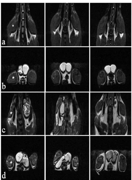
At clinical examination, aged (twenty-month-old) rats showed an evident reduction of skeletal muscle tissue. Fig. 1 shows coronal and axial slices acquired for three rats be-longing to the young and old group. This figure clearly shows that old rats showed less muscle tissue and more adipose tissue. This finding is confirmed by quantitative data shown in Table 1 and 2 where the average volume (expressed in cm3) of the muscle and adipose tissue tissue
measured in the hindlimbs are reported. No significant difference between left and right leg is observed for both young and old rats. On the contrary, the difference be-tween the same leg of the young and old group is highly significant (P < 0.05) for both muscle and adipose tissue.
No significant difference of the relaxation time T2 of the muscle was found between these two groups (four-month-old rats: 34.6 ± 1.6 ms in the left muscle and 33.9
± 1.1 ms in the right one; twenty-month-old rats: 33 ± 0.5 ms in the left and 33.2 ± 1 ms in the right muscle).
Figure 1
values than those found in senescent rats. In aged rats, the analysis of the enhancement curves shows a clear altera-tion of the first phase, which mainly expresses a retard of contrast medium arrival in the tissue. The maximal differ-ence between young and senescent rats occurred at about 98 sec after injection of the contrast agent (scan 14). In Ta-ble I we have reported the enhancement of the signal in-tensity measured at the point of maximal difference (scan 14). Data obtained from the legs of each rat showed high correlation in young rats (r2 = 0.98) and a worse
correla-tion (r2 = 0.59) in senescent rats. Unpaired t-test
per-formed on these data confirms a significant difference (P < 0.05) between old and young rats on both legs.
Discussion
Magnetic resonance imaging (MRI) has the ability to dis-criminate between various soft tissues in vivo [18]. Both cross-sectional and longitudinal studies on human have shown age-related changes in body composition: with ad-vancing age, elderly men and women tend to became more obese, their amount of visceral fats tends to increase, and their skeletal muscle mass declines [19–21].
Table 1: Average volume (cm3) of the muscle of the hindlimb.
4-MONTH-OLD 20-MONTH-OLD
LEFT RIGHT LEFT RIGHT
1 27,6 30,5 19,7 21,4 2 28,9 30,1 15,5 16,0 3 28,2 34,5 22,3 20,5 4 32,8 32,5 21,3 20,0 5 32,1 34,6 23,0 23,0 6 29,1 30,7 21,1 22,1
MEAN 29,7 32,1 20,4 20,5
SD 1,9 1,8 2,4 2,2
Table 2: Average volume (cm3) of the adipose tissue of the hindlimb.
4-MONTH-OLD 20-MONTH-OLD
LEFT RIGHT LEFT RIGHT
1 6,0 5,0 12,0 13,0 2 5,0 6,0 13,0 12,0 3 5,0 4,0 10,0 11,0 4 7,0 6,0 8,0 11,0 5 5,0 5,0 12,0 11,0 6 4,0 5,0 18,0 17,0
MEAN 5,3 5,1 12,1 12,5
SD 0,9 0,6 3,0 2,1
Table 3: Dynamic percentage of enhancement at about 2 minutes after contrast injection.
4-MONTH-OLD 20-MONTH OLD
LEFT RIGHT LEFT RIGHT
1 82,4 80,9 21,9 28,9 2 42,4 41,1 48,1 38,3 3 50,4 49,7 45,8 50 4 50,9 46,6 24,4 32,5 5 49,5 48,8 28 25,7 6 63,4 67,6 45,8 29,1
MEAN 56,5 55,8 35,7 34,1
SD 14,4 15,2 12,1 8,9
CORRELATION LEFT-RIGHT
0,98 0,59
Figure 2
Dynamic percentage of enhancement ∆S(t) = 100{[SI(t)-SIpre]/Sipre} on the ROIs selected on the gastrocnemius of the 4-moth-old (solid line) and on the 20-moth-old (dotted line). Values and error-bars represent mean ± SEM of both legs. Each dynamic scan was 7.4 sec long. The maximal differ-ence between the two groups was around the 14th scan (at about 2 minutes after contrast injection).
sec
37 74 111 148
0 10 20 30 40 50 60 70
Mean ±SEM
Our quantitative data show that MRI can detect body composition change with age, in particular hindlimb composition change, with increase in adipose tissue and decline in skeletal muscle, also in small laboratory spe-cies. Increase in adipose tissue is probably due to a process of differentiation of myoblasts into adipocyte [22]. Previ-ous study shows that myoblasts isolated from mPrevi-ouse hindlimb skeletal muscle demonstrate increased adipo-genic potential as a function of age. This change suggests that a default program may be activated in mesenchimal cells with increasing age resulting in a more adipogenic-like phenotype. However, whether this change in differen-tiation potential contributes to the increased adiposity in muscle with age remains to be determined [23].
Furthermore, muscle modifications we found are quanti-tative as well as qualiquanti-tative.
Data of CE-MRI show that the enhancement of the signal intensity in muscle is significantly higher in young rats that in old rats. This finding has never been described in the literature and demonstrates that a modification of the vasculo-stromal component of the muscle occurs in elder-ly. The tissue determinants of such modification are not clear because several parameters may change MRI-signal enhancement: the blood volume, the blood flow, the per-meability of the microcirculatory bed, the axial area or number of microvessels and the extracellular volume. In the sarcopenic muscle, all this parameters are probably al-tered. Data about age-related changes in vascular permea-bility of the rat skeletal muscle have not been reported yet; however data obtained on humans demonstrated high amount of water in the skeletal muscle of elderly popula-tion [16]. About blood volume, a meta-analysis of litera-ture suggests that a certain degree of reduction in capillarization can exist in elderly also if data are discord-ant. Reduced capillary density and reduced blood flow have been reported in some studies [7,24]. Alteration in heart rate and cardiac output with age such as the decrease of basal metabolic rate (BMR) acquired with aging could be factors affecting the hemodynamic pattern, while an age-related increased collagen concentration could modi-fy the MRI characteristics of the muscle. It has been found that the anaesthesia could also induce a lower blood flow in many tissues of the elderly rat compared to the adult, but data for the skeletal muscle are not in trend with the other organs [25].
The above reported considerations show that tissue mod-ification of the sarcopenic muscle is very complex and, taken together, they seem to suggest that the age-related impairment of the signal enhancement is a multifactorial event. However, in aged rats, the analysis of the enhance-ment curves shows a clear alteration of the first phase, which mainly expresses a retard of contrast arrival in the
tissue. Therefore, in vivo data, also if not conclusive, seem to suggest a reduction in blood flow in the sarcopenic muscle, whose possible origin from macroangiopathy should be excluded since the dynamics are quite different from those we have recently described in experimental ar-terial occlusion [17]. Instead, the pattern that we have found in aged animals is quite similar to that described in an experimental model of myocardium ischemia [26], where neo-angiogenesis by VEGF gene transfer significant-ly increased functional performances of the organ and magnetic resonance mapping demonstrated benefits of VEGF-induced myocardial angiogenesis.
Data obtained from the two different legs of each animal showed a greater homogeneity in young with respect to senescent rats. This finding seems to be in accordance with the existence of a microangiopathy, which may dif-ferently involve the left and right hindlimb.
Conclusion
Our functional MRI-findings suggest the existence of an alteration of the microcirculatory function in skeletal muscle of aged animals. Further studies, using MRI on longitudinal observation, will allow to show if the micro-circulatory alteration we described are foregoing or subse-quent to the process of differentiation of myoblasts into adipocyte.
Moreover, further studies will be necessary to evaluate the tissue determinants of CE-MRI modification that we have detected in the muscle of aged animals. It could be hy-pothesised that such a modification could be the expres-sion of multiple biological processes involving the sarcopenic skeletal muscle. This work clearly demon-strates that these events are quantifiable in vivo and that CE-MRI could represent a further tool for following up tis-sue modification and therapeutic intervention both in pa-tients with sarcopenia and in experimental models of this pathology.
Author's contribution
Author1: Conceived of the study, carried out the experi-ments and drafted the manuscript.
Author 2: Participated in analysis of data and manuscript writing.
Author 3: Veterinary advice.
Author 4: Conceived of the study, set up MRI sequences.
Author 6: Laboratory sponsor who participated in experi-ment design. Wrote the final version of the manuscript.
Author 7: Laboratory sponsor who conceived and coordi-nated the overall study.
Competing interests
None declared.
Acknowledgements
This work was supported by Ministero dell'Università e della Ricerca Scien-tifica e Tecnologica, MURST grant 9805406803, to Francesco Osculati.
References
1. Nair KS: Age-related changes in muscle. Mayo Clin Proc 2000, 75(Suppl):S14-18
2. Roubenoff R: Sarcopenia and its implications for the elderly.
Eur J Clin Nutr 2000, 54(Suppl 3):S40-47
3. Short KR, Nair KS: Mechanisms of sarcopenia of aging.J Endocri-nol Invest 1999, 22(5):95-105
4. Navarro A, Lopez-Cepero JM, Sànchez del Pino MJ: Skeletal muscle and aging.Front Biosci 2001, 6:26-44
5. Roubenoff R, Hughes VA: Sarcopenia: current concepts.J Geron-tol A Biol Sci Med Sci 2000, 55(12):716-724
6. Irion GL, Vasthare US, Tuma RF: Preservation of skeletal muscle hyperemic response to contraction with aging in female rats.
Exp Gerontol 1988, 23:183-188
7. McCully KK, Posner JD: The application of blood flow measure-ments to the study of aging muscle. J Gerontol Ser A 1995, 50A:130-136
8. Denis C, Chatard JC, Dormois D, Linossier MT, Geyssant A, Lacour JR: Effects of endurance training on capillary supply of human skeletal muscle on two age groups (20 and 60 years).J Physiol Paris 1986, 81:379-383
9. Grimby G, Danneskiold-Samsoe B, Hvid K, Saltin B: Morphology and enzymatic capacity in arm and leg muscles in 78–81 year old men and women.Acta Physiol Scand 1982, 115:125-134 10. Jakobsson F, Borg K, Edstrom L: Fibre-type composition,
struc-ture and cytoskeletal protein location of fibres in anterior tibial muscle. Comparison between young adults and physi-cally active aged humans.Acta Neuropathol (Berl) 1990, 80: 459-468
11. Coggan AR, Spina RJ, King DS, et al: Histochemical and enzymatic comparison of the gastrocnemius muscle of young and eld-erly men and women.J Gerontol 1992, 47:B71-76
12. Frontera WR, Hughes VA, Fielding RA, Fiatarone MA, Evans WJ, Roubenoff R: Aging of skeletal muscle: a 12-yr longitudinal study.J Appl Physiol 2000, 88:1321-1326
13. Parizkova J, Eiselt E, Sprynarova S, Wachtlova M: Body composi-tion, aerobic capacity and density of muscle capillaries in young and old men.J Appl Physiol 1971, 31:323-325
14. Davidson YS, Clague JE, Horan MA, Pendleton N: The effect of ag-ing on skeletal muscle capillarization in a murine model.J Gerontol A Biol Sci Med Sci 1999, 54(10):B448-451
15. Heymsfield SB, Gallagher D, Visser M, Nunez C, Wang ZM: Meas-urement of skeletal muscle: laboratory and epidemiological methods.J Gerontol A Biol Sci Med Sci 1995, 50(Spec No):23-29 16. Tsubahara A, Chino N, Akaboshi K, Okajima Y, Takahashi H:
Age-related changes of water and adipose tissue content in mus-cles estimated by magnetic resonance (MR) imaging.Disabil Rehabil 1995, 17(6):298-304
17. Asperio RM, Nicolato E, Marzola P, Farace P, Lunati E, Sbarbati A, Os-culati F: Delayed muscle injuries in arterial insufficiency: a contrast-enhanced MRI and 31P spectroscopy study in rats.
Radiology 2001, 220:413-419
18. Tang H, Vasselli JR, Wu EX, Boozer CN, Gallagher D: High resolu-tion magnetic resonance imaging tracks changes in organ and tissue mass in obese and aging rats.Am J Physiol Regul Integr Comp Physiol 2002, 282(3):R890-9
19. Santana H, Zoico E, Turcato E, Tosoni P, Bissoli L, Olivieri M, Bosello O, Zamboni M: Relation between body composition, fat
distri-bution, and lung function in elderly men.Am J Clin Nutr 2001, 73:827-31
20. Kyle Ug, Genton L, Hans D, Karsegard VL, Michel JP, Slosman DO, Pichard C: Total body mass, fat mass, fat-free mass, and skel-etal muscle in older people: c-ross sectional differences in 60-year-old persons.J Am Geriatr Soc 2001, 49(12):1633-40 21. Ryan AS, Nicklas BJ: Age-related changes in fat deposition in
mid-thigh muscle in women: relationships with metabolic cardiovascular disease risk factors.Int J Obes Relat Metab Disord
1999, 23(2):126-32
22. Ross SE, Hemati N, Longo KA, Bennet CN, Lucas PC, Erickson RL, MacDougald OA: Inhibition of adipogenesis by Wnt signalin.
Science 2000, 289:950-953
23. Taylor-Jones JM, McGehee RE, Rando TA, Lecka-Czernik B, Lipschitz DA, Peterson CA: Activation of an adipogenic program in adult myoblasts with age.Mech Ageing Dev 2002, 123(6):649-61 24. Irion GL, Vasthare US, Tuma RF: Age-related change in skeletal
muscle blood flow in the rat.J Gerontol 1987, 42:660-665 25. Tuma RF, Irion GL, Vashtare US, Heinel LA: Age-related changes
in regional blood flow in the rat.Am J Physiol 1985, 249:H485-491 26. Pearlman JD, Hibberd MG, Chuang ML, Harada K, Lopez JJ, Gladstone SR, Friedman M, Sellke FW, Simons M: Magnetic resonance map-ping demonstrates benefits of VEGF-induced myocardial an-giogenesis.Nat Med 1995, 1(10):1085-1089
Pre-publication history
The pre-publication history for this paper can be accessed here:
http://www.biomedcentral.com/1471-2342/2/2/prepub
Publish with BioMed Central and every scientist can read your work free of charge
"BioMedcentral will be the most significant development for disseminating the results of biomedical research in our lifetime."
Paul Nurse, Director-General, Imperial Cancer Research Fund
Publish with BMCand your research papers will be:
available free of charge to the entire biomedical community
peer reviewed and published immediately upon acceptance
cited in PubMed and archived on PubMed Central
yours - you keep the copyright
editorial@biomedcentral.com Submit your manuscript here:
http://www.biomedcentral.com/manuscript/
![Figure 2Dynamic percentage of enhancement ∆S(t) = 100{[SI(t)-SIpre]/Sipre} on the ROIs selected on the gastrocnemius ofthe 4-moth-old (solid line) and on the 20-moth-old (dottedline)](https://thumb-us.123doks.com/thumbv2/123dok_us/488072.1543493/4.612.318.550.107.524/figure-dynamic-percentage-enhancement-sipre-selected-gastrocnemius-dottedline.webp)
