Open Access
Research
Theory of sampling and its application in tissue based diagnosis
Klaus Kayser*
1, Holger Schultz
2, Torsten Goldmann
2, Jürgen Görtler
3,
Gian Kayser
4and Ekkehard Vollmer
2Address: 1UICC-TPCC, Institute of Pathology, Charite, Berlin, Germany, 2Clin. & Exp. Pathology, Research Center Borstel, Borstel, Germany, 3Deep Computing, IBM, Amsterdam, the Netherlands and 4Institute of Pathology, University of Freiburg, Freiburg, Germany
Email: Klaus Kayser* - [email protected]; Holger Schultz - [email protected]; Torsten Goldmann - [email protected]; Jürgen Görtler - [email protected]; Gian Kayser - [email protected]; Ekkehard Vollmer - [email protected] * Corresponding author
Abstract
Background: A general theory of sampling and its application in tissue based diagnosis is presented. Sampling is defined as extraction of information from certain limited spaces and its transformation into a statement or measure that is valid for the entire (reference) space. The procedure should be reproducible in time and space, i.e. give the same results when applied under similar circumstances. Sampling includes two different aspects, the procedure of sample selection and the efficiency of its performance. The practical performance of sample selection focuses on search for localization of specific compartments within the basic space, and search for presence of specific compartments.
Methods: When a sampling procedure is applied in diagnostic processes two different procedures can be distinguished: I) the evaluation of a diagnostic significance of a certain object, which is the probability that the object can be grouped into a certain diagnosis, and II) the probability to detect these basic units. Sampling can be performed without or with external knowledge, such as size of searched objects, neighbourhood conditions, spatial distribution of objects, etc. If the sample size is much larger than the object size, the application of a translation invariant transformation results in Kriege's formula, which is widely used in search for ores. Usually, sampling is performed in a series of area (space) selections of identical size. The size can be defined in relation to the reference space or according to interspatial relationship. The first method is called random sampling, the second stratified sampling.
Results: Random sampling does not require knowledge about the reference space, and is used to estimate the number and size of objects. Estimated features include area (volume) fraction, numerical, boundary and surface densities. Stratified sampling requires the knowledge of objects (and their features) and evaluates spatial features in relation to the detected objects (for example grey value distribution around an object). It serves also for the definition of parameters of the probability function in so – called active segmentation.
Conclusion: The method is useful in standardization of images derived from immunohistochemically stained slides, and implemented in the EAMUS™ system http://www.diagnomX.de. It can also be applied for the search of "objects possessing an amplification function", i.e. a rare event with "steering function". A formula to calculate the efficiency and potential error rate of the described sampling procedures is given. Published: 16 February 2009
Diagnostic Pathology 2009, 4:6 doi:10.1186/1746-1596-4-6
Received: 27 January 2009 Accepted: 16 February 2009
This article is available from: http://www.diagnosticpathology.org/content/4/1/6
© 2009 Kayser et al; licensee BioMed Central Ltd.
Diagnostic Pathology 2009, 4:6 http://www.diagnosticpathology.org/content/4/1/6
Introduction
Diagnostic surgical pathology or tissue – based diagnosis is confronted with remarkable changes in its environment and workflow. The technological progress has led to a broad application of molecular biological methods such as Fluorescent in Situ Hybridization (FISH), and other DNA – sequence amplification techniques [1,2]. Com-mercially available slide scanners digitize a complete glass slide within a few minutes, and permit the implementa-tion of completely digitized images into routine diagnos-tics [3,4]. In other words, the workload of a pathologist increases steadily not only by increase of material, but, in addition, due to the mandatory introduction of new, still tissue – based diagnostic technologies. Thus, the question arises: How can the availability of and access to digitized histological slides (virtual slides) be used to release the diagnostic pathologist from time consuming work steps in order to make the pathologist's work more effective and disease related?
In the early days of telepathology, which can be consid-ered to be the "mother of the digital pathologist's world", several authors reported on the diagnostic accuracy of viewing digitized slides in comparison to conventional microscopy [4-8]. The results were clear: the diagnostic accuracy viewing at a digitized (or virtual) slide is indistin-guishable to that of conventional microscopy; however, the required time is essentially longer [9,10]. The non appropriate and more time consuming search for appro-priate fields of view or the performed sampling procedure are obviously one reason of these constraints. To our knowledge, the theory of sampling in cytology and his-topathology has not been described in detail, and is nearly unknown in the environment of diagnostic pathologists. In this article we want to explain the main theoretical aspects and the derivatives of sampling which are per-formed in routine tissue – based diagnostics. The derived formulas will allow interested pathologists or scientists to search for applications that can diminish the sampling time in virtual slides.
Basic aspects of sampling in digitized histological slides (virtual slides)
Surgical pathology is a medical discipline that "extracts" information from human tissue and classifies the infor-mation in distinct terms that are called diagnoses. The common performance is to screen an organ or a tissue sec-tion for those spaces or areas that contain the most signif-icant information, and try to classify this information seen in the specific field of view. Thus, tissue – based diagnosis is based upon a procedure to search for small samples that allow to derive information that is valid for the whole (or even patient). In other words, an appropriate sampling procedure is a precondition to evaluate accurate and reproducible diagnoses [2,4,11-15]. Therefore, a detailed
definition and accurate description of the sampling method is a necessity if we want to further evaluate the diagnostic algorithms. This statement induces the defini-tion of sampling as follows: Sampling is a method to derive information from a limited (small) compartment of a large (even unlimited) system that is valid for the entire (basic) system. The system can be a space, a func-tion or set of funcfunc-tions, a body, an organ, a slide, or a DNA sequence.
The definition includes the term information, which has again to be defined: Information is a property that is exchanged between a sender and a receiver. Information is a property that can be understood by, and allows the receiver to react in an adequate, i.e., predictive manner. This definition of sampling includes two different aspects, which depend upon each other:
1. the method of sampling, and
2. the aim of the sampling procedure, i.e., which informa-tion should be extracted.
Different aims can require different methods of sampling, or at least different parameters of the same algorithm. The inclusion of an "aim" or "goal" to be assessed introduces the calculation of efficiency, or a cost/benefit estimation.
The most frequently used sampling goals are
➢ search for localization of specific items within the basic space, with the knowledge or assumption, that the space under consideration contains such items, and
➢ search for presence of specific items (tumour cells, ores, lobster, etc.), where the exact localisation of these items is of minor interest (for example localization of tumour cells in a cytological smear).
The prepositions to apply an adequate sampling proce-dure in tissue – based diagnosis include that number and size of the samples are limited. In addition, the detectable information has to be known. This information com-monly depends upon additional (external) factors, and can be translated into diagnostic features that allow the detection and identification of a probe within the sam-pling space. These features can depend upon the size of the probes, their number, and their position within the collective, or even within the sampling space.
I) the evaluation of a diagnostic significance of a certain object or "basic unit" which is the probability that the object can be grouped into a certain diagnosis, and
II) the probability to detect these basic units within the entire space.
The detection probability of a wanted object depends then upon the size of the basic space A (filed of view, organ, nucleus, etc), the number and size of the samples (diag-nostic frames) D, and number and the sizes of the detected objects [Ci], as demonstrated in figure 1. Each detected object Ci possesses a certain probability to con-tribute to the diagnosis I which can be obtained by a map-ping (Ci, D) on ID. ID is the probability to state the
diagnosis I within the frame D by the object i, or ID = s(Ci, D). Basic examples are given in figure 2, figure 3, and in figure 4. In principle, the differentiation of the mapping of
(Ci, D) on ID into the two different procedures, namely a) the detection (geometric significance) and into the diag-nostic contribution reflects to the applied segmentation algorithms. These distinguish between areas (pixels) that contributed to the object and those that do not. Measure-ments of objects can only be done if the object is com-pletely covered by the sample frame. The spatial selection of the samples can either be performed randomly, or dependent upon the localization of already segmented objects (stratified sampling). Stratified sampling is based upon the general law of self organization, i.e., similar bio-logical systems tend to be localized in a neighbourhood relation. In other words, to detect a cancer cell is more likely in the neighbourhood of an already identified can-cer cell than elsewhere. The function of diagnostic signifi-cance is often of exponential nature, and in light microscopy related to three different image properties,
Survey of sampling algorithm
Figure 1
Diagnostic Pathology 2009, 4:6 http://www.diagnosticpathology.org/content/4/1/6
namely the texture, the object, and the object – associated structure [12,16-19].
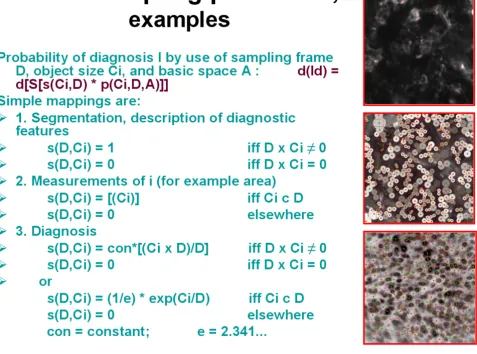
Sampling aims, applications and examples
Sampling is basically an information detection and trans-formation procedure, and thus undertaken to reach a cer-tain final aim, for example to state a diagnosis, or to identify the presence or absence of certain objects. A time and space invariant translation of the sampling procedure can be assumed as long as we want to obtain reproducible results (figure 3). Such a translation permits a separation of the object detection likelihood from the diagnostic sig-nificance of the segmented objects, and allows us to com-pute both properties separately. Assuming a digitized image, each point (pixel x, y) within the basic image is either a presentation of an object or not. All object fea-tures can be reduced to a function that represents the object pixels in relation to sample and basic image size
(figure 4). The introduction of an exponential diagnosis function then gives us the well known formula of Krige, which is commonly used to detect ores, oil fields, or underground water reservations [20]. Furthermore, the application of specific mappings (dilation, erosion) per-mits us to "increase the magnification of an object within its sampling frame, or to define the center of gravity in an object in order to compute the image structure. One will obtain a so – called order of structures if these procedures are repetitively applied to light microscopy [21].
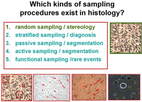
Random and stratified sampling procedures
The prerequisite of any (random, stratified) sampling is at least a binary image, i.e. a foreground defining the basic units and a background have to exist. Any sampling pro-cedure can be performed either as random or stratified sampling figure 5 and figure 6. In addition, an active, pas-sive, and a functional sampling can be distinguished.
Examples of different sampling procedures
Figure 2
Random sampling is the selection of biological meaning-ful units (nuclei, cells, proteins, etc.) at random. It is used to measure
➢ the frequency of the analyzed units in relation to each other or to the basic space (structure);
➢ certain features of the biological units in relation to the basic space (to further identify and classify the objects).
No information about the basic (reference) space is needed. The detection of biological meaningful units is then equivalent to the segmentation of the image and analysis of randomly chosen segmented elements. This procedure forms the basis of numerous investigations since the 1950s. It is commonly called stereology [22-26]. In principle, a grid consisting of regular lines (points) with identical length (and distance in between) is over-layed to the image, and the number of hits (intersections) is counted. From the number of intersections the volume – adjusted frequency, size, surface can be derived, inde-pendent from the orientation and shape of the elements. In a binary image the pixels (binary x, y points) can be used as a grid. Random chosen are the cutting angle (plane/volume), and the start point of the grid (pixel).
Stratified are the selection of the grid (all pixels) and the count of intersections. Thus, any random sampling is pro-vided by the start of the procedure, for example by ran-dom selection of the upper right position (x, y) coordinates of the sample space. From the relation x/A
(number of hits x/reference area A) two-dimensional (and also three – dimensional) parameters can be derived. These include the area density (Aa), the volume density (Vv), the boundary density (Ba), the numerical density (Na), and the surface density (Sv). It should be noted, that this quite easily applied procedure permits the estimation of significant three – dimensional object features without any sophisticated three dimensional reconstruction [23,27,18,29].
Stratified sampling, in contrast, is provided by a specific selection of intersections (objects). Its objective is the detection of specific objects (of known features), and the measurement of features of known specific objects, or the estimation of objective-associated reference volumes, for example the density of proliferating cells related to dis-tance from the nearest vessel [3,9,18,21]. Using again a grid as a measurement tool, the cutting angle (plane/vol-ume) and the start point of the grid (pixel) are also ran-domly chosen. Stratified are the selection of the grid (all
Examples of probability calculations in various sampling procedures
Figure 3
Diagnostic Pathology 2009, 4:6 http://www.diagnosticpathology.org/content/4/1/6
pixels), and the count of specific intersections (for exam-ple large cells) only. Thus: stratified sampling is provided by specific a selection of intersections (objects). A classic example is its application in cytology, i.e. to find the diag-nosis-relevant cell (tumor cell) within a large number of "normal" cells. One could try to analyze
1. only those areas which contain features of (any) cell (gray value selection at low magnification)
2. within these areas only those cells which seem to be abnormal (gray value, size, moderate magnification)
3. within these cells those with abnormal nuclear size (DNA content), high magnification.
4. terminate the procedure once the diagnosis – signifi-cant information has been obtained
All other items are disregarded or neglected. The imple-mentation of such an algorithm can speed up the time required "to screen a slide" significantly [5,12,30].
Stratified sampling requires some external knowledge in order to detect the biological meaningful events such as cancer cells. The image features of a cancer cell have to be known if one would like to detect this event by stratified sampling. The alternative algorithm would be to "sample" all cells, and start, if possible, a statistical analysis. This would then try to evaluate the rare events (supposing that cancer cells are rare to normal cells). Again, some external knowledge would be necessary. Obviously, this is related to the diagnosis function s(Ci, D).
Stratified sampling requires an accurate segmentation of objects with known features. Independent upon the
Examples of sampling procedures in segmentation and diagnosis classification
Figure 4
actual segmentation procedure the sampling can be per-formed as active and passive sampling.
Active and passive sampling
Any segmentation procedure has to accurately define the area of an object, which is equivalent to detect its bound-ary. Each pixel has to be distinguished either to belong to the object or not, which can be written: f(x, y, meaning) = [1,0], with f(x, y, object) = [1], and f(x, y, backgound) = [0] This approach is called passive sampling, as it discrim-inates the object area by a simple yes – no function [14]. In other words, passive sampling is provided by a constant relation between the objects and the grid (intersections). The intersection has the probability function p(i) = [1].
Active sampling is a different approach. It is provided by an objective-specific relation between the objects and the grid (intersections). The probability that a pixel belongs to an object ranges between [1,0]. The intersection has a
probability function p(i, o), i.e., the probability to detect the pixels that belong to a certain object depends on the object itself and its neighborhood [20]. For example, a pixel displays a probability of 0.7 that it belongs to the object. This probability can increase or decrease depend-ent upon additional parameters, such as size, oridepend-entation, or shape of neighboring objects. Naturally, the probability value of 0.7 itself might be used to define whether it is an "object" – or a "background" pixel.
The probability function p(i, o) can be calculated if we separate p(i, o) in its two components: p(i, o) = gr(x, v) * af(gr, v).
gr(x, y) is the frequency distribution of different objects in the reference space v,
af(gr, v) is the detection probability in the space v.
Derivatives of basic sampling procedures in separating the diagnosis function s from the sampling function p
Figure 5
Diagnostic Pathology 2009, 4:6 http://www.diagnosticpathology.org/content/4/1/6
If we assume that af(gr, v) = const in the reference space v, we can estimate p(i, o) by a set of measurements in differ-ent sample spaces and transform p(i, o) = [1] if gr(x, y) > const, and p(I, o) = [0] elsewhere.
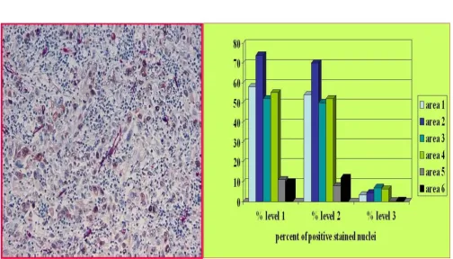
Active sampling has been reported to be an effective method to correct the variation in immunohistochemistry staining, for example to compute the threshold of positive staining intensity [2]. The classic problem is: At which staining intensity (color level) can an "immunohisto-chemically analyzed cell" be grouped into the positive class? The active sampling attempt is to measure the rela-tion positive/negative cells at different threshold levels in several randomly selected sample areas. We then select the discrimination threshold which results in (number of positive objects/number of negative objects) = const for all selected samples. A characteristic application is dem-onstrated in figure 7. It displays a Mib_1 stain for
prolifer-ating nuclei in a lung carcinoma, and the relative number of positive nuclei dependent upon three different thresh-olds measured in five different sample areas. Threshold number three is obviously too high, and the threshold number 1 too low. The threshold number two should be chosen as discriminating threshold, i.e., to calculate the relative number of proliferating nuclei. This algorithm has been successfully implemented in the automated immu-nohistochemistry measurement system (EAMUS™) [2,3,9,16,17].
Functional sampling
The idea of functional sampling focuses on the interpreta-tion of rare events [12,14,15,31-33]. The quesinterpreta-tion arises: Do there exist certain rare cells within a cellular society (tissue) that possess a high functional power similar to catalysts in chemistry. If yes, how can they be identified? Therefore, functional sampling is defined as the search for
Survey of different sampling procedures
Figure 6
a specific (key) function of rare biological objects within a different (majority) population. As the function to be analyzed might be unknown and we cannot observe the proposed function directly, we have to state the following prerequisites for proper analysis:
1. The specific object (cell) is rare within the basic popu-lation.
2. It has to possess regular neighborhood relations to objects of the basic population.
3. It has to be randomly distributed within the reference space.
The proposed algorithm tries to evaluate the distance properties between the rare events and the frequent events, and the general distribution of rare events within the reference space as follows:
1. We perform a random sampling of the specific (rare) object (O) within the basic population Ni (to estimating
O [Ni]).
2. We perform a stratified sampling "around" each detected specific object (to estimating Ni(0)).
3. If Ni(O) = constant we can assume a specific function of the object (cell) within the basic population (for example
Example of an active sampling to determine the discrimination threshold of a Mib-1 PAP stained undifferentiated large cell car-cinoma (in association to the distance of the nearest vessel (proliferating nuclei are stained brown, vessels are stained red)
Figure 7
Diagnostic Pathology 2009, 4:6 http://www.diagnosticpathology.org/content/4/1/6
cellular immune competence, functional activation of cells, etc.).
An example is shown in figure 8 which displays the bind-ing capacities of labeled galectin-1 in an undifferentiated lung carcinoma. Only one large intensively stained cell is present in this sample area (marked by an arrow). Its fre-quency in all samples is < 5%, and the mean distance between these cells measures 245 ± 198 μm, i.e. these cells do not express a constant distance in between. The oppo-site, however, holds true for the distance between these large cells and their nearest (majority) cells as well as between the other cells within this tissue. From these fig-ures we can derive, that the rare large cells probably posses a significant biological function for the whole tissue (according to biochemical investigations galectin-1
belongs to a family of galactoside binding proteins that has growth regulatory and immunomodulatory proper-ties [21].
Sampling efficiency
As we have defined sampling, it is a procedure that wants to describe space and time-related properties in surgical pathology, i.e. in tissue – based diagnosis. Such an inves-tigation can be performed in different manners, which can be of different efficiencies. How can sampling efficiency be measured? Obviously, any sampling efficiency is closely related to the spatial distribution of the events searched for in the reference space. If the spatial distribu-tion is known we can adjust the sampling procedure cor-respondingly. However, its spatial distribution is often not known, and we have to start with a random selection
Example of expanded functional sampling: A rare event (galectin-1 binding large cancer cell) within a large cell anaplastic lung carcinoma displays a regular distance to its nearest neighbouring cells and a random distribution within the whole slide
Figure 8
of certain compartments (samples). We can then measure the spatial distribution (frequency) p = s(N)/v, and the variation of p is the error E(p), which should be small in order to have an efficient sampling procedure. Usually, we can state that the reference space v >> s(Ne) (size of ele-ment e). The error E(p) can then be calculated according to
E(p) = Σ[E2(Ne) + E2(B(n)) + E2(Ne/sv)](1/2)
with
E(Ne) = error of detecting an individual event (i.e., prob-ability of identification/missing a tumor cell)
E(B(n)) = error of measuring all elements in the reference space (i.e., related to the biological variance of the tissue, dependent upon N)
E(Ne/v) = error of measuring the size of events e in rela-tion to size of sampling space S (frequency of e in sample space sv).
We can derive the following statements from this formula:
1. We obtain the smallest sampling error if we select the reference volume as sample size, and if we are dealing with regular tissue (small biological variance).
2. The smaller the sample sizes in relation to the size of events, the bigger is the sampling error, as long as the error to segment (identify) per event is not increasing.
3. The sampling error is increasing if we choose different sizes of the samples.
Discussion
To take and to analyze samples of a broad variety of tis-sues is a basic procedure in surgical pathology, or in tissue – based diagnosis. All diagnostic algorithms depend upon a correct and reliable sampling procedure, and extensive training in surgical pathology addresses to identify and sample those tissue compartments that probably contain the most significant information to classify the disease present [7,19,34-38]. The majority of investigations addresses to an optimum sampling procedure, for exam-ple. How many sentinel lymph nodes should be investi-gated in relation to the stage of breast cancer [29,31], or "optimizing sampling of tomato fruit for carotenoid con-tent, or how to perform endometrial sampling in patients with trophoblastic disease after suction curettage [39,40]. In the early days of stereology several authors took atten-tion on the sampling procedures, as the results of count-ing interceptions are closely associated to the nature of the used sampling method [22,23]. Recently, sampling has
returned to the focus of investigations, especially in live imaging [41]. Most of the investigations try to optimize the sampling, which is equivalent to evaluate the "best" stratified sampling method.
In addition to medical applications, sampling plays a dominant role in geology, especially mining. In fact, Krige's sampling analysis can be considered to be the first approach to develop a "sampling theory" [12,20].
In this article we want to derive a scheme of sampling that permits a principle view of sampling, its different meth-ods, and to calculate the efficiency of the used sampling method. In principle, two different algorithms exist, the random sampling and the stratified sampling [12,9]. Ran-dom sampling has to be performed, if no knowledge of the information searched for exists. It is the appropriate technique to measure features of biological units such as chromosomes, DNA fragments, nuclei, cells, vessels, etc. Its accuracy (error rate) can be predefined by number and size of the chosen samples in relation to the expected size of events and to the reference space. Its results can be implemented in additional classification algorithms, such as diagnostic procedures. The sampling can be terminated if a certain classification can be performed with a prede-fined accuracy, i.e, a diagnosis can be assessed with high certainty. The accurate measurement of events' features is a prerequisite, but not the aim of stratified sampling. Its implementation requires additional (external) informa-tion, and numerous investigations have been performed to "speed up" the procedure (or to make it more efficient) using spatial structures within the reference space. When an exponential event probability distribution is given, Krige's formula can be derived from stratified sampling.
In addition to the discussed principle differences between random and stratified sampling procedures, passive and active sampling plays a major role in image segmentation algorithms. The common principle of active sampling associates neighbourhood knowledge (i.e. knowledge derived from general external observations) to the object under investigation, for example to accurately define its boundaries [18]. Especially in measuring accurate thresh-olds for grading purposes in immunohistochemistry this approach has been proven to be successful [2]. A further-more derived application is the functional sampling, which is again a stratified sampling in principle. This pro-cedure can assist to investigate in the "biological impor-tance" of rare events, which is widely not known to our experience.
Diagnostic Pathology 2009, 4:6 http://www.diagnosticpathology.org/content/4/1/6
of major importance that all diagnostic investigations start with appropriate sampling.
Competing interests
The authors declare that they have no competing interests.
Authors' contributions
KK and EV initialized the study and drafted the paper. HS, TG, JG and GK were involved in generating and evaluating the data and in the writing of the manuscript.
Acknowledgements
The financial support of the international Academy of Telepathology e.V., and of the Verein zur Förderung des biologisch-technologischen Fort-schritts in der Medizin e.V. are gratefully acknowledged.
References
1. Harewood GC: Endoscopic tissue diagnosis of cholangiocarci-noma. Current opinion in gastroenterology 2008, 24:627-630. 2. Kayser G, Radziszowski D, Bzdyl P, et al.: Theory and
implemen-tation of an electronic, automated measurement system for images obtained from immunohistochemically stained slides. Anal Quant Cytol Histol 2006, 28:27-38.
3. Kayser K, Molnar B, Weinstein R: Virtual Microscopy: Funda-mentals, Applications, Perspectives of Electronic Tissue-based Diagnosis. Berlin: VSV Interdisciplinary Medical Publishing; 2006.
4. Molnar B, Berczi L, Diczhazy C, et al.: Digital slide and virtual microscopy based routine and telepathology evaluation of routine gastrointestinal biopsy specimens. J Clin Pathol 2003,
56:433-438.
5. Kayser K, Kayser G: Basic aspects of and recent developments in telepathology in Europe, with specific emphasis on quality assurance. Anal Quant Cytol Histol 1999, 21:319-328.
6. Kayser K, Kayser G, Radziszowski D, et al.: New developments in digital pathology: from telepathology to virtual pathology laboratory. Stud Health Technol Inform 2004, 105:61-69.
7. Leong FJ, McGee JO: Automated complete slide digitization: a medium for simultaneous viewing by multiple pathologists.
J Pathol 2001, 195:508-514.
8. Leong FJ, Nicholson AG, McGee JO: Robotic telepathology: effi-cacy and usability in pulmonary pathology. J Pathol 2002,
197:211-217.
9. Kayser K, Kayser G: Virtual Microscopy and Automated Diag-nosis. In Virtual Microscopy and Virtual Slides in Teaching, Diagnosis and Research Edited by: Gu J, Ogilvie R. Boca Raton: Taylor & Francis; 2005.
10. Florell SR, Smoller BR, Boucher KM, et al.: Sampling of melano-cytic nevi for research purposes: a prospective, pilot study to determine effect on diagnosis. Journal of the American Academy of Dermatology 2008, 59:814-821.
11. Kayser K, Kayser G, Becker HD, et al.: Telediagnosis of trans-bronchial fine needle aspirations--a feasibility study. Anal Cell Pathol 2000, 21:207-212.
12. Kayser K, Hufnagl P, Kayser G, et al.: Stratified sampling: Basic ideas and application in pathology. Elec J Pathol Histol 1999,
5:994-906.
13. Mavrodieva V, Levy L, Gabriel DW: Improved sampling methods for real-time polymerase chain reaction diagnosis of citrus canker from field samples. Phytopathology 2004, 94:61-68. 14. Trpkov K, Thompson J, Kulaga A, et al.: How much tissue
sam-pling is required when unsuspected minimal prostate carci-noma is identified on transurethral resection? Archives of pathology & laboratory medicine 2008, 132:1313-1316.
15. Winters R, Waters BL: What is adequate sampling of extrapla-cental membranes?: a randomized, prospective analysis.
Archives of pathology & laboratory medicine 2008, 132:1920-1923. 16. Kayser K, Gortler J, Goldmann T, et al.: Image standards in
Tis-sue-Based Diagnosis (Diagnostic Surgical Pathology). Diagn Pathol 2008, 3:17.
17. Kayser K, Gortler J, Metze K, et al.: How to measure image qual-ity in tissue-based diagnosis (diagnostic surgical pathology).
Diagn Pathol 2008, 3(Suppl 1):S11.
18. Kayser K, Hoshang SA, Metze K, et al.: Texture and object related automated information analysis in histological still images of various origins. Analytical and Quantitative Cytology and Histology
2008, 30:323-35.
19. Kayser K, Radziszowski D, Bzdyl P, et al.: Towards an automated virtual slide screening: theoretical considerations and practi-cal experiences of automated tissue-based virtual diagnosis to be implemented in the Internet. Diagn Pathol 2006, 1:10. 20. Krige D, DS Rand: Some basic considerations in the application
of gepstatistics to the valuation of ore in South African gold mines. J South Afr Inst Min Metal 1976, 77:383-391.
21. Kayser K, Gabius HJ: Graph theory and the entropy concept in histochemistry. Theoretical considerations, application in histopathology and the combination with receptor-specific approaches. Prog Histochem Cytochem 1997, 32:1-106.
22. Gundersen HJ: Stereology of arbitrary particles. A review of unbiased number and size estimators and the presentation of some new ones, in memory of William R. Thompson. Jour-nal of microscopy 1986, 143:3-45.
23. Gundersen HJ, Jensen EB: Stereological estimation of the vol-ume-weighted mean volume of arbitrary particles observed on random sections. Journal of microscopy 1985, 138:127-142. 24. Knust J, Ochs M, Gundersen HJ, et al.: Stereological estimates of
alveolar number and size and capillary length and surface area in mice lungs. Anat Rec (Hoboken) 2009, 292:113-122. 25. Vesterby A, Gundersen HJ, Melsen F: Unbiased stereological
esti-mation of osteoid and resorption fractional surfaces in trabecular bone using vertical sections: sampling efficiency and biological variation. Bone 1987, 8:333-337.
26. Vesterby A, Kragstrup J, Gundersen HJ, et al.: Unbiased stereologic estimation of surface density in bone using vertical sections.
Bone 1987, 8:13-17.
27. Mayhew TM: A stereological perspective on placental mor-phology in normal and complicated pregnancies. Journal of anatomy 2008 in press.
28. Mayhew TM, Muhlfeld C, Vanhecke D, et al.: A review of recent methods for efficiently quantifying immunogold and other nanoparticles using TEM sections through cells, tissues and organs. Ann Anat 2008 in press.
29. Michel SC, Low R, Singer G, et al.: [Stereotactic Mammotome breast biopsy: routine clinical experience and correlation with BI-RADS--classification and histopathology]. Praxis 2007,
96:1459-1474.
30. Kennedy MP, Jimenez CA, Bruzzi JF, et al.: Endobronchial ultra-sound-guided transbronchial needle aspiration in the diagno-sis of lymphoma. Thorax 2008, 63:360-365.
31. Atula T, Shoaib T, Ross GL, et al.: How many sentinel nodes should be harvested in oral squamous cell carcinoma? Eur Arch Otorhinolaryngol 2008, 265(Suppl 1):S19-23.
32. Trotz M, Weber MA, Jacques TS, et al.: Disseminated langerhans cell histiocytosis-related sudden unexpected death in infancy. Fetal and pediatric pathology 2009, 28:39-44.
33. Zong JC, Arav-Boger R, Alcendor DJ, et al.: Reflections on the interpretation of heterogeneity and strain differences based on very limited PCR sequence data from Kaposi's sarcoma-associated herpesvirus genomes. J Clin Virol 2007, 40:1-8. 34. Bartels P, Weber J, Duckstein L: Machine learning in quantitative
histopathology. Anal Quant Cytol Histol 1988, 10:299-306. 35. Bartels PH: Computer-generated diagnosis and image
analy-sis. An overview. Cancer 1992, 69:1636-1638.
36. Oger M, Belhomme P, Klossa J, et al.: Automated region of inter-est retrieval and classification using spectral analysis. Diagnos-tic Pathology 2008, 3(Suppl 1):S17.
37. Permuth-Wey J, Boulware D, Valkov N, et al.: Sampling strategies for tissue microarrays to evaluate biomarkers in ovarian cancer. Cancer Epidemiol Biomarkers Prev 2009, 18:28-34.
38. van Diest PJ, Kayser K, Meijer GA, et al.: Syntactic structure anal-ysis. Pathologica 1995, 87:255-262.
Publish with BioMed Central and every scientist can read your work free of charge
"BioMed Central will be the most significant development for disseminating the results of biomedical researc h in our lifetime."
Sir Paul Nurse, Cancer Research UK
Your research papers will be:
available free of charge to the entire biomedical community
peer reviewed and published immediately upon acceptance
cited in PubMed and archived on PubMed Central
yours — you keep the copyright
Submit your manuscript here:
http://www.biomedcentral.com/info/publishing_adv.asp
BioMedcentral 40. Lertkhachonsuk R, Treratanachat S: Endometrial sampling in
patients with trophoblastic disease after suction curettage.
The Journal of reproductive medicine 2008, 53:634-638.
41. Lundstrom C, Ljung P, Persson A, et al.: Uncertainty visualization in medical volume rendering using probabilistic animation.
IEEE transactions on visualization and computer graphics 2007,