RESEARCH
Effect of blood contamination
of cerebrospinal fluid on amino acids, biogenic
amines, pterins and vitamins
Marta Batllori
1, Mercedes Casado
1,4, Cristina Sierra
1, Maria del Carmen Salgado
1, Laura Marti‑Sanchez
1,
Joan Maynou
2, Guerau Fernandez
2, Angels Garcia‑Cazorla
3,4, Aida Ormazabal
1,4, Marta Molero‑Luis
1*and Rafael Artuch
1,4Abstract
Background: Cerebrospinal fluid (CSF) metabolomic investigations are a powerful tool for studying neurometa‑ bolic diseases. We aimed to assess the effect of CSF contamination with blood on the concentrations of selected biomarkers.
Methods: CSF samples were spiked in duplicate with increasing volumes of whole blood under two conditions: (A) pooled CSF spiked with fresh whole blood and frozen to cause red blood cell (RBC) lysis; (B) pooled CSF spiked with fresh blood and centrifuged (the supernatant with no RBCs was frozen until the moment of analysis). CSF concen‑ trations of amino acids, biogenic amines, pterins, and vitamins were analysed by HPLC coupled with tandem mass spectrometry, electrochemical and fluorescence detection.
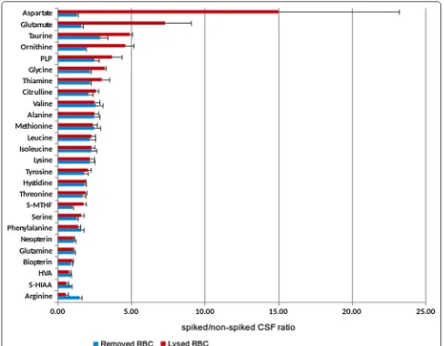
Results: Aspartate, glutamate, taurine, ornithine, glycine, citrulline, pyridoxal 5´‑phosphate, 5‑methyltetrahydrofolate, and thiamine showed higher values when RBCs were lysed when compared with those of CSF with no RBC, while arginine, 5‑hydroxyindoleacetic and homovanillic acids showed lower values. When RBCs were removed from CSF, only some amino acids, thiamine and pyridoxal 5´‑phosphate showed moderately higher values when compared with the non‑spiked CSF sample.
Conclusions: CSF‑targeted metabolomic analysis is feasible even when substantial RBC contamination of CSF has occurred since CSF centrifugation to remove RBC prior to freezing eliminated most of the interferences observed. Keywords: Cerebrospinal fluid, Amino acids, Biogenic amines, Pterins, Vitamins, Blood contamination
© The Author(s) 2019. This article is distributed under the terms of the Creative Commons Attribution 4.0 International License (http://creat iveco mmons .org/licen ses/by/4.0/), which permits unrestricted use, distribution, and reproduction in any medium, provided you give appropriate credit to the original author(s) and the source, provide a link to the Creative Commons license, and indicate if changes were made. The Creative Commons Public Domain Dedication waiver (http://creativecommons.org/ publicdomain/zero/1.0/) applies to the data made available in this article, unless otherwise stated.
Background
Cerebrospinal fluid (CSF) is a biological fluid that is mainly produced by the choroid plexus, which constitutes the interface between blood vessels and CSF [1, 2]. The composition of CSF is also controlled by the blood–brain barrier, which separates blood from the brain paren-chyma [3]. Both structures deliver substrates for brain cell metabolism and remove the corresponding waste [1,
4, 5]. In general, the blood–brain barrier greatly restricts
the influx of most molecules, including amino acids and other compounds [6]. Amino acids, with few exceptions (e.g., glutamine), show lower values in CSF when com-pared with those of plasma [7, 8]. For other metabolites, biosynthetic pathways are compartmentalized in the brain, and similar concentrations may be observed in CSF and blood since no transport from blood to CSF is expected; this is the case for biogenic amines and pterins [9, 10]. In contrast, some vitamins have to be transported into the brain through central nervous system barriers by specific transporters, and differences between vitamin concentrations in CSF and blood samples are noticeable [11, 12]. While folate is one of the few molecules more concentrated in CSF when compared to plasma, other
Open Access
*Correspondence: mmolerol@sjdhospitalbarcelona.org
1 Clinical Biochemistry Department, Hospital Sant Joan de Déu, Barcelona,
Spain
vitamins such as thiamine and pyridoxine display lower values in CSF when compared to those of blood [12, 13].
CSF metabolomic investigations have been dem-onstrated to be a powerful tool for studying specific neurometabolic pathways and related diseases and for exploring metabolic transport from the blood into the brain [14]. Several neurogenetic conditions are caused by specific disturbances in these processes (Table 1). In recent decades, targeted metabolomic approaches have been used for the study of these neurogenetic conditions [14]. Owing to the important differences in the metabolite concentrations between blood and CSF,
contamination of CSF with blood may cause dramatic effects in the measured concentrations of most of the above-mentioned metabolites [15–18]. CSF is collected by lumbar puncture, which is an invasive method. Since blood/plasma contamination can be frequently observed by different causes (traumatic lumbar punc-tures, impaired blood–brain barrier permeability or intraventricular bleeding) [19–21], a misinterpretation of metabolic profiles is a problem that should be mini-mized to avoid repeated lumbar puncture procedures and diagnostic errors.
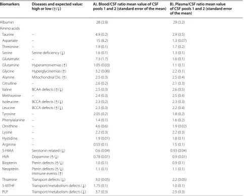
Table 1 CSF biomarkers and their semiological value for different diseases (left columns)
Blood/CSF ratios for the two experimental conditions: A) Red blood cells (RBC) lysed in CSF and B) RBC removed from CSF. CSF samples were spiked with 20% of whole blood. A total of 20 CSF aliquots coming from 50 CSF samples were analysed (see details in Additional file 2: Figure S1)
When CSF blood contamination occurs, the most critical metabolites for data interpretation can be glycine (the ratio blood/CSF glycine values is very high) and vitamins such as pyridoxine, thiamine and folate: In genetic diseases leading to brain pyridoxine, folate, and thiamine deficiencies, the blood concentrations of these vitamins can be normal, while CSF values may be near undetectable. Thus, blood contamination could mask the CSF vitamin deficiency. The monoamines HVA and 5-HIAA are sensitive to haemoglobin oxidation
BCAA Branched chain amino acids
Biomarkers Diseases and expected value:
high or low (↑/↓) A). Blood/CSF ratio mean value of CSF pools 1 and 2 (standard error of the mean) B). Plasma/CSF ratio mean value of CSF pools 1 and 2 (standard error of the mean)
Albumin 28 (3.9) 29 (3.2)
Amino acids
Taurine – 4.9 (0.2) 2.9 (0.5)
Aspartate – 15 (8.2) 1.3 (0.07)
Threonine – 1.9 (0.1) 1.7 (0.2)
Serine Serine deficiency (↓) 1.6 (0.1) 1.3 (0.1)
Glutamate – 7.3 (1.7) 1.6 (0.1)
Glutamine Hyperamoniemias (↑) 1.05 (0.03) 1.1 (0.1)
Glycine Hyperglycinemias (↑) 3.2 (0.06) 2.2 (0.1)
Alanine Mitochondrial Dis. (↑) 2.5 (0.3) 2.5 (0.4)
Citrulline – 2.6 (0.2) 2.1 (0.3)
Valine BCAA defects (↑/↓) 2.5 (0.3) 2.6 (0.5)
Methionine – 2.4 (0.3) 2.5 (0.4)
Isoleucine BCCA defects (↑/↓) 2.3 (0.2) 2.3 (0.3)
Leucine BCCA defects (↑/↓) 2.3 (0.3) 2.2 (0.4)
Tyrosine – 2.05 (0.2) 1.8 (0.2)
Phenylalanine – 1.4 (0.1) 1.6 (0.2)
Ornithine – 4.6 (0.6) 1.9 (0.02)
Lysine – 2.2 (0.3) 2.2 (0.3)
Hystidine – 1.9 (0.01) 1.8 (0.1)
Arginine – 0.55 (0.1) 1.5 (0.1)
5‑HIAA Serotonin related (↓) 0.6 (0.04) 0.93 (0.04)
HVA Dopamine (↑/↓) 0.78 (0.01) 0.9 (0.01)
Biopterin Pterin defects (↑/↓) 1.0 (0.1) 0.9 (0.1)
Neopterin Pterin defects (↑/↓)
Immune events (↑) 1.1 (0.1) 1.1 (0.1)
Thiamine Transport defects (↓) 3.0 (0.05) 2.2 (0.05)
5‑MTHF Transport/metabolism defects (↓) 1.75 (0.1) 1.0 (0.1)
With this background, we aimed to assess the effect of CSF contamination with blood on the concentrations of selected molecules which are biomarkers for the study of different neurometabolic conditions.
Methods Samples
CSF samples were collected from patients where lumbar puncture was done to rule out meningoencephalitis, and stored at − 80 °C, following a previously reported proto-col [22]. The remnants of 50 CSF anonymized samples with no red blood cell (RBC) contamination (assessed by light microscopy as less than 5 RBC per field) were thawed, pooled (25 samples for pool 1 and the other 25 for pool 2), reaching a final volume of 10 mL for each pool. The pooled samples were divided into 1 mL ali-quots, which were spiked with different volumes of whole blood (at that moment, a fresh blood sample was withdrawn from a healthy volunteer). Non-spiked CSF samples and four spiking conditions were prepared in duplicate in the 2 CSF pools. The CSF pools were spiked with increasing volumes of whole blood: 2.5%, 5%, 10%, and 20%, in 2 different conditions: (A) CSF samples spiked with fresh whole blood and then frozen at − 80 °C to cause RBC lysis. (B). CSF samples spiked with fresh blood, then centrifuged at 1500×g 10 min at 4 °C, with the clear supernatant frozen at − 80 °C. Details of the pro-tocol are stated in Additional file 1: Table S1 and Addi-tional file 2: Figure S1. The total sample preparation time spent was 45 min (all samples were frozen at the same time). With these conditions we could assess the effect of whole blood interference (RBC can increase metabolite concentrations in CSF and can cause oxidative/catabolic effects on some of the metabolites studied (condition A) when compared with plasma contamination (condition B), where an increase in metabolites which are more con-centrated in plasma than CSF is expected).
Initially, to identify a cut-off value at which blood con-tamination can cause substantial interference in the measurement of the above-mentioned metabolites, CSF was spiked with whole blood volume range from 1 to 0.02%. No relevant effects were detected in most metabo-lite concentrations studied under these conditions (data not shown).
Methods
The concentration of albumin in CSF, used as a surro-gate biomarker of CSF RBC contamination or impaired blood–brain and blood-CSF barriers, was analysed using an Abbot automated analyser (Architect c8000) by spec-trophotometric procedures. Other biomarkers of blood contamination such as haemoglobin concentration were analysed by an automated procedure (Advia 2120,
Siemens Diagnostics). CSF amino acids were analysed by UHPLC coupled to tandem mass spectrometry detec-tion in a Xevo QT Waters system, as previously reported [23]. Biogenic amines (5-hydroxyindoleacetic (5-HIAA) and homovanillic (HVA) acids) and pterins (biopterin and neopterin) were analysed as biomarkers of serotonin and dopamine deficiencies (and in the case of neopterin, also as a biomarker of neuroinflammatory conditions) by HPLC with electrochemical and fluorescence detec-tion as previously reported [22]. The vitamins thiamine, thiamine-diphosphate (TDP), 5-methyltetrahydrofolate (5-MTHF) and pyridoxal 5´-phosphate (PLP) were ana-lysed by HPLC with fluorescence detection as reported [14, 22, 24]. Typical chromatograms of these procedures are presented in Additional file 3: Figure S2.
Data analysis
The precision of the different techniques was initially cal-culated using the coefficient of variation (CV = standard deviation/average × 100%) from 20 replicates and was below 10% for all of the metabolites studied, as previ-ously reported [22–24]. Thus, we considered that the effect of blood contamination on CSF samples was not significant when it was lower than 10% when compared with the value obtained in the non-spiked CSF samples. CSF parameters studied here are accredited by the ENAC (ISO 15,189 norm) and certified by AENOR agencies (ISO 9001 norm). CSF amino acids, pterins, and biogenic amines are subjected to external quality control schemes from ERNDIM (data of the results available on request).
Ethical issues
CSF anonymized samples from remnants were collected in our Hospital following our diagnostic protocols, and the study was conducted only once such investigations were concluded. In every case, informed consent was obtained from each patient before performing the lum-bar puncture and CSF collection. The Ethical commit-tee of Sant Joan de Déu Hospital approved the study. All samples from the patients were obtained following the 2013 revised Helsinki Declaration of 1964.
Results
differences stated in Table 1. Aspartate, glutamate, tau-rine, ornithine, glycine, and citrulline had higher val-ues when RBC were lysed when compared with RBC removed from CSF before freezing. PLP, 5-MTHF, and thiamine also showed this tendency. Arginine, 5-HIAA, and HVA had lower values when RBC were lysed in the CSF samples, while the rest of metabolites studied were consistent between the two different conditions.
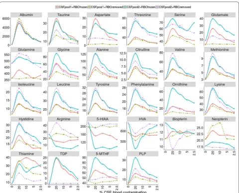
In Table 2 and Fig. 1 data from the different spiking conditions (represented as the percentage of variation and absolute values when compared with non-spiked CSF samples, respectively) are shown. The 2.5% spiking condition A (lysed RBC) still had percentages of varia-tion moderately higher than 10% for most metabolites. However, in the spiking condition B (RBC removed
from CSF), only some amino acids, thiamine and PLP had variations higher than 10% when compared with the non-spiked CSF sample values. As expected, albu-min was still highly elevated under all conditions. TDP, an intracellular form of thiamine, was only increased under the spiking condition A, but undetectable when RBC were removed from CSF by centrifugation. While most metabolites displayed changes in concentrations when RBCs were added to CSF, arginine, 5-HIAA and HVA had decreased values. Pterins and glutamine con-centrations did not vary under the different spiking conditions.
Results of the surrogate biomarkers of CSF blood con-tamination (albumin and haemoglobin) are stated in Additional file 1: Table S1 and Additional file 2: Figure S1.
Discussion
CSF metabolomic analysis is a good analytical tool for the study of the neurometabolic conditions stated here [14]. Furthermore, in such diseases, the quantifica-tion of these metabolites in blood/urine is not reliable, because they usually display normal or even paradoxi-cal results [25]. This is especially true when the meta-bolic pathways studied are highly active in the brain, or the genetic blood–brain barrier transport diseases related to vitamins and other metabolites.
CSF RBC contamination is frequent and has been recognized as a substantial confounding factor for proper interpretation of CSF analysis data describing concentrations of amino acids and other molecules [26,
27]. However, literature regarding blood contamination
effects on biogenic amines, pterins and vitamins is scarce [28]. The main causes of CSF blood contami-nation are traumatic lumbar punctures or spontane-ous intrathecal bleeding, which can occur in several situations, especially in newborns. Moreover, impaired blood–brain barrier permeability can occur under dif-ferent conditions, such as in asphyxia and epilepsy [19–21]. Thus, having an estimation of when a misin-terpretation of the metabolic profile can occur due to RBC/plasma contamination is important, considering that lumbar puncture is an invasive intervention, that it is difficult to perform, and that the final volume col-lected is sometimes low in paediatric patients.
Albumin, a protein synthesized in the liver, is a good surrogate biomarker for compromised permeability of
the blood–brain barrier and also for blood contami-nation. However, since its concentration in the blood largely exceeds that of the CSF (by approximately 100-fold), its concentration may remain elevated even in the case of low RBC/plasma contamination (in our hands around 1% of blood contamination; data not shown). Haemoglobin measures are an alternative surrogate marker when RBC lysis has occurred, and in our hands, values around 0.35 g/dL of haemoglobin may be a sig-nal for cautious interpretation of the data presented here (Table 2), since it corresponds to a CSF blood con-tamination approximately from 2.5%, which is the limit
where some metabolites may display artefactual results after haemolysis.
With regards to amino acids, several reports have indicated differences between blood and CSF compart-ments [8], but to our knowledge precise definitions of the limits under which RBC contamination can cause a misinterpretation of the metabolic profiles were not established. While lysed RBCs affected the concentra-tions of most amino acids at 2.5% of blood contamina-tion, centrifugation of the spiked CSF samples to remove RBC resolved the problem in most cases. In any case, the amino acids that had higher values were aspartate,
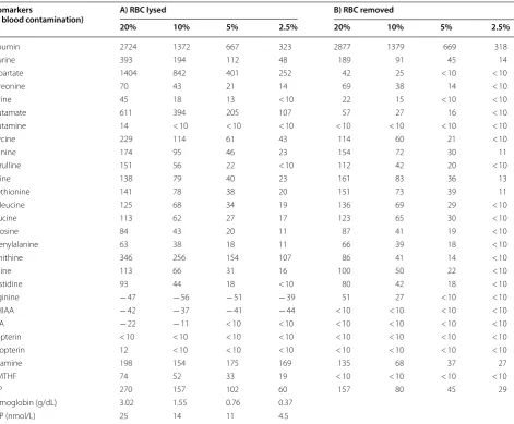
Table 2 Percentage of variation of the different biomarkers measured according to the different volumes of blood spiked into the CSF (expressed as percentage) when compared with the non-spiked CSF samples
Data are expressed as the mean value of the 2 CSF pools from the 2 conditions: A) spiked CSF frozen containing lysed-RBC cells (left columns), and B) spiked CSF with RBC removed by centrifugation prior freezing (right columns). A percentage of variation below 10% (comparing spiked CSF samples to the non-spiked CSF samples) was considered as the cut-off value for a proper interpretation of the results, considering the coefficient of variation for every biomarker measured when procedures were standardized. Haemoglobin and TDP values are expressed in g/dL and nmol/L, respectively since their values in condition B were not detectable. A total of 20 CSF aliquots coming from 50 CSF samples were analysed (Additional file 2: Figure S1)
Biomarkers
(% blood contamination) A) RBC lysed B) RBC removed
20% 10% 5% 2.5% 20% 10% 5% 2.5%
Albumin 2724 1372 667 323 2877 1379 669 318
Taurine 393 194 112 48 189 91 45 14
Aspartate 1404 842 401 252 42 25 < 10 < 10
Threonine 70 43 21 14 69 38 14 < 10
Serine 45 18 13 < 10 22 15 < 10 < 10
Glutamate 611 394 205 107 57 27 16 < 10
Glutamine 14 < 10 < 10 < 10 < 10 < 10 < 10 < 10
Glycine 229 114 61 43 114 60 21 < 10
Alanine 174 95 46 23 154 72 30 11
Citrulline 151 56 22 < 10 112 42 20 < 10
Valine 138 79 40 23 161 83 36 13
Methionine 141 78 38 20 151 73 39 11
Isoleucine 125 68 34 19 136 69 29 < 10
Leucine 113 62 27 17 123 65 30 < 10
Tyrosine 84 43 20 11 87 41 19 < 10
Phenylalanine 63 38 18 11 66 39 18 < 10
Ornithine 346 256 154 107 86 41 14 < 10
Lysine 113 66 31 16 100 50 22 < 10
Hystidine 93 44 18 < 10 80 42 18 < 10
Arginine − 47 − 56 − 51 − 39 51 27 < 10 < 10
5‑HIAA − 42 − 37 − 41 − 44 < 10 < 10 < 10 < 10
HVA − 22 − 11 < 10 < 10 < 10 < 10 < 10 < 10
Biopterin < 10 < 10 < 10 < 10 < 10 < 10 < 10 < 10
Neopterin 12 < 10 < 10 < 10 < 10 < 10 < 10 < 10
Thiamine 198 154 175 169 135 68 37 27
5‑MTHF 74 52 33 19 < 10 < 10 < 10 < 10
PLP 270 157 102 60 157 80 45 29
Hemoglobin (g/dL) 3.02 1.55 0.76 0.37
glutamate, threonine, ornithine, glycine and citrulline. The explanation for this is that some of these amino acids display higher concentration in RBC when compared with plasma (aspartate, glutamate and threonine) [8]. Amongst the other amino acids, glycine has the high-est plasma/CSF ratio [29]. Regarding ornithine, arginase activity is high in RBC, and this would explain our obser-vation that arginine values were lower when RBC lysis occurred while ornithine values increased, as it is the product of the reaction catalysed by arginase [8]. Once again, centrifugation of the samples prevented all of these contamination biases. Some of the results in Table 2
were striking, however. No precise correlations with the expected values were observed for some amino acids considering the different percentages of blood contami-nation. A possible explanation is that some amino acids are present in CSF at very low concentrations physi-ologically (close to the quantification limits) and for other amino acids, the matrix effect, common in UPLC-MS/ MS technology, could contribute to such differences [22]. Biogenic amines and pterins are synthesized peripher-ally in some tissues but also in the brain, and no substan-tial transport has been documented between blood and brain (only OAT3 transporters efflux both 5-HIAA and HVA from CSF to blood [2]). Since their concentrations in blood are similar to those of CSF [9, 10], no significant changes were observed after CSF blood contamination. Interestingly, both 5-HIAA and HVA had lower values when RBC lysis was caused. Autoxidation of these mol-ecules by haemoglobin/free radicals is a potential mech-anism explaining this observation [30], and thus, one should be cautious when interpreting data when CSF has not been centrifuged prior freezing, since low 5-HIAA and HVA values are surrogate biomarkers of serotonin and dopamine deficiencies and may be an indication of therapeutic intervention [31]. In any case, centrifugation and RBC removal prior to freezing corrected the results when compared with non-spiked CSF.
Vitamins displayed unpredictable results, except for folate. Folate forms (especially 5-MTHF) are highly con-centrated in RBC when compared with plasma and this would explain the positive interference observed when RBC lysis occurred, but not after RBC removal. Only thiamine and PLP had increased values when compar-ing spiked CSF samples at 2.5% with non-spiked samples under both experimental conditions, although it was less remarkable when RBC removal was performed. These effects were minimized when the CSF blood contami-nation was 1% (data not shown). Regarding thiamine, active conversion of free thiamine, TMP and TDP occur inside cells (RBCs have a high activity of either thiamine phosphokinase, which phosphorylates thiamine to form TDP, or thiamine phosphatases, which convert TDP to
TMP and thiamine) [32]. This would explain the plateau results observed when RBC lysis occurred, results that were minimized when RBCs were removed. In any case, thiamine values are higher in blood than in CSF, and thus, the results should be cautiously interpreted when RBC contamination occurs [12]. TDP, a strictly intracel-lular thiamine vitamer [32], would be a good surrogate biomarker of RBC lysis in CSF samples since undetect-able amounts of TDP were observed when RBCs were removed from CSF. With PLP, the observations were similar, and although less significant, even when RBCs were removed from CSF, PLP displayed higher concen-trations in the spiked CSF samples. As with thiamine, a complex intracellular metabolic pathway accounts for the synthesis of the different pyridoxine-related vitamers [13]. Moreover, some of these vitamins can be degraded by nucleophiles and oxygen-derived free-radicals, as CSF has low concentrations of other molecules that can react with these compounds [13]. Thus, results should be ana-lysed cautiously concerning to these two vitamins, since either in thiamine or pyridoxine related disorders, which cause severe neurological phenotypes, diagnostic hall-marks are low CSF thiamine and PLP values [12, 13, 33].
Conclusions
CSF-targeted metabolomic analysis is feasible even when remarkable RBC CSF contamination occurs since CSF centrifugation to remove RBC prior to freezing prevents most of the biases observed. However, data should be cautiously interpreted, especially for some metabolites. CSF albumin, haemoglobin, and TDP can be used as sur-rogate biomarkers of the potential confounding effect of CSF plasma/RBC contamination.
Supplementary information
Supplementary information accompanies this paper at https ://doi. org/10.1186/s1298 7‑019‑0154‑5.
Additional file 1: Table S1. Percentage of blood contamination, albumin and haemoglobin levels from CSF spiked with increasing amounts of blood.
Additional file 2: Figure S1. CSF blood spiking protocol. A total of 20 CSF aliquots were analysed. In the picture, the colours of the 5 CSF sample are presented. Even in the 2.5% spiking condition, the red colour was intense. The median CSF blood contamination observed in our laboratory typically ranged from 0.01 to 0.035 g/dL of a haemoglobine, which is lower than the 0.35 g/dL observed in the 2.5% blood contamination condition. Additional file 3: Figure S2. Typical chromatograms of the different metabolites analysed in non‑spiked CSF samples: (1) Amino acids. (2) Biogenic amines. (3) Pterins. (4) 5‑methyltetrahydrofolate. (5) Pyridoxal 5´‑phosphate. (6) Thiamine.
Abbreviations
thiamine‑diphosphate; 5‑MTHF: 5‑methyltetrahydrofolate; PLP: pyridoxal 5´‑phosphate; BCAA : branched‑chain amino acids.
Acknowledgements
The Department of Clinical Biochemistry is part of the CIBERER‑ISCIII and ‘Centre Daniel Bravo de Diagnòstic I Recerca en Malalties Minoritàries’. “We are indebted to the “Biobanc de l’Hospital Infantil Sant Joan de Déu per a la Inves‑ tigació” integrated into the Spanish Biobank Network of ISCIII for the sample and data procurement.”
Authors’ contributions
MB contributed to conception and design, acquisition, analysis and interpreta‑ tion of data, drafted the initial manuscript and approved the final manuscript as submitted. MC, CS, MCS, LMS, JM, GF, AGC and AO contributed to analysis and acquisition of data, reviewed and revised the manuscript, and approved the final manuscript as submitted. MML and RA contributed to conception and design, analysis and interpretation of data, reviewed and supervised the manuscript, and approved the final manuscript as submitted. Every one of the authors has participated sufficiently in the study, meeting the appropriate authorship criteria, and each has seen, reviewed and approved this version of the manuscript and takes full responsibility for it. We all agree to its submission for publication. Nobody who qualifies for authorship has been omitted from the list of authors. All the authors have complete access to the study data. All authors read and approved the final manuscript.
Funding
This work was supported by Grants from the Instituto de Salud Carlos III (ISCIII‑FIS PI15/01082 and PI18/00111), the FEDER Funding Program from the European Union and CIBERER‑ISCIII.
Availability of data and materials
The datasets used and/or analysed during the current study are available from the corresponding author on reasonable request.
Ethics approval and consent to participate
Informed consent was obtained from each patient before performing the lumbar puncture and CSF collection or genetic study. The Ethical committee of Sant Joan de Déu Hospital approved the study.
Consent for publication Not applicable.
Competing interests
The authors declare that they have no competing interests.
Author details
1 Clinical Biochemistry Department, Hospital Sant Joan de Déu, Barcelona,
Spain. 2 Molecular Genetics, Hospital Sant Joan de Déu, Barcelona, Spain. 3 Pediatric Neurology Department, Institut de Recerca Sant Joan de Déu,
Barcelona, Spain. 4 CIBERER‑Instituto de Salud Carlos III, Barcelona, Spain.
Received: 9 September 2019 Accepted: 2 November 2019
References
1. Hladky SB, Barrand MA. Fluid and ion transfer across the blood‑brain and blood‑cerebrospinal fluid barriers; a comparative account of mechanisms and roles. Fluids Barriers CNS. 2016;13:19 (Review).
2. Spector R. Nature and consequences of mammalian brain and CSF efflux transporters: four decades of progress. J Neurochem. 2010;112:13–23. 3. Redzic Z. Molecular biology of the blood‑brain and the blood‑cerebrospi‑
nal fluid barriers: similarities and differences. Fluids Barriers CNS. 2011;8:3. 4. Nedergaard M. Garbage truck of the brain. Science. 2013;340:1529–30. 5. Akaishi T, Onishi E, Abe M, Toyama H, Ishizawa K, Kumagai M, et al. The
human central nervous system discharges carbon dioxide and lactic acid into the cerebrospinal fluid. Fluids Barriers CNS. 2019;16:8.
6. Mann GE, Yudilevich DL, Sobrevia L. Regulation of amino acid and glucose transporters in endothelial and smooth muscle cells. Physiol Rev. 2003;83:183–252.
7. Kornhuber ME, Kornhuber J, Kornhuber AW, Hartmann GM. Positive correlation between contamination by blood and amino acid levels in cerebrospinal fluid of the rat. Neurosci Lett. 1986;69:212–5.
8. Duran M. Amino acids. In: Blau N, Duran M, Gibson KM, editors. Labora‑ tory guide to the methods in biochemical genetics. Berlin: Springer; 2008. p. 53–90.
9. Coppus AW, Fekkes D, Verhoeven WM, Tuinier S, Egger JI, van Duijn CM. Plasma amino acids and neopterin in healthy persons with Down’s syndrome. J Neural Transm (Vienna). 2007;114:1041–5.
10. Mori S, Takanaga H, Ohtsuki S, Deguchi T, Kang YS, Hosoya K, et al. Rat organic anion transporter 3 (rOAT3) is responsible for brain‑to‑blood efflux of homovanillic acid at the abluminal membrane of brain capillary endothelial cells. J Cereb Blood Flow Metab. 2003;23:432–40.
11. Steinfeld R, Grapp M, Kraetzner R, Dreha‑Kulaczewski S, Helms G, Dechent P, et al. Folate receptor alpha defect causes cerebral folate transport defi‑ ciency: a treatable neurodegenerative disorder associated with disturbed myelin metabolism. Am J Hum Genet. 2009;85:354–63.
12. Ortigoza‑Escobar JD, Molero‑Luis M, Arias A, Oyarzabal A, Darín N, Serrano M, et al. Free‑thiamine is a potential biomarker of thiamine transporter‑2 deficiency: a treatable cause of Leigh syndrome. Brain. 2016;139:31–8.
13. Footitt EJ, Heales SJ, Mills PB, Allen GF, Oppenheim M, Clayton PT. Pyri‑ doxal 5′‑phosphate in cerebrospinal fluid; factors affecting concentration. J Inherit Metab Dis. 2011;34:529–38.
14. Ormazabal A, Molero‑Luis M, Garcia‑Cazorla A, Artuch R. Biomarkers for the study of catecholamine and serotonin genetic diseases. In: Garg U, Smith LD, editors. Biomarkers in inborn errors of metabolism. Cambridge: Clinical aspects and laboratory determination; 2017. p. 301–29. 15. Akiyama T, Kobayashi K, Higashikage A, Sato J, Yoshinaga H. CSF/plasma
ratios of amino acids: reference data and transports in children. Brain Dev. 2014;36:3–9.
16. MacNeill AL, Andre BG, Zingale Y, Packer RA, McGrath S. The effects of iatrogenic blood contamination on total nucleated cell counts and protein concentrations in canine cerebrospinal fluid. Vet Clin Pathol. 2018;47:464–70.
17. Srinivasan L, Shah SS, Abbasi S, Padula MA, Harris MC. Traumatic lumbar punctures in infants hospitalized in the neonatal intensive care unit. Pediatr Infect Dis J. 2013;32:1150–2.
18. McFarlin KE, Kruesi MJ, Nadi NS. RBC contamination and amino acid concentration in the CSF of children. Psychiatry Res. 1990;32:99–101. 19. Tan R, Traylor M, Rutten‑Jacobs L, Markus H. New insights into mecha‑
nisms of small vessel disease stroke from genetics. Clin Sci (Lond). 2017;131:515–31.
20. Krueger M, Mages B, Hobusch C, Michalski D. Endothelial edema precedes blood‑brain barrier breakdown in early time points after experi‑ mental focal cerebral ischemia. Acta Neuropathol Commun. 2019;7:17. 21. Klebe D, McBride D, Krafft PR, Flores JJ, Tang J, Zhang JH. Posthemor‑
rhagic hydrocephalus development after germinal matrix hemorrhage: established mechanisms and proposed pathways. J Neurosci Res. 2019. https ://doi.org/10.1002/jnr.24394 ([Epub ahead of print] Review). 22. Batllori M, Molero‑Luis M, Ormazabal A, Casado M, Sierra C, García‑Cazorla
A, et al. Analysis of human cerebrospinal fluid monoamines and their cofactors by HPLC. Nat Protoc. 2017;12:2359–75.
23. Casado M, Sierra C, Batllori M, Artuch R, Ormazabal A. A targeted metabo‑ lomic procedure for amino acid analysis in different biological specimens by ultra‑high‑performance liquid chromatography‑tandem mass spec‑ trometry. Metabolomics. 2018;14:76.
24. Ormazabal A, García‑Cazorla A, Pérez‑Dueñas B, Gonzalez V, Fernández‑ Alvarez E, Pineda M, et al. Determination of 5‑methyltetrahydrofolate in cerebrospinal fluid of paediatric patients: reference values for a paediatric population. Clin Chim Acta. 2006;371:159–62.
25. Wassenberg T, Willemsen MA, Geurtz PB, Lammens M, Verrijp K, Wilmer M, et al. Urinary dopamine in aromatic l‑amino acid decarboxylase defi‑
ciency: the unsolved paradox. Mol Genet Metab. 2010;101:349–56. 26. Aasebø E, Opsahl JA, Bjørlykke Y, Myhr KM, Kroksveen AC, Berven FS.
Effects of blood contamination and the rostro‑caudal gradient on the human cerebrospinal fluid proteome. PLoS ONE. 2014;9:e90429. 27. Krishnamurthy V, Nabil N, Reddy SM, Doreswamy SM. Dilemma in diag‑
•fast, convenient online submission •
thorough peer review by experienced researchers in your field • rapid publication on acceptance
• support for research data, including large and complex data types •
gold Open Access which fosters wider collaboration and increased citations maximum visibility for your research: over 100M website views per year •
At BMC, research is always in progress.
Learn more biomedcentral.com/submissions
Ready to submit your research? Choose BMC and benefit from: 28. Verbeek MM, Blom AM, Wevers RA, Lagerwerf AJ, van de Geer J, Wil‑
lemsen MA. Technical and biochemical factors affecting cerebrospinal fluid 5‑MTHF, biopterin and neopterin concentrations. Mol Genet Metab. 2008;95:127–32.
29. Swanson MA, Coughlin CR Jr, Van Hove JL. Corrigendum: Swanson MA, Coughlin CR Jr, Scharer GH, et al: Biochemical and molecular predictors for prognosis in nonketotic hyperglycinemia. Ann Neurol 2015;78:606– 618. Ann Neurol. 2016;79:505.
30. Kato Y, Oki K, Suga N, Ono S, Ishisaka A, Miura Y, et al. A novel quinone derived from 5‑hydroxyindoleacetic acid reacts with protein: possible participation of oxidation of serotonin and its metabolite in the develop‑ ment of atherosclerosis. Free Radic Biol Med. 2016;101:500–10. 31. Ng J, Papandreou A, Heales SJ, Kurian MA. Monoamine neurotransmit‑
ter disorders–clinical advances and future perspectives. Nat Rev Neurol. 2015;11:567–84.
32. Collie JTB, Greaves RF, Jones OAH, Lam Q, Eastwood GM, Bellomo R. Vitamin B1 in critically ill patients: needs and challenges. Clin Chem Lab Med. 2017;55:1652–68.
33. Zeng WQ, Al‑Yamani E, Acierno JS Jr, Slaugenhaupt S, Gillis T, MacDon‑ ald ME, Ozand PT, Gusella JF. Biotin‑responsive basal ganglia disease maps to 2q36.3 and is due to mutations in SLC19A3. Am J Hum Genet. 2005;77:16–26.
Publisher’s Note