Original Research Article.
Role of Slit Skin Smear Examination and rK39 for Diagnosis of
Post Kala-Azar Dermal Leishmaniasis
Pratulya Nandan
1*, Spriha Smriti
2, Babita Kumari
2, Anita Sundi
3, Swapnil Keshav
31*Assistant Professor, 2Tutor, 3Post Graduate Student,
Department of Microbiology, Patna Medical College, Patna, Bihar, India.
ABSTRACT
Introduction: Post Kala-azar dermal leishmaniasis is a complication of visceral leishmaniasis characterised by multiple hypopigmented, macular, maculopapular or nodular rash on the face, trunk or other part of the body. It occurs mainly in the East Asia and the Indian Subcontinent behaving as major reservoir of parasite in areas of anthroponotic transmission.
Materials and Methods: The study was aimed to analyse the
role of Slit skin smear (SSS) examination and rK39 in the diagnosis of PKDL, in the Department of Microbiology, Patna Medical College, Patna. The study was conducted on 60 patients for the period of 1 year. Specimen was obtained from the site of lesion and material obtained was smeared uniformly on the microscopic slide. Fresh peripheral whole blood was drawn and serum was used for the rK39 test. Rapid test kit (InBios International Kala-azar detection kit) was used. Results: Out of 60 suspected cases of PKDL, on SSS examination 16 cases (26.67%) were positive for LD bodies while, on performing rK39 rapid kit test, 25 cases (41.67%) were found positive.
Conclusion: Initial screening by serology (rK39 kit test)
followed by confirmatory diagnosis of PKDL with SSS examination is simple and minimally invasive procedure for epidemiological studies and assessment of cure in PKDL.
Key Words:PKDL (Post Kalazar Dermal Leishmaniasis), SSS
(Slit Skin Smear), VL (Visceral leishmaniasis), LD Bodies, rK39.
*Correspondence to:
Dr. Pratulya Nandan, Flat no. M-6/46, Block No. 8, Patliputra Path,
Rajendra Nagar, Patna, Bihar. Article History:
Received: 22-10-2017, Revised: 16-11-2017, Accepted: 22-12-2017
Access this article online
Website: www.ijmrp.com
Quick Response code
DOI:
10.21276/ijmrp.2018.4.1.022
INTRODUCTION
PKDL or Dermal leishmanoid is non-ulcerative cutaneous complication of visceral leishmaniasis, characterized by a macular, maculopapular or nodular rash on the face, trunk or other parts of the body.1
PKDL was first described by Brahmchari in 1922 in cured VL patients with eruptions and plaque in the skin, confirmed by demonstration of LD bodies (LDB) in slit skin smear and termed as Dermal leishmanoid.2 It develops in about 10% (5 – 15 %) of
kala-azar patients often one or two years after treatment for visceral leishmaniasis (VL).3,4 Recently, it has also been reported
in individuals without prior history of VL as well as those undergoing treatment for VL and in cases of spontaneously cured kala-azar.5
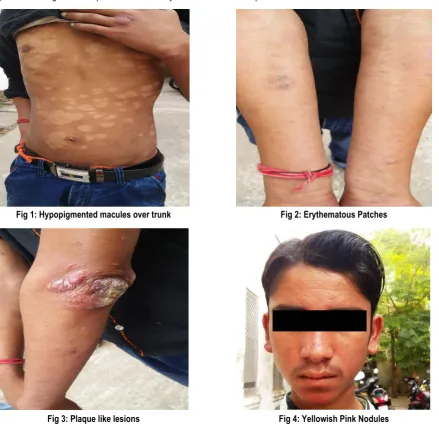
PKDL may be of three types depigmented macules (Fig 1), erythematous patches (Fig 2) and yellowish pink nodules (Fig 3,4). Other unusual variants include the annular, warty, papillomatous growth and presence of lesions in uncommon site such as eyelids, palms and perionychium. Complications can result when mucous membranes are affected, the most serious being blindness due to corneal involvement.3
It occurs mainly in East Africa and Indian subcontinent. In India, Kala Azar (KA) is endemic in the North- eastern states like Bihar, Jharkhand, West Bengal, Assam and Plains of eastern Uttar Pradesh. In Bihar, Vaishali, Saran, Patna and Muzaffarpur districts are most affected. In Jharkhand, Sahebganj is the worst affected district.
Pathogenesis of PKDL is still not clearly understood. There is increasing evidence that the pathogenesis is largely immunologically mediated; high concentrations of interleukin 10 in the peripheral blood of VL patients predict the development of PKDL. During VL, interferon gamma is not produced by peripheral blood mononuclear cells (PBMC). After treatment of VL, PBMC start producing interferon gamma, which coincides with the appearance of PKDL lesions due to interferon-gamma-producing cells causing skin inflammation as a reaction to persisting parasites in the skin.
immune reconstitution, while at the same time there is dissociation of the immune response between the skin and the viscera. This causes the typical skin rash in the absence of systemic illness. In PKDL the Th2 response shows the presence and persistence of IL-10 in the skin that was already present during VL, while systemically the Th1 response that was induced after VL therapy persists with IFN-γ production.
In the skin, as typically the PKDL lesions occur in sun exposed areas of the skin (face, neck), the immune response is thought to be related to the influence of UV light; this causes damage to dendritic cells resulting in a Th2 type of response by inhibition of regulatory T cells.
Post kalaazar dermal leishmaniasis is considered to be an important reservoir for VL.4,6,7 In Indian Subcontinent, untreated
cases of VL and PKDL are considered to be the sole reservoir of the causative parasite (Leishmania donovani) in the absence of zoonotic transmission.1,5,8,9 Phlebotomous argentipes, the vector
transmitting VL in India, when allowed to feed on PKDL patients, became infected and developed promastigote in the midgut, seeming capable of transmitting the disease.7,10
PKDL is a challenge for clinicians and researchers, because its burden is poorly investigated and pathogenesis is disputable. Misdiagnosis can occur with patients showing less specific clinical manifestations. The hypopigmented form of PKDL has been often misdiagnosed as vitiligo since the parasite load is scanty and not
always proportional to the extent of dermal lesions while its nodular form may be easily confused with a number of dermatological conditions among which leprosy is the most important.3,11 Recently, nerve involvement in Indian PKDL has
been documented which is common in Sudan PKDL, showing that PKDL may simulate leprosy both clinically and pathologically.11,12
Asymptomatic cases in the community may remain undetected and may act as potential reservoirs in VL transmission areas, so, diagnostics for PKDL should have a sufficiently high sensitivity. Accurate diagnosis of PKDL is also important due to the long (upto 4 to 6months) and toxic treatment with antileishmanial drugs, which exposes the patient to toxic drugs and also leads to a waste of medical and economic resources, so, a testing regime with a high specificity is essential to avoid false positive results. Demonstration of parasite in the slit skin smear ((SSS) examination or by culture of the dermal tissueis considered the gold standard.
rK39 dipstick test is a non-invasive, nitrocellulose dipstick immunoassay that uses a recombinant leishmania antigen, K39 (K39 is an antigen coded by a kinesin related gene and contains a repeatitive epitope of 39 amino acid residue conserved on amastigotes of Leishmania species), for the detection of IgG antibodies. The study was aimed to analyse the role of Slit skin smear (SSS) examination and rK39 in the diagnosis of PKDL in our set up.
Fig 1: Hypopigmented macules over trunk Fig 2: Erythematous Patches
MATERIAL AND METHODS
The study was conducted in the Department of Microbiology, PMCH, Patna on 60 patients for a period of 1 year. These patients
were clinically suspected as PKDL?/ Hansen’s disease?
A short history of patients were taken, they were enquired about past history of dermatological diseases, history of kala-azar or family history of kala-azar. HIV test was done for all patients and those testing positive were excluded from the study, in view of the fact that immuno-deficient state may lead to a false negative result in rK39 test
Slit Skin Smear Examination
Specimen was taken from the site of lesion taking all aseptic precautions. After cleaning the skin with spirit, skin was pinched to minimise bleeding, a cut about 5mm long and 2 – 3 mm deep was made, the blade was then turned at right angle to the cut and tissue fluid and cells obtained which were smeared uniformly on
two sets of slides. One set of slides was used for Leishman’s
staining and the other was stained with ZN stain to demonstrate
LD bodiesand leprae bacilli respectively. rK39 Dipstick Test
Fresh peripheral whole blood was drawn from patients and serum was added on the nitrocellulose strip of the dipstick and inserted in the 3ml test tube, held vertically, with two drops of chase buffer solution (supplied with the dipstick kit).
The test kit used was Rapid test kit (InBios International Kala-azar detection kit). The appearance of two visible red bands means one control band (the test was valid) and one test band (rK39 antibody was present) – indicated that the test was positive. The test was negative if only the control band appeared.
The results were read after five minutes and if still negative, after 10 minutes. The test was repeated if the control line remained negative after 10 minutes. Even a weak band in the test region was considered as a positive result. The red color in the test region will vary depending on the concentration of anti-Leishmanial antibodies present. Weakly positive samples are those with low affinity or low titre antibodies against the recombinant test antigen.
RESULTS
A total of 60 patients were analysed in our study. The mean age of the patients was 26.5 years (Range- 12- 41 years). 65% patients (39 out of 60) were male (Fig 5). 51 patients out of 60 ie 85% gave a history of kala-azar (Fig 6). Out of 60 suspected cases 16 cases (26.67%) were positive for LD bodies on SSS examination. On performing rK39 rapid kit test, 25 cases (41.67%) were found positive (Fig 7). Two patients were positive for lepra bacilli on ZN staining.
Fig 5: Sex distribution of PKDL
Fig 6: PKDL patients with and without history of Kala azar
Male Female
0 10 20 30 40 50 60
Fig 7: PKDL patients showing positive result in sss examination and rK39 test
DISCUSSION
In our study, 39 out of 60 patients (65%) were male, which is in accordance with the report of a meeting of the WHO expert committee on the control of leishmaniasis, Geneva 2010 which shows, leishmaniasis is reported more in males.13 The age group
affected in our study was the most productive group ie 12- 41 years (mean 26.5 years). The age group affected and the male preponderance may be due to the fact that they are involved in outdoor activities and chances of sandfly bite are more.
Slit skin smear were positive for 26.67% cases (16 out of 60 patients analysed). Work done by Das ML et al in 2007 showed 40% positive cases in SSS.14 They also showed that positivity
varied in different clinical forms: nodular (70%), maculopapular (20%), macular (20%),14 which is similar to our study on macular
lesions. Ramesh V et al showed that 32 – 36% cases were positive by slit skin microscopy.3 In 2004, Mathur P et al concluded
that 100% cases were positive by SSS microscopy.15
On performing rK39 rapid kit test, 25 cases (41.67%) were found positive. According to Das ML et al it is 96% sensitive and 100% specific.14 Study done on VL by Kiros YK et al in 2015 showed
80.6% positive cases in rK39- ICT test.16 Chappuis et al (2003)
found it to be 97% sensitive and 71% specific for VL. The study by Chappuis et al (2003) was hospital based, and the specificity decreased, as the dipstick was also found to react positive to malaria, enteric fever, and disseminated TB.17 Similar reports are
available from Southern Europe and Brazil.18,19
In India, rK39 has been widely used for the diagnosis of VL as well as PKDL, with sensitivity of the test ranging from 95 -100 %.20,21 In
a comprehensive study with 88 Indian patients of PKDL, it was seen that specificity of rK39 was 94% and sensitivity 94.5%.20
Various studies have shown that specificity of rK39 in general is quite variable in different set upseg- in one meta-analysis study specificity ranged from 66.8 to 97.9%, which is quite wide.22
CONCLUSION
PKDL is considered a public health problem in VL endemic areas because of its possible role in transmission during interepidemic period. PKDL is not a static or uniform disease and each patient may be different in terms of clinical presentation. There is no standard guideline for diagnosis and treatment of PKDL.
Slit skin smear is invasive, less sensitive (58%) and difficult to perform in field conditions. As the number of parasites in skin smear is often low, it requires prolonged searches by an expert on routine microscopy but the advantage of microscopy is its acknowledged high specificity, which leads to low numbers of patients unnecessarily treated with antileishmanial drugs. The recombinant K39 immunochromatographic test (rK39 ICT) offers a simple, non-invasive and rapid test with increased patient compliance, but positive serological tests may be due to persisting antibody from past infection. False positive rK39 is seen in patients with rheumatoid factor +ve and patients suffering from malaria. False negative test is seen in HIV positive patients and other immunodeficient states and the crux is, relapse of VL has been reported in immunodeficient PKDL patients.
In India, Leishmaniasis and HIV co-infection has been reported from Bihar, Sub- Himalayan region and other North Indian states. Various studies reported the prevalence of co-infection to be around 2 – 6%.
Thus, we conclude that for epidemiological and clinical assessment of patients both slit skin smear examination and rK39 dipstick test should be done simultaneously. This will help clinicians to make an accurate diagnosis and save patients from unnecessary exposure to toxic drugs and community as a whole from this silent reservoir of kala-azar. It is our responsibility to help clinicians to reach an accurate diagnosis leading to decrease in the misuse of resources, containment of the disease and betterment of the patients and as far as diagnosis of PKDL in our set up is concerned simultaneous use of slit skin smear examination and rK39 dipstick test serves the purpose.
ACKNOWLEDGEMENTS
We would like to thank Dr S N Singh, Prof and Head, Department of Microbiology, PMC Patna and our lab technician for extending their support in conduct of this study.
REFERENCES
1. Thakur CP, Kumar K. Post kala-azar dermal leishmaniasis: a neglected aspect of kala-azar control programmes. Ann Trop Med Parasitol 1992; 86: 355-9.
0 5 10 15 20 25
SSS Positive
2. Brahmachari UN. A new form of cutaneous leishmaniasis-dermal leishmanoid. Indian Med Gaz 1922; 57: 125.
3. Ramesh V, Mukherjee A. Post-kala-azar dermal leishmaniasis. Int J Dermatol 1995; 34: 85-91.
4. Rahman KM, Islam S, Rahman MW, Kenah E, Galive CM, Zahid MM, et al. Increasing incidence of post kala-azar dermal leishmaniasis in a populationbased study in Bangladesh. Clin Infect Dis. 2010; 50: 73–76. pmid:19951168
5. Desjeux P. The increase in risk factors for leishmaniasis worldwide. Trans R Soc Trop Med Hyg 2001; 95: 239-43. 6. Zijlstra EE, Musa AM, Khalil EA, el-Hassan IM, el-Hassan AM. Post kala- azar dermal leishmaniasis. Lancet Infect Dis. 2003; 3: 87–98. pmid:12560194
7. Addy M, Nandy A. Ten years of kala-zar in West Bengal, Part I. Did post kala-azar dermal leishmaniasis initiate the outbreak in 24-Parganas? Bull World Health Organ. 1992; 70: 341–346. pmid:1638662
8. Dhiman RC, Prasad LSN. Studies on the possible role of zoonosis in the transmission of kalaazar in Bihar. Indian J Parasitol 1986; 10:171-2
9. Birley MH. An historical review of malaria, kala-azar and filariasis in Bangladesh in relation to the flood action plan. Ann Trop Med Parasitol 1993; 87: 319-34.
10. Dinesh DS, Kar SK, Kishore K, Palit A, Verma N, Gupta AK, et al. Screening sandflies for natural infection with Leishmania donovani using a non-radioactive probe based on the total DNA of the parasite. Ann Trop Med Parasitol 2000; 94: 447-51.
11. El Hassan AM, Ghalib HW, Zijlstra EE, Eltoum IA, Satti M, Ali MS, et al. Post kala-azar dermal leishmaniasis in the Sudan: clinical features, pathology and treatment. Trans R Soc Trop Med Hyg 1992; 86: 245-8.
12. Khandpur S, Ramam M, Sharma VK, Salotra P, Singh MK, Malhotra A. Nerve involvement in Indian post kala-azar dermal leishmaniasis. Acta Derm Venereol 2004; 84: 245-6.
13. Control of the leishmaniases. Report of a meeting of the WHO expert committee on the control of leishmaniasis, Geneva, 22–26 March 2010.
14. ML Das, M Deb, BMS Karki, M Sarif, B Khanal, SK Bhattacharya, S Agrawal and S Koirala. Use of rk39 for diagnosis of post kala-azar dermal leishmaniasis in nepalsoutheast asian j trop med public health. 38 no. 4 july 2007 619-625.
15. Mathur P, Samantaray J & Chauhan N K. Evaluation of a rapid immunochromatographic test for diagnosis of kala-azar & post kala-azar dermal leishmaniasis at a tertiary care centre of north India. Indian J Med Res 122, December 2005, pp 485-490.
16. Kiros YK, Regassa BF. The role of rk39 serologic test in the diagnosis of visceral leishmaniasis in a Tertiary Hospital, Northern Ethiopia BMC Res Notes. 2017 Apr 26;10(1):169. doi: 10.1186/s13104-017- 2490-3.
17. Chappuis F, Rijal S, Singh R, Acharya P, Karki BMS, Das ML. Prospective evaluation and comparison of the direct agglutination test and rK39 antigen based dipstick test for the diagnosis of suspected kala- azar in Nepal. Trop Med Int Health 2003; 8: 277-85.
18. Jelinek T, Eichenlaub S, Loscher T. Sensitivity and specificity of a rapid immunochromatographic test for diagnosis of visceral leishmaniasis. Eur J Clin Microbiol Infect Dis 1999; 18: 669-70. 19. Carvalho SF, Lemos EM, Corey R, Dietze R.Performance of recombinant K39 antigen in the diagnosis of Brazilian visceral leishmaniasis. Am J Trop Med Hyg 2003; 68: 321-4.
20. Salotra P, Sreenivas G, Nasim AA, Subba Raju BV, Ramesh V. Evaluation of enzyme-linked immunosorbent assay for diagnosis of post- kala-azar dermal leishmaniasis with crude or recombinant K39 antigen. Clin Diagn Lab Immunol 2002;9:370-3. 21. Singh S, Gilman-Sachs A, Chang KP, Reed SG. Diagnostic and prognostic value of K39 recombinant antigen in Indian leishmaniasis. J Parasitol 1995;81:1000-3 .
22. Chappuis F, Rijal S, Soto A, Menten J, Boelaert M. A meta-analysis of the diagnostic performance of the direct agglutination test and rK39 dipstick for visceral leishmaniasis. BMJ. 2006; 333(7571):723. doi:10.1136/bmj. 38917.503056.7C.
[
Source of Support: Nil.
Conflict of Interest: None Declared.
Copyright: © the author(s) and publisher. IJMRP is an official publication of Ibn Sina Academy of Medieval Medicine & Sciences, registered in 2001 under Indian Trusts Act, 1882. This is an open access article distributed under the terms of the Creative Commons Attribution Non-commercial License, which permits unrestricted non-commercial use, distribution, and reproduction in any medium, provided the original work is properly cited.