International Journal of Medical Science and Current Research (IJMSCR)
Available online at: www.ijmscr.com
Volume2, Issue 6,Page No: 249-253
November-December 2019
249
Medicine ID-101739732
Nasal Foreign Bodies- Our Experience
Anchal Gupta1*, Amit Mahajan2, Padam Singh Jamwal3 1
Senior Resident, 2 Lecturer, 3 Professor
1,3
Department of ENT, Head and Neck Surgery, SMGS Hospital, Government Medical College
2
Department of Shalakya, Government Ayurvedic Medical College Jammu, Jammu and Kashmir, India
*Corresponding Author:
Anchal Gupta
Department of ENT, Head and Neck Surgery, SMGS Hospital, Government Medical College
Type of Publication: Original Research Paper
Conflicts of Interest: Nil
ABSTRACT
Background: Early diagnosis and intervention in nasal foreign body is required to prevent complications.
Materials and methods: This study was conducted at Department of ENT, SMGS Hospital, Government Medical College, Jammu, for a period of two years. The study population included the patients with nasal foreign body who presented in the Outpatient Department (OPD) and emergency of the ENT department of the hospital. The data were obtained from the hospital record books. Anterior rhinoscopy was done to diagnose nasal foreign body. Xray nose was done in cases where there was suspicion of metallic foreign body as told by attendants of the patients. Most of the nasal foreign bodies were removed in Outpatient Department; some were admitted for removal under general anaesthesia.
Results: Out of 120 patients, there were 68(56.67%) males and 52(43.33%) female patients with M:F ratio of 1.3:1. 94(78.3% ) patients were in the age group of 2-6 years. Most common symptom was unilateral nasal discharge(45%) followed by asymptomatic(35.8%),foul odor in nose(8.3%).The most common foreign bodies seen in our study were eraser(27.5%) and seeds(24.16%). The most common site was right nasal cavity followed by left nasal cavity. 5(4.16%) patients presented with foreign bodies in bilateral nasal cavities.18 (15%) patients were admitted and removal of foreign body was required general anaesthesia. In all cases, removal was done with direct instrumentation either by extraction or by suction. Conclusion: Nasal foreign bodies are a frequent accident in medical practice, especially in young children.They are generally harmless, but may incur complications if overlooked. The best treatment, however, remains prevention.
Keywords: foreign body, nose, extraction.
INTRODUCTION
Nasal foreign bodies are frequently encountered, especially in children. When children start moving by themselves, they have access to many objects that have to be explored. This process can cause the placement of objects in orifices [1].
The circumstances are usually accidental, with a foreign body trapped or incarcerated in one or both nasal cavities by the anterior (vestibular) or more rarely posterior (choanal) route [2]
Nasal cavity foreign bodies are among those with the richest symptoms. After a few days in the nasal
foul odor [3]. It is a classic axiom that these two symptoms, specially if unilateral in children, until proven contrary, are highly suggestive of foreign bodies. Nasal obstruction and epistaxis may also occur. Diagnosis is carried out through anterior rhinoscopy, which shows most foreign bodies [4].
e
250
e
250
e250
e
250
e250
e
250
e250
e
250
e250
e
250
e
250
e
250
e
250
e250
e
250
e250
e
250
e250
e
250
e250
e
250
chronicization of the resultant irritation, with a real risk of superinfection. The present study reports epidemiological, clinical and therapeutic aspects of nasal foreign bodies encountered in our set up.
MATERIALS AND METHODS
This retrospective observational study was conducted at Department of ENT, SMGS Hospital, Government Medical College, Jammu, for a period of two years from October 2017 to September 2019. The study population included the patients with nasal foreign body who presented in the Outpatient Department (OPD) and emergency of the ENT department of the hospital. The data were obtained from the hospital record books. Anterior rhinoscopy was done to diagnose nasal foreign body. Xray nose was done in cases where there was suspicion of metallic foreign body as told by attendants of the patients. Most of the
nasal foreign bodies were removed in Outpatient Department; some were admitted for removal under general anaesthesia. Instruments such as Nasal foreign body hook, Jobson Horne probe, Tilley forceps were used in foreign body removal from the nose.
OBSERVATIONS AND RESULTS:
120 patients were studied. The following observations were made.
Age and sex distribution of patients
Out of 120 patients, there were 68(56.67%) males and 52(43.33%) female patients with M:F ratio of 1.3:1.[ Figure 1]
94(78.3%) patients were in the age group of 2-6 years. The most common age group involved in both the sexes was 2-4 years.[Figure 2]
Figure 1: Sexwise distribution of patients.
Figure 2: Agewise and sexwise distribution of patients.
68 52
males
females
3
27
25
8
4
1 1
24
18
7
1 1
0 5 10 15 20 25 30
0--2 2--4 4--6 6--8 8--10 >10
n
o
. o
f p
atien
ts
Age group(in years)
males
e
251
e
251
e251
e
251
e251
e
251
e251
e
251
e251
e
251
e
251
e
251
e
251
e251
e
251
e251
e
251
e251
e
251
e251
e
251
Signs and symptoms
The incident was reported by a family member or the actual child in 81 patients (67.5%), and or discovered following nasal symptoms in 39 (32.5%). Most common symptom was unilateral nasal discharge (45%) followed by asymptomatic (35.8%), foul odor in nose (8.3%), nasal obstruction (6.7%), bleeding from nose (2.5%) and discomfort (1.7%).[Figure 3]
Figure 3: Distribution of patients according to signs and symtoms.
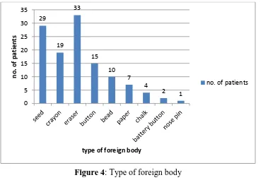
Type of foreign body
The most common foreign bodies seen in our study were eraser (27.5%) and seeds(24.16%). Other foreign bodies seen were crayons, buttons, beads, paper, chalk, battery button and nose pins. Their distribution is shown in Figure 4.
Figure 4: Type of foreign body
54
43
10 8
3 2
0 10 20 30 40 50 60
n
o
. o
f p
atien
ts
signs and symptoms
no. of patients
29
19 33
15
10 7
4
2 1
0 5 10 15 20 25 30 35
n
o
. o
f p
atien
ts
type of foreign body
e
252
e
252
e252
e
252
e252
e
252
e252
e
252
e252
e
25
2
e
252
e
252
e
252
e252
e
252
e252
e
252
e252
e
252
e252
e
252
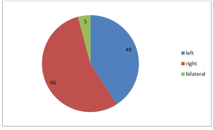
Site of foreign body and method of removal
The most common site was right nasal cavity followed by left nasal cavity. 5(4.16%) patients presented with foreign bodies in bilateral nasal cavities.[Figure 5]
Out of 120 cases of nasal foreign bodies, 102 (85%) were removed in outpatient department or Emergency department with or without local anaesthesia, rest 18(15%) were admitted and removal of foreign body were required general anaesthesia. In all cases, removal was done with direct instrumentation either by extraction or by suction.
Figure 5: Site of foreign body
DISCUSSION
Nasal foreign bodies are common problems in the pediatric age group [4-6] encountered in our daily practice.
In our study,out of 120 patients, there were 68(56.67%) males and 52(43.33%) female patients with M:F ratio of 1.3:1. 94(78.3% ) patients were in the age group of 2-6 years. The most common age group involved in both the sexes was 2-4 years. It is similar to findings by Kadish HA et. al.[6] and Gregori D. et. al.[7]
In present study, the incident was reported by a family member or the actual child in 81 patients (67.5%), and or discovered following nasal symptoms in 39 (32.5%). Most common symptom was unilateral nasal discharge (45%) followed by asymptomatic (35.8%),foul odor in nose(8.3%), nasal obstruction(6.7%), bleeding from nose(2.5%) and discomfort(1.7%). In one large series with 1559 cases of nasal foreign body by Alberto Chinskiet. al.[8], the most frequent symptoms were cacosmia (96, 6.16%) and rhinorrea (59, 3.78%), however, in
the majority of cases (1342, 86.08%) children were asymptomatic which is similar to us. Our findings correspond to the findings in another series by Ogunleye AO et al.[9]
The most common foreign bodies seen in our study were eraser (27.5%) and seeds(24.16%). Other foreign bodies seen were crayons, buttons, beads, paper, chalk, battery button and nose pins. In a large series of nasal foreign body by Alberto Chinski et al.[8] found Pearls (399, 25.59%), Pins, nails, screws, floats (119, 7.63%), Paper (93, 5.97%), Stones (92, 5.90%), Rubber (82,5.26%), Seeds (63, 4.04%) mainly with only one cases of battery was found (0.06%). In
another series by Ogunleye AO et al.[9] the type of foreign body were found as the most common nasal foreign bodies were seeds 34(32.1%). Button batteries are not uncommon as nasal foreign body. Children always choose it because of its shape, size and shiny character thinking like a toy. Prompt identification and rapid removal of these foreign bodies is recommended [10].
49
66
5
left
right
e
253
e
253
e253
e
253
e253
e
253
e253
e
253
e253
e
253
e
253
e
253
e
253
e253
e
253
e253
e
253
e253
e
253
e253
e
253
The most common site in our study was right nasal cavity followed by left nasal cavity. 5(4.16%) patients presented with foreign bodies in bilateral nasal cavities. These findings coincide with the findings by Alberto Chinski et al.[8] and Ogunleye AO et al[9].This may be explained by right handedness of maximum patients.
Out of 120 cases of nasal foreign bodies in our study,102 (85%) were removed in outpatient department or Emergency department with or without local anaesthesia, rest 18(15%) were admitted and removal of foreign body were required general anaesthesia. In all cases, removal was done with direct instrumentation either by extraction or by suction. In a study by Okoye BC et. al. 6 (4.48%) cases out of 134 required general anaesthesia[11].
CONCLUSION
Nasal foreign bodies are seen most commonly in the children in age group of 2-6 years. Parents/caretaker should not allow children to play with toys, household objects or other small objects to prevent the risk of insertion of foreign body in natural orifices.
Funding: No funding sources
Conflict of interest: None declared
REFERENCES
1. Kharoubi S. Corps étrangers des fosses nasales : étude de 700 cas et revue de lalittérature. J Pediatr Puericult 2010;23:314–21.
2. H.Rodrý´guez, G.C. Passali, D. Gregori, A. Chinski, C. Tiscornia, H. Botto, et al., Management of foreign bodies in the airway and oesophagus, Int. J. Pediatr. Otorhinolaryngol.14 (76 Suppl. 1) (2012)
3. Hungria H. Corpos Estranhos. Epistaxe. Imperfuração Coanal. In: Hungria H. Otorrinolaringologia. 6ª edição. Rio de Janeiro: Ed. Guanabara Koogan; 1992. p. 92-3.
4. Lopes Filho O, Campos CH. Inflamações Agudas das Fossas Nasais. In: Lopes Filho O. Tratado de Otorrinolaringologia. 3ª Edição. São Paulo: Editora Roca; 1994. p. 274-82.
5. Botma M, Bader R, Kubba H. ‘A parent’s kiss’: evaluating an unusual methodfor removing nasal foreign body in children.
6. Kadish HA, Corneli HM. Removal of nasal foreign bodies in paediatric population. Am J Emerg Med 1997;J LaryngolOtol 2000;114:598– 60015:54–6.
7. Gregori D, Salerni L, Scarinzi C et al. Foreign bodies in the nose causing complications and requiring hospitalization in children 0–14 age: Results from the European survey of foreign bodies injuries study. Rhinology 2008; 46: 28–33. 8. Alberto Chinskiet. al.,Francesca Foltran, Dario Gregori, Desiderio Passaliand Luisa Bellussi. Nasal foreign bodies: the experience of the Buenos Aires pediatric otolaryngology clinic. Pediatrics International (2011) 53, 90–93.
9. Ogunleye AO, Sogebi OA.Nasal foreign bodies in the African children.Afr. J. Med. Sci.2004;33: 225-8.
10.Capo JM, Lucente FE. Alkaline battery foreign bodies of the ear and nose. Arch. Otolaryngol. Head Neck Surg. 1986; 112: 562–3.