R E S E A R C H
Open Access
Development and application of a
next-generation-sequencing (NGS) approach to detect known and
novel gene defects underlying retinal diseases
Isabelle Audo
1,2,3,4,5*, Kinga M Bujakowska
1,2,3, Thierry Léveillard
1,2,3, Saddek Mohand-Saïd
1,2,3,4,
Marie-Elise Lancelot
1,2,3, Aurore Germain
1,2,3, Aline Antonio
1,2,3,4, Christelle Michiels
1,2,3, Jean-Paul Saraiva
6,
Mélanie Letexier
6, José-Alain Sahel
1,2,3,4,7,8, Shomi S Bhattacharya
1,2,3,5,9and Christina Zeitz
1,2,3*Abstract
Background:
Inherited retinal disorders are clinically and genetically heterogeneous with more than 150 gene
defects accounting for the diversity of disease phenotypes. So far, mutation detection was mainly performed by
APEX technology and direct Sanger sequencing of known genes. However, these methods are time consuming,
expensive and unable to provide a result if the patient carries a new gene mutation. In addition, multiplicity of
phenotypes associated with the same gene defect may be overlooked.
Methods:
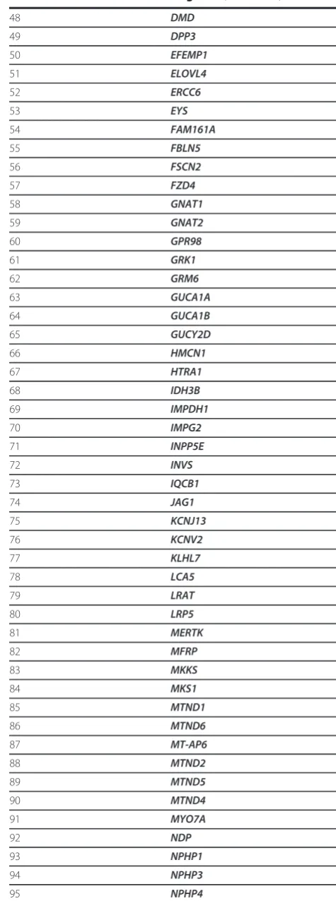
To overcome these challenges, we designed an exon sequencing array to target 254 known and
candidate genes using Agilent capture. Subsequently, 20 DNA samples from 17 different families, including four
patients with known mutations were sequenced using Illumina Genome Analyzer IIx next-generation-sequencing
(NGS) platform. Different filtering approaches were applied to identify the genetic defect. The most likely disease
causing variants were analyzed by Sanger sequencing. Co-segregation and sequencing analysis of control samples
validated the pathogenicity of the observed variants.
Results:
The phenotype of the patients included retinitis pigmentosa, congenital stationary night blindness, Best
disease, early-onset cone dystrophy and Stargardt disease. In three of four control samples with known genotypes
NGS detected the expected mutations. Three known and five novel mutations were identified in
NR2E3, PRPF3, EYS,
PRPF8, CRB1, TRPM1
and
CACNA1F
. One of the control samples with a known genotype belongs to a family with
two clinical phenotypes (Best and CSNB), where a novel mutation was identified for CSNB. In six families the
disease associated mutations were not found, indicating that novel gene defects remain to be identified.
Conclusions:
In summary, this unbiased and time-efficient NGS approach allowed mutation detection in 75% of
control cases and in 57% of test cases. Furthermore, it has the possibility of associating known gene defects with
novel phenotypes and mode of inheritance.
Keywords:
NGS, retinal disorders, diagnostic tool.
Background
Inherited retinal disorders affect approximately 1 in 2000
individuals worldwide [1]. Symptoms and associated
phe-notypes are variable. In some groups the disease can be
mild and stationary such as in congenital stationary night
blindness (CSNB) or achromatopsia (ACHM), whereas
other disorders are progressive leading to severe visual
impairment such as in rod-cone dystrophies, also known as
retinitis pigmentosa (RP) or cone and cone-rod
dystro-phies. The heterogeneity of these diseases is reflected in the
number of underlying gene defects. To date more than 150
genes have been implicated in different forms of retinal
dis-orders http://www.sph.uth.tmc.edu/Retnet/home.htm and
yet in a significant proportion of patients the disease
caus-ing mutation could not be identified, suggestcaus-ing additional
novel genes that remain to be discovered. Furthermore,
recent studies have outlined that distinct phenotypes can
* Correspondence: [email protected]; [email protected]
1INSERM, U968, Paris, F-75012, France
Full list of author information is available at the end of the article
be related to the dysfunction of the same gene [2-4].
Furthermore, there may be additional phenotype-genotype
associations that are still not recognized. The
state-of-the-art phenotypic characterization including precise family
history and functional as well as structural assessment (i.e.
routine ophthalmic examination, perimetry, color vision,
full field and multifocal electroretinography (ERG), fundus
autofluorescence (FAF) imaging and optical coherence
tomography (OCT)) allows targeted mutation analysis for
some disorders. However, in most cases of inherited retinal
diseases, similar phenotypic features can be due to a large
number of different gene defects.
Various methods can be used for the identification of
the corresponding genetic defect. All these methods have
advantages and disadvantages. Sanger sequencing is still
the gold-standard in determining the gene defect, but due
to the heterogeneity of the disorders it is time consuming
and expensive to screen all known genes. Mutation
detec-tion by commercially available APEX genotyping
microar-rays (ASPER Ophthalmics, Estonia) [5,6] allows the
detection of only known mutations. In addition, a separate
microarray has been designed for each inheritance pattern,
which tends to escalate the costs especially in simplex
cases, for which inheritance pattern cannot be
predeter-mined. Indirect methods with single nucleotide
poly-morphism
(SNP)
microarrays
for
linkage
and
homozygosity mapping are also powerful tools, which has
proven its reliability in identifying novel and known gene
defects [7-12]. However, in case of homozygosity mapping
the method can only be applied to consanguineous
families or inbred populations. To overcome these
chal-lenges, we designed a custom sequencing array in
colla-boration with a company (IntegraGen, Evry, France) to
target all exons and part of flanking sequences for 254
known and candidate retinal genes. This array was
subse-quently applied through NGS to a cohort of 20 patients
from 17 families with different inheritance pattern and
clinical diagnosis including RP, CSNB, Best disease,
early-onset cone dystrophy and Stargardt disease.
Methods
Clinical investigation
The study protocol adhered to the tenets of the
Declara-tion of Helsinki and was approved by the local Ethics
Committee (CPP, Ile de France V). Informed written
consent was obtained from each study participant. Index
patients underwent full ophthalmic examination as
described before [13]. Whenever available, blood
sam-ples from affected and unaffected family members were
collected for co-segregation analysis.
Previous molecular genetic analysis
Total genomic DNA was extracted from peripheral blood
leucocytes according to manufacturer
’
s recommendations
(Qiagen, Courtaboeuf, France). DNA samples from some
patients with a diagnosis of RP were first analyzed and
excluded for known mutations by applying commercially
available microarray analysis (arRP and adRP ASPER
Ophthalmics, Tartu, Estonia). In some cases, pathogenic
variants in
EYS, C2orf71, RHO, PRPF31, PRPH2
and
RP1
were excluded by direct Sanger sequencing of the coding
exonic and flanking intronic regions of the respective
genes [13-17]. Conditions used to amplify
PRPH2
can be
provided on request.
Molecular genetic analysis using NGS
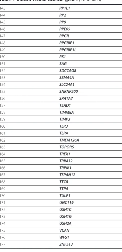
Table 1 Known retinal disease genes
Number Gene name
1 ABCA4
2 ABCC6
3 ADAM9
4 AHI1
5 AIPL1
6 ALMS1
7 ARL6
8 ARMS2
9 ATXN7
10 BBS10
11 BBS12
12 BBS2
13 BBS4
14 BBS5
15 BBS7
16 BBS9
17 BEST1
18 C1QTNF5
19 C2
20 C2orf71
21 C3
22 CA4
23 CABP4
24 CACNA1F
25 CACNA2D4
26 CC2D2A
27 CDH23
28 CDH3
29 CEP290
30 CERKL
31 CFB
32 CFH
33 CHM
34 CLN3
35 CLRN1
36 CNGA1
37 CNGA3
38 CNGB1
39 CNGB3
40 CNNM4
41 COL11A1
42 COL2A1
43 COL9A1
44 CRB1
45 CRX
46 CYP4V2
47 DFNB31
Table 1 Known retinal disease genes
(Continued)
48 DMD
49 DPP3
50 EFEMP1
51 ELOVL4
52 ERCC6
53 EYS
54 FAM161A
55 FBLN5
56 FSCN2
57 FZD4
58 GNAT1
59 GNAT2
60 GPR98
61 GRK1
62 GRM6
63 GUCA1A
64 GUCA1B
65 GUCY2D
66 HMCN1
67 HTRA1
68 IDH3B
69 IMPDH1
70 IMPG2
71 INPP5E
72 INVS
73 IQCB1
74 JAG1
75 KCNJ13
76 KCNV2
77 KLHL7
78 LCA5
79 LRAT
80 LRP5
81 MERTK
82 MFRP
83 MKKS
84 MKS1
85 MTND1
86 MTND6
87 MT-AP6
88 MTND2
89 MTND5
90 MTND4
91 MYO7A
92 NDP
93 NPHP1
94 NPHP3
silent, nonsense etc.). For each position, the exomic
fre-quencies (homozygous and heterozygous) were
deter-mined from all the exomes already sequenced by
IntegraGen and the exome results provided by HapMap
project.
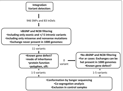
Investigation of annotated sequencing data
We received the annotated sequencing data in the form
of excel tables. On average 946 SNPs and 83 insertions
and deletions were identified for each sample (Figure 1).
By using the filtering system, we first investigated
var-iants (nonsense and missense mutations, intronic
Table 1 Known retinal disease genes
(Continued)
96 NR2E3
97 NRL
98 NYX
99 OAT
100 OFD1
101 OPA1
102 OPA3
103 OPN1LW
104 OPN1MW
105 OPN1Sw
106 OTX2
107 PANK2
108 PAX2
109 PCDH15
110 PCDH21
111 PDE6A
112 PDE6B
113 PDE6C
114 PDE6G
115 PDZD7
116 PEX1
117 PEX2
118 PEX7
119 PGK1
120 PHYH
121 PITPNM3
122 PRCD
123 PROM1
124 PRPF3
125 PRPF31
126 PRPF8
127 PRPH2
128 RAX2
129 RB1
130 RBP3
131 RBP4
132 RD3
133 RDH12
134 RDH5
135 RGR
136 RGS9
137 RGS9BP
138 RHO
139 RIMS1
140 RLBP1
141 ROM1
142 RP1
Table 1 Known retinal disease genes
(Continued)
143 RP1L1
144 RP2
145 RP9
146 RPE65
147 RPGR
148 RPGRIP1
149 RPGRIP1L
150 RS1
151 SAG
152 SDCCAG8
153 SEMA4A
154 SLC24A1
155 SNRNP200
156 SPATA7
157 TEAD1
158 TIMM8A
159 TIMP3
160 TLR3
161 TLR4
162 TMEM126A
163 TOPORS
164 TREX1
165 TRIM32
166 TRPM1
167 TSPAN12
168 TTC8
169 TTPA
170 TULP1
171 UNC119
172 USH1C
173 USH1G
174 USH2A
175 VCAN
176 WFS1
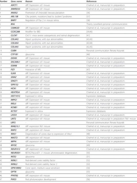
Table 2 Candidate genes for retinal disorders
Number Gene name Reason References
1 ADCY1 diff. Expression rd1 mouse Chalmel et al., manuscript in preparatiom
2 ANKRD33 diff. Expression rd1 mouse Chalmel et al., manuscript in preparatiom
3 ANXA2 Promotion of choroidal neovascularization [36]
4 ARL13B Cilia protein, mutations lead to Joubert Syndrome [37]
5 BMP7 Regulation of Pax 2 in mouse retina [38]
6 BSG - Thierry Leveillard personal commmunication
7 CAMK2D diff. Expression rd1 mouse Chalmel et al., manuscript in preparatiom
8 CCDC28B Modifier for BBS [39,40]
9 CLCN7 Cln7-/- mice severe osteopetrosis and retinal degeneration [41]
10 COL4A3 Alport syndrome, with eye abnormalities [42,43]
11 COL4A4 Alport syndrome, with eye abnormalities [42,44]
12 COL4A5 Alport syndrome, with eye abnormalities [42,45]
13 CUBN - Personal communication Renata Kozyraki
14 CYP1B1 glaucoma [46]
15 DOHH diff. Expression rd1 mouse Chalmel et al., manuscript in preparatiom
16 DSCAML1 diff. Expression rd1 mouse Chalmel et al., manuscript in preparatiom
17 ESRRB diff. Expression rd1 mouse Chalmel et al., manuscript in preparatiom
18 FIZ1 Interactor ofNRL [47]
19 GJA9 diff. Expression rd1 mouse Chalmel et al., manuscript in preparatiom
20 GNAZ diff. Expression rd1 mouse Chalmel et al., manuscript in preparatiom
21 GNGT1 diff. Expression rd1 mouse Chalmel et al., manuscript in preparatiom
22 GPR152 diff. Expression rd1 mouse Chalmel et al., manuscript in preparatiom
23 HCN1 diff. Expression rd1 mouse Chalmel et al., manuscript in preparatiom
24 HEATR5A diff. Expression rd1 mouse Chalmel et al., manuscript in preparatiom
25 HIST1H1C Expressed in retina Expression databases
26 IMPG1 diff. Expression rd1 mouse Chalmel et al., manuscript in preparatiom
27 INSL5 diff. Expression rd1 mouse Chalmel et al., manuscript in preparatiom
28 KCNB1 diff. expression rd1 mouse Chalmel et al., manuscript in preparatiom
29 KCTD7 Expressed in retina Expression databases
30 LASS4 diff. expression rd1 mouse Chalmel et al., manuscript in preparatiom
31 LRIT2 diff. expression rd1 mouse Chalmel et al., manuscript in preparatiom Rd1 mouse
32 LRP2 - Personal communication Renata Kozyraki
33 MAB21L1 diff. expression Rd1 mouse Chalmel et al., manuscript in preparatiom
34 MAP2 diff. expression rd1 mouse Chalmel et al., manuscript in preparatiom
35 MAS1 Degeneration of cones due to expression ofMas1 [48]
36 MAST2 diff. expression rd1 mouse Chalmel et al., manuscript in preparatiom
37 MPP4 diff. expression rd1 mouse Chalmel et al., manuscript in preparatiom
38 MYOC glaucoma [49]
39 NDUFA12 diff. expression rd1 mouse Chalmel et al., manuscript in preparatiom
40 NEUROD1 BETA2/NeuroD1 -/- mouse: photoreceptor degeneration [50]
41 NOS2 glaucoma [51]
42 NXNL1 Rod-derived cone viability factor [52]
43 NXNL2 Rod-derived cone viability factor 2 [53]
44 OPN1MW2 Cone opsin, medium-wave-sensitive2 [54]
45 OPTN glaucoma [55]
46 PFKFB2 diff. expression rd1 mouse Chalmel et al., manuscript in preparatiom
variants located +/- 5 apart from exon), which were
absent in dbSNP and NCBI databases http://ncbi.nlm.
nih.gov/. In the absence of known gene defects or
puta-tive pathogenic variants (see below) in the first step, we
selected known genes, which were previously clinically
associated including variants present in dbSNP and
NCBI databases (Figure 1). Each predicted pathogenic
variant was confirmed by Sanger sequencing.
Assessment of the pathogenicity of variants
Following criteria were applied to evaluate the
patho-genic nature of novel variations identified by NGS: 1)
stop/frameshift variants were considered as most likely
to be disease causing; 2) co-segregation in the family; 3)
absence in control samples; 4) for missense mutations
amino acid conservation was studied in the UCSC
Gen-ome Browser http://genGen-ome.ucsc.edu/ across species
from all different evolutionary branches. If the amino
acid residue did not change it was considered as
“
highly
conserved
”
. If a different change was seen in fewer than
five species and not in the primates then it was
consid-ered as
“
moderately conserved
”
and if a change was
pre-sent in 5-7, it was considered as
“
weakly conserved
”
,
otherwise the amino acid residue was considered as
“
not
conserved
”
, 5) pathogenicity predictions with
bioinfor-matic tools (Polyphen:
Poly
morphism
Phen
otyping,
http://genetics.bwh.harvard.edu/pph/ and SIFT: Sorting
Intolerant From Tolerant, http://blocks.fhcrc.org/sift/
SIFT.html) if at least one of the program predicted the
variant to be possibly damaging, it was considered to be
pathogenic; 6) presence of the second mutant allele in
the case of autosomal recessive inheritance. Mutations
were described according to the HGVS website http://
www.hgvs.org/mutnomen. In accordance with this
nomenclature, nucleotide numbering reflects cDNA
numbering with +1 corresponding to the A of the ATG
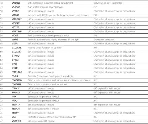
Table 2 Candidate genes for retinal disorders
(Continued)
48 PKD2L1 Diff. expression in human retinal detachment Delyfer et al. 2011 submitted
49 PLEKHA1 Age-related macular degeneratiom [57]
50 PPEF2 diff. expression rd1 mouse Chalmel et al., manuscript in preparatiom
51 RAB8A Interacts with RPGR, role in cilia biogenesis and maintenance [58]
52 RABGEF1 diff. expression rd1 mouse Chalmel et al., manuscript in preparatiom
53 RCVRN diff. expression rd1 mouse Chalmel et al., manuscript in preparatiom
54 RGS20 diff. expression rd1 mouse Chalmel et al., manuscript in preparatiom
55 RNF144B diff. expression rd1 mouse Chalmel et al., manuscript in preparatiom
56 RORB Rod photoreceptor development in mice [59]
57 RXRG Retinoic acid receptor, highly expressed in the eye Expression databases
58 SGIP1 diff. expression rd1 mouse Chalmel et al., manuscript in preparatiom
59 SLC16A8 Altered visual function in ko-mice [60]
60 SLC17A7 diff. expression rd1 mouse Chalmel et al., manuscript in preparatiom
61 STAM2 diff. expression rd1 mouse Chalmel et al., manuscript in preparatiom
62 STK35 diff. expression rd1 mouse Chalmel et al., manuscript in preparatiom
63 STX3 diff. expression rd1 mouse Chalmel et al., manuscript in preparatiom
64 SV2B diff. expression rd1 mouse Chalmel et al., manuscript in preparatiom
65 TBC1D24 diff. expression rd1 mouse Chalmel et al., manuscript in preparatiom
66 THRB Essential for M-cone development in rodents [61]
67 TMEM216 Cilia protein, mutations lead to Joubert and Meckel syndrome [62]
68 TMEM67 Cilia protein, mutations lead to Joubert [63]
69 TRPC1 diff. expression rd1 mouse diff. expression Rd1 mouse
70 UHMK1 diff. expression rd1 mouse diff. expression Rd1 mouse
71 VSX1 Stimulator for promoter NXNL1 [64]
72 VSX2 Stimulator for promoter NXNL1 [64]
73 WDR17 diff. expression rd1 mouse diff. expression Rd1 mouse
74 WDR31 diff. expression Nxnl1-/- mouse [65]
75 WISP1 diff. expression rd1 mouse Chalmel et al., manuscript in preparatiom
76 XIAP Protects photoreceptors in animal models of RP [66]
translation initiation codon in the reference sequence.
The initiation codon is codon 1. The correct
nomencla-ture for mutation was checked applying Mutalyzer
http://www.lovd.nl/mutalyzer/.
Results
The overall sequencing coverage of the captured regions
was 98.4% and 90.4% for a 1× and a 10× coverage
respec-tively. The overall sequencing depth was > 120×. The
number of reference and variant sequences detected by
NGS, reflected the correct zygosity state of the variant;
on average if 50% of the sequences represented the
var-iant, then a heterozygous state was called, while if 100%
of the sequences represented the variant, then a
homozy-gous or hemizyhomozy-gous state was annotated by IntegraGen.
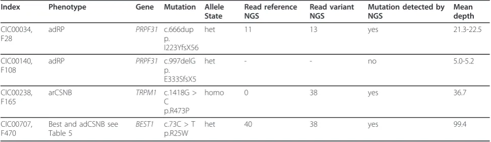
Validation of the novel genetic testing tool for retinal
disorders
To validate the novel genetic testing tool for retinal
dis-orders, we used four DNA samples from families, in
which we had previously identified different types of
mutations by Sanger sequencing: one 1 bp duplication
and one 1 bp deletion in
PRPF31
and missense
muta-tions in
TRPM1
and
BEST1
(Table 3). Three of the four
mutations were detectable by NGS, whereas the deletion
in
PRPF31
was not identified. To validate if this was due
to a technical problem of deletion detection in general
or low coverage at this position, the sequencing depth
was investigated in detail. Indeed the coverage at this
position reflected by the mean depth was only ~1-6 for
all samples. This indicates that although the coverage in
Ϭ
ǀĂƌŝĂŶƚ
/ŶƚĞŐƌĂ'ĞŶ
sĂƌŝĂŶƚĚĞƚĞĐƚŝŽŶ͗
•
Ěď^EWĂŶĚE/ĨŝůƚĞƌŝŶŐ
•/ŶĐůƵĚŝŶŐŽŶůLJĞdžŽŶŝĐĂŶĚнͬͲϱŝŶƚƌŽŶŝĐǀĂƌŝĂŶƚƐ
•
/ŶĐůƵĚŝŶŐŽŶůLJŵŝƐƐĞŶƐĞĂŶĚŶŽŶƐĞŶƐĞŵƵƚĂƚŝŽŶƐ
•džĐŚĂŶŐĞŶĞǀĞƌƉƌĞƐĞŶƚŝŶϭϬϬϬŐĞŶŽŵĞƐ͗
•<ŶŽǁŶŐĞŶĞĚĞĨĞĐƚ͍
•
ŵŽĚĞŽĨŝŶŚĞƌŝƚĂŶĐĞ
•
ƉƌŽƚĞŝŶĨƵŶĐƚŝŽŶ
•ƉŽůLJƉŚĞŶ͕ƐŝĨƚ͗
•ŽŶĨŽƌŵĂƚŝŽŶďLJ^ĂŶŐĞƌƐĞƋƵĞŶĐŝŶŐ
•
ŽͲƐĞŐƌĞŐĂƚŝŽŶĂŶĂůLJƐŝƐ
•džĐůƵƐŝŽŶŝŶĐŽŶƚƌŽůƐĂŵƉůĞƐ
ϵϰϲ^EWƐĂŶĚϴϯ/ŶĞůƐ
ϭϭǀĂƌŝĂŶƚƐ
•EŽĚď^EWĂŶĚE/ĨŝůƚĞƌŝŶŐ
•&ŽƌĂƌĐĂƐĞƐ͗džĐŚĂŶŐĞƐĐĂŶďĞ
ŚĞƚƉƌĞƐĞŶƚŝŶϭϬϬϬŐĞŶŽŵĞƐ
•<ŶŽǁŶŐĞŶĞĚĞĨĞĐƚ͍
ϭͲϱǀĂƌŝĂŶƚƐ
ϭͲϱǀĂƌŝĂŶƚƐ
general was very good, specific probes used here need to
be redesigned to improve the capture for specific exons.
Detection of known and novel mutations
Some of the patients from the 14 families with no
known gene defect were previously excluded for known
mutations using microarray analysis and by Sanger
sequencing in the known genes
EYS, C2orf71, RHO,
PRPF31, PRPH2
and
RP1
. Other samples were never
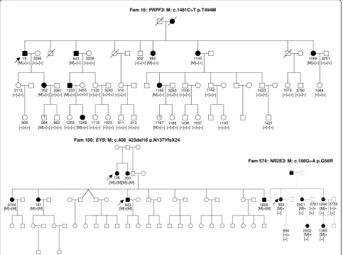
genetically investigated. In four DNA samples known
mutations were detected (Table 4) from three different
families with autosomal dominant (ad) or recessive (ar)
RP. All mutations co-segregated with the phenotype
(Figure 2). In seven samples, novel mutations in known
genes were identified. These mutations co-segregated
with the phenotype from five different families with
adCSNB, linked incomplete CSNB, adRP, arRP and
x-linked RP (Table 5, Figures 3 and 4). One of the cases
from these five families was also used as a control for
Best disease carrying a known
BEST1
mutation (Table
3). In addition to the Best phenotype, ERG-responses of
this patient resembled those of complete CSNB, i.e.
showing selective ON-bipolar pathway dysfunction. This
phenotype was independent of the Best phenotype
(Fig-ure 3). The most likely disease causing mutation
detected by NGS was a novel heterozygous
TRPM1
mutation (Table 4, Figure 3).
Unsolved cases
In six of the 14 families with Stargardt disease, adRP,
adCD with postreceptoral defects, arRP, early onset
arCD with macrocephaly and mental retardation
described in affected sister and x-linked cCSNB, the
dis-ease associated mutations remain to be elucidated or
validated (Table 6, Figure 5).
Discussion
By using NGS in 254 known and candidate genes we
were able to detect known and novel mutations in 57%
of families tested. In order to achieve this goal, we
applied a rigorous protocol (Figure 1). To our
knowl-edge, this is the first report using NGS to investigate all
inherited retinal disorders at once. In a study restricted
to adRP, Bowne and co-workers used a similar approach
including 46 known and candidate genes for adRP [18].
All their cases had previously been screened and
excluded for most of the known genes underlying adRP.
The authors were able to identify known or novel
muta-tions in five out of 21 cases in genes not included in a
pre-screening [18]. This added five patients to their
Table 3 Patients with known mutations used to validate the novel genetic approach for retinal disorders
Index Phenotype Gene Mutation Allele State
Read reference NGS
Read variant NGS
Mutation detected by NGS
Mean depth
CIC00034, F28
adRP PRPF31 c.666dup
p. I223YfsX56
het 11 13 yes 21.3-22.5
CIC00140, F108
adRP PRPF31 c.997delG
p. E333SfsX5
het - - no 5.0-5.2
CIC00238, F165
arCSNB TRPM1 c.1418G >
C p.R473P
homo 0 38 yes 36.7
CIC00707, F470
Best and adCSNB see Table 5
BEST1 c.73C > T p.R25W
het 40 38 yes 99.4
Table 4 Detection of known mutations by using the novel genetic approach for retinal disorders
Index Phenotype Pre-screening Gene Mutation Allele State
Read reference NGS
Read variant NGS
Reference Mutation verified by Sanger and co-segregation
CIC00019, F16
adRP Linkage,RHO,
PRPF31, PRPH2, RP1
PRPF3 c.1481C > T p.T494M
het 25 22 [67] yes
CIC0000893, F574
adRP RHO, PRPF31,
PRPH2, RP1 NR2E3
c.166G > A p.G56R
het 5 3 [68] yes
CIC000128, F100
arRP, consang.
- EYS c.408_423del
p.N137VfsX24
homo - 179 [13,69] yes
CIC0000943, F100
arRP, consang
- EYS c.408_423del p.
N137VfsX24
adRP cohort with known gene defects, indicating that
64% of their patients show known mutations with new
genes still to be discovered in the remaining 36%. The
current study provides a more exhaustive tool, since it
incorporates screening of 254 genes implicated in
var-ious retinal disorders of different inheritance patterns
and additional candidate genes for these phenotypes.
With this approach a cohort of both pre-screened and
unscreened samples, was investigated. The mutation
detection rate of 57% is high and was never obtained
before by high throughput screening methods.
Further-more, this approach is probably less time consuming
and expensive than existing methods such as direct
sequencing of all known genes or microarray analysis.
Of note however is one of the variants detected with the
NGS approach (i.e. p.V973L exchange in
GUCY2D
),
which was not confirmed by direct Sanger sequencing,
suggesting the possibility of false positive using the high
throughput screening. Verification by direct Sanger
sequencing of most likely pathogenic variants is
there-fore essential to validate NGS data, although the false
positive rate is assumed to be low (in our study 1/28
verified sequence variants represented a false positive).
Overall, the study of 20 subjects from 17 families by
NGS showed that most of the targeted regions are well
covered (more than 98%). However, some of the regions
showed a lower coverage (GC-rich regions) or were not
captured (repetitive regions). This was for instance the
case for two genes underlying cCSNB, (i.e.
NYX
and
GRM6
) and the repetitive region of ORF15 of
RPGR
.
For GC-rich regions the capture design could be
improved in the future by modifying NGS chemistry, as
Fam 16: PRPF3: M: c.1481C>T p.T494M
? ?
352 19 3266
3113
909 984 3361
983 1259 1248 1119 1023 911 913 1167 1166 1036 1037 1143 1421 843 3239
1250 3455 1120 3240 910 1165 3263
932 982 1145 1069 3251
1084 3780 1073 1423
1142 1035
[=]+[=] [=]+[=] [=]+[=] [=]+[=] [=]+[=] [=]+[=] [=]+[=] [=]+[=] [=]+[=] [=]+[=] [=]+[=] [=]+[=]
[=]+[=] [=]+[=]
[=]+[=] [=]+[=]
[=]+[=] [=]+[=] [=]+[=] [=]+[=] [=]+[=] [=]+[=] [=]+[=] [=]+[=] [=]+[=] [=]+[=] [=]+[=] [=]+[=] [=]+[=]
[M]+[=] [M]+[=] [M]+[=] [M]+[=] [M]+[=]
[M]+[=] [M]+[=]
[M]+[=]
[M]+[=] [M]+[=]
Fam 100: EYS: M: c.408_423del16 p.N137VfsX24
128 [M]+[M]
203 [M]+[M]
3744
[M]+[M] [M]+[M]181 [M]+[M]943 [M]+[=]
Fam 574: NR2E3: M: c.166G>A p.G56R
[M]+ [=]
893 2501 [M]+ [=]
2761 [=]+ [=]
1394 [M]+ [=]
2733 [=]+ [=]
2502 1395 894
[M]+ [=]
[M]+ [=] [=]+
[=] 1808 [M]+[M]
it was successfully achieved for Sanger sequencing using
different additives, which improved the amplification
and subsequent sequencing. If repetitive regions like
ORF15 of
RPGR
remain problematic for sequencing by
NGS, direct Sanger sequencing of these targets might be
the first screening of choice; in particular for disorders
caused only by a few gene defects such as CSNB, and
xl-RP.
By applying NGS sequencing to our retinal panel,
known and novel mutations were detected in different
patients. We believe that our diagnostic tool is
particu-larly important for heterogeneous disorders like RP, for
which many gene defects with different prevalence have
been associated to one phenotype. It also allows the
rapid detection of novel mutations in minor genes
which are often not screened as a priority by direct
San-ger sequencing. This was the case in our study for three
individuals from one family with adRP in which NGS
detected a novel
PRPF8
mutation in both affected and
one unaffected family member (Table 4, Figure 4). In
this family, the RP phenotype is mild and therefore it is
possible that the unaffected member may develop
symp-toms later in life or alternatively it may be a case of
incomplete penetrance as reported for another splicing
factor gene,
PRPF31
and recently for
PRPF8
as well
[19-22]. Interestingly, a novel
TRPM1
mutation was
identified in a patient with adCSNB, a gene previously
only associated with arCSNB [23-26]. This is the first
report of a
TRPM1
mutation co-segregating with ad
Schubert-Bornschein type complete CSNB. Since the
location of this mutation is not different compared to
other mutations leading to arCSNB, it is not quite clear
how
TRPM1
mutations might lead to either ad or
arCSNB. Functional investigations are needed to validate
the pathogenicity of this variant. Furthermore, this
find-ing suggests that
TRPM1
heterozygous mutation carriers
from arCSNB families should be investigated by
electro-retinography to determine whether they display similar
retinal dysfunction as in affected members of the
pre-sented adCSNB family. Detection of a novel
RPGR
splice
site mutation in family 146 presented a challenge. The
actual disease causing change was concealed under a
wrongly annotated rs62638633, which had previously
been clinically associated to RP by a German group
http://www.ncbi.nlm.nih.gov/sites/varvu?gen-e=6103&rs=62638633, (personal communication,
Mar-kus Preising). These observations indicate that the
stringent filtering we applied initially can mask those
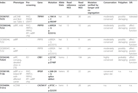
Table 5 Detection of novel mutations by using the novel genetic approach for retinal disorders
Index Phenotype Pre-screening
Gene Mutation Allele State Read reference NGS Read variant NGS Mutation verified by Sanger and co-segregation
Conservation Polyphen Sift
CIC00707,
F470
adCSNB and Best see Table 3
RHO, PDE6B, GNAT1 TRPM1 c.1961A > C p.H654P
het 39 38 yes moderately
conserved possibly damaging tolerated CIC000348, F232
adRP, mild RHO, PRPF31, PRPH2, RP1, adRP chip
PRPF8 c.6992A > G p. E2331G
het 13 10 yes moderately
conserved possibly damaging affect protein function CIC000346, F232
adRP - PRPF8 c.6992A
> G p.E2331G
het 5 9 yes moderately
conserved possibly damaging affect protein function CIC000347, F232 as adRP
- PRPF8 c.6992A
> G p.E2331G
het 15 17 yes moderately
conserved possibly damaging affect protein function CIC04240, F2025 arRP, consang., detailed clinic in [70]
RS1 CRB1 c.2219C > T p.S740F
homo 2 194 yes highly
conserved probably damaging affect protein function CIC00199, F146
adRP or x-linked RP with affected carrier RHO, PRPF31, PRPH2, RP1, adRP chip
RPGR c.248-2A > G splice defect
hetero 30 22 yes conserved
splice site
n.a. n.a.
CIC04094,
F1915
icCSNB - CACNA1F c.973C >
T p.Q325X
referenced disease causing variants. Bearing this in mind
one can still first investigate unknown variants, but
should then examine dbSNP for referenced variants
either described to be disease causing, having a low
minor allele frequency or present in interesting
candi-date genes. An accurate discrimination of
non-patho-genic
polymorphisms
versus
disease
causing
polymorphism in SNP databases is warranted to resolve
this challenge.
In six families from the investigated cohort the disease
causing mutations still remain to be identified. In the
Stargardt patient with no pathogenic
ABCA4
mutations
two variants in
CFH
were detected, one of which
(rs1061170) had previously been reported to predispose
to age related macular degeneration (AMD) [27-29]. The
second
CFH
change is a novel variant, affecting a highly
conserved residue, not found in NGS data from the other
19 samples and never associated with a disease. The
variants co-segregated in the only available family
mem-bers, which were the patient
’
s parents. Apart from the
association with AMD,
CFH
mutations have been
pre-viously associated with renal diseases, the most common
being membranoproliferative glomerulonephritis and
hemolytic uremic syndrome, which can be also associated
with an eye phenotype [30,31]. No renal dysfunction was
present in our patient. To validate if the two variants
identified in
CFH
are indeed disease causing, the DNA
samples from other available family members for
co-seg-regation analysis as well as characterization of functional
consequences of the novel variant are needed. One
patient with complete CSNB had an affected nephew and
thus x-linked inheritance was assumed. However, neither
Sanger nor NGS detected a mutation in the only known
x-linked gene,
NYX
, causing cCSNB. To exclude
reces-sive inheritance
TRPM1
and
GRM6
were investigated in
detail. Indeed the patient carried a novel heterozygous
Fam 470: BEST1: M1: c.73C>T p.R25W; TRPM1: M2:c.1961A>C p.H654P707 [M1]+[=]
705
[M1]+[=] [M1]+[=] 706 [=]+[=] 2715 708 [=]+[=]
Best disease cCSNB
707 [M2]+[=]
705
[M2]+[=] [=]+[=] 706 [M2]+[=] 2715 708 [M2]+[=]
OD
OS
Arden ratio Arden ratio
a b
c d
e
TRPM1
variant, which affects a highly conserved amino
acid and was not identified in the other 19 samples
inves-tigated here (Table 6). However, direct Sanger
sequen-cing of lower covered regions did not identify a second
mutation in this gene. Similarly no mutations in
GRM6
were identified. These findings outline the need for
addi-tional family members to determine, through
co-segrega-tion, the pathogenicity of the numerous variants
identified by NGS. This was also true for two other
families with nonsense mutations in
CUBN
(Fam795)
and
RP1L1
(Fam761) (Table 6). The nonsense mutation
in
CUBN
, co-segregated with the phenotype in most of
the family members (Figure 5). Had we not had access to
additional family members, we might have retained this
gene defect as the underlying cause for adCD and
consid-ered
CUBN
as a new gene involved in adCD. None of the
other putatively pathogenic mutations identified in
CUBN, TRPM1
and
GUCY2D
co-segregated with the
phenotype in this family (Table 6, Figure 5).
RP1L1
was
already a candidate for adRP [32] but was previously
associated with occult macular dystrophy [33]. In our
study, this variant did not co-segregate with the
pheno-type in other affected family members (data not shown).
This NGS study ended with six genetically unresolved
families, which can be further investigated with whole
exome sequencing. Although, no clear information
about the actual percentage of missing gene defects
underlying each group of inherited retinal disorders
exists, previous studies have reported that in many cases
the genetic cause still needs to be determined [18,34].
Whole exome sequencing approaches allow the
detec-tion of both, novel and known gene defects, but also
generate numerous variants and therefore require the
inclusion of more than one DNA sample for each family
to rapidly exclude non-pathogenic variants. Due to the
higher costs of exome sequencing for one sample
com-pared to targeted sequencing, we propose to initially
perform targeted sequencing in the index patient and
proceed only after exclusion of a known gene defect to
whole exome sequencing.
Fam 2025: CRB1: M: c.2219C>T p.S2740F
Fam 146: RPGR: M: c.248-2A splice defect
Fam 1915: CACNA1F: M: c.973C>T p.Q325X
? 346 [M]+[=]
348 [M]+[=]
347 [M]+[=]
4241
[M]+[M] [M]+[M]4240 4499
[M]+[=] 4242 [M]+[=]
4094 [M]
4248 [M]+[=]
Fam 232: PRPF8: M: c.6992A>G p.E2331G
[M]+[=] 219
[M]+[=]200 [M]+[=]220 [M]+[=] 199
?
[M]+[=]222 [M] 221
Table 6 Patients with unsolved genotype and unlikely disease causing mutations
Index Phenotype Pre-screening
Gene Mutation Allele State
Read reference NGS
Read variant NGS
Mutation verified by Sanger and co-segregation
Comment
CIC03282,
F1388
Stargardt ABCA4
microarray
ABCA4 c.1268A > G p.H423R
het 77 61 yes but reported as
polymorphism [71]
c.6764G > T p.S2255I no additional variants in lower covered exons
het 2 7 yes but reported
as polymorphism [72]
CFH c.3482C > A p.P1161Q
het 77 52 yes conserved,
probably damaging
c.1204C > T p.H402Y
het 94 87 yes AMD
CIC01269, F761
adRP - RP1L1 c.5959C > T
p.Q1987X
het 145 150 yes, did not co-segregate pass to whole
exome sequencing
CIC01312,
F795
adCD with post-receptoral defects
RHO, PDE6B, GNAT1 adRP chip
CUBN c.127C > T p.R43X
het 139 102 yes, did not co-segregate pass to whole
exome sequencing
CUBN c.9340G > A p.G3114S
het 61 44 yes, did not co-segregate
GUCY2D c.1499C > T p.P500L
het 41 34 yes, did not co-segregate
TRPM1 c.3904T > C p.C1302R
het 102 99 yes, did not co-segregate
CIC03225,
F1362
arRP consang. arRP chip PROM1 c.314A > G
p.Y105C
het 120 115 yes, but no additional
mutation
no homo, no compound hets, pass to whole exome sequencing
GUCY2D c.2917G > A p.V973L
het 6 2 false positive, not found
by Sanger
DSCAML1 c.592C > T p.R198C
het 70 81 yes, but no additional
mutation
TBC1D24 c.641G > A p.R214H
het 27 12 yes, but no additional
mutation
TMEM67 c.1700A > G p.Y567C
het 80 58 yes, but no additional
mutation
CIC04757 F2364
Index and affected sister early onset arCD, macro-cephaly and mental retardation in affected sister consang.
- IMPG2 c.3439C > T
p.P1147S
homo 0 140 no Polyphen and Sift
benign, not conserved
PKD2L1 c.1027C > T p.R343C
het 63 68
c.1202T > G p.V401G
het 25 19 appeared also
Conclusions
In summary, our diagnostic tool is an unbiased time
efficient method, which not only allows detecting known
and novel mutations in known genes but also potentially
associates known gene defects with novel phenotypes.
This genetic testing tool can now be applied to large
cohorts of inherited retinal disorders and should rapidly
deliver the prevalence of known genes and the
percen-tage of cases with missing genetic defect for underlying
forms of retinal disorders.
List of abbreviations
ad: autosomal dominant; ar: autosomal recessive; as: asymptomatic; het: heterozygous; homo: homozygous; hemi: hemizygous; - not noted; consang.:
Table 6 Patients with unsolved genotype and unlikely disease causing mutations
(Continued)
DFNB31 c.1943C > A p.S648Y
het 7 7 yes affected sister
also both variants but both come from father, no other variant in lower covered region.
c.2644C > A p.R882S
het 27 14 yes
EYS c.7597A > G p.K2533E
het 151 149 yes Affected sister
does not carry this variant
RPGRIP1 c.2417C > T p.T806I
het 138 132 no not conserved
CIC04152, F1955
male x-linked cCSNB, has affected nephew
NYX TRPM1 c.470C > T p.S157F
het 118 130 yes, no other het
mutation.
x-linked inheritance and phenotype verification
Index patients and respective gene defect are highlighted in bold. In some cases also family members were used for NGS.
Fam 795:
M1: CUBN: c.127C>T p.R43X M2: CUBN: c.9340G>A p.G311S M3: GUCY2D: c.1499C>T p.P500L M4: TRPM1: c.3904T>C p.C1302R
?
1396 1402
+
+ + + +
?
2771 2819
?
2911 1312 1371
1369 1370
* * *
2566
3013 ? ?
[=]+[=]
[M3]+[=]
[=]+[=] [M3]+[=] [=]+[=]
[=]+[=]
*
[=]+[=] [M4]+[=] [M4]+[=] [M4]+[=]
[M4]+[=] [M4]+[=]
[M1]+[M2] [M1]+[M2]
[=]+[=] [=]+[=] [M1]+[M2] [M1]+[M2]
5008
4921
[=]+[=] [M4]+[=] [M1]+[M2] [=]+[=]
[=]+[=] [=]+[=]
[=]+[=] [=]+[=] [=]+[=]
?
[=]+[=] [=]+[=] [=]+[=]
[=]+[=] [=]+[=] [=]+[=]
[=]+[=] [=]+[=] [=]+[=]
[=]+[=] [=]+[=] [=]+[=]
consanguinity was reported; n.a.: not applicable; CSNB: congenital stationary night blindness; RP: retinitis pigmentosa:
Acknowledgements
The authors are grateful to the families described in this study, Dominique Santiard-Baron and Christine Chaumeil for their help in DNA collection and to clinical staff. The project was financially supported by GIS-maladies rares (CZ), Agence Nationale de la Recherche (ANR, SSB), Foundation Voir et Entendre and BQR, Foundation Fighting Blindness (IA, FFB Grant # CD-CL-0808-0466-CHNO and the CIC503 recognized as an FFB center, FFB Grant # C-CMM-0907-0428-INSERM04), Ville de Paris and region Ile de France.
Author details
1
INSERM, U968, Paris, F-75012, France.2CNRS, UMR_7210, Paris, F-75012, France.3UPMC Univ Paris 06, UMR_S 968, Department of Genetics, Institut
de la Vision, Paris, F-75012, France.4Centre Hospitalier National
d’Ophtalmologie des Quinze-Vingts, INSERM-DHOS CIC 503, Paris, F-75012, France.5UCL-Institute of Ophthalmology, London, UK.6IntegraGen SA, Genopole CAMPUS 1 bat G8 FR-91030 EVRY France.7Fondation
Ophtalmologique Adolphe de Rothschild, Paris, France.8Académie des
Sciences-Institut de France, 75006 Paris, France.9Department of Celular
Therapy and Regenerative Medicine, Andalusian Molecular Biology and Regenerative Medicine Centre (CABIMER), Isla de Cartuja, Seville, Spain.
Authors’contributions
IA was involved in the study design, participated in the choice of genes, interpreted the NGS data, clinically investigated patients, collected DNA samples, and has been involved in drafting the manuscript. KB participated in the choice of genes, interpreted the NGS data and has been involved in drafting the manuscript. TL was involved in the study design, participated in the choice of genes and has been involved in drafting the manuscript. SM-S clinically investigated patients and collected DNA samples. M-EL confirmed the NGS data by Sanger sequencing, performed control and co-segregation analysis. AG extracted DNA, confirmed the NGS data by Sanger sequencing, and performed control and co-segregation analysis. AA extracted DNA, confirmed the NGS data by Sanger sequencing, and performed control and co-segregation analysis. CM confirmed the NGS data by Sanger sequencing, and performed control and co-segregation analysis. J-PS performed NGS. ML performed the bioinformatic interpretation of NGS. J-AS clinically
investigated patients and participated in the study design. SSB participated in the study design and has been involved in drafting the manuscript. CZ has made the study design, participated in the choice of genes, interpreted the NGS data and wrote the manuscript. All authors read and approved the final manuscript.
Competing interests
The authors declare that they have no competing interests.
Received: 12 September 2011 Accepted: 25 January 2012 Published: 25 January 2012
References
1. Sohocki MM, Daiger SP, Bowne SJ, Rodriquez JA, Northrup H,
Heckenlively JR, Birch DG, Mintz-Hittner H, Ruiz RS, Lewis RA, Saperstein DA, Sullivan LS:Prevalence of mutations causing retinitis pigmentosa and other inherited retinopathies.Hum Mutat2001,17:42-51.
2. Boon CJ, den Hollander AI, Hoyng CB, Cremers FP, Klevering BJ, Keunen JE:
The spectrum of retinal dystrophies caused by mutations in the peripherin/RDS gene.Prog Retin Eye Res2008,27:213-235.
3. Boon CJ, Klevering BJ, Leroy BP, Hoyng CB, Keunen JE, den Hollander AI:
The spectrum of ocular phenotypes caused by mutations in the BEST1 gene.Prog Retin Eye Res2009,28:187-205.
4. Schorderet DF, Escher P:NR2E3 mutations in enhanced S-cone sensitivity syndrome (ESCS), Goldmann-Favre syndrome (GFS), clumped
pigmentary retinal degeneration (CPRD), and retinitis pigmentosa (RP).
Hum Mutat2009,30:1475-1485.
5. Jaakson K, Zernant J, Kulm M, Hutchinson A, Tonisson N, Glavac D, Ravnik-Glavac M, Hawlina M, Meltzer MR, Caruso RC, Testa F, Maugeri A, Hoyng CB, Gouras P, Simonelli F, Lewis RA, Lupski JR, Cremers FP, Allikmets R:
Genotyping microarray (gene chip) for the ABCR (ABCA4) gene.Hum Mutat2003,22:395-403.
6. Zeitz C, Labs S, Lorenz B, Forster U, Uksti J, Kroes HY, De Baere E, Leroy BP, Cremers FP, Wittmer M, van Genderen MM, Sahel JA, Audo I, Poloschek CM, Mohand-Said S, Fleischhauer JC, Huffmeier U, Moskova-Doumanova V, Levin AV, Hamel CP, Leifert D, Munier FL, Schorderet DF, Zrenner E, Friedburg C, Wissinger B, Kohl S, Berger W:Genotyping microarray for CSNB-associated genes.Invest Ophthalmol Vis Sci2009,50:5919-5926. 7. Wang H, den Hollander AI, Moayedi Y, Abulimiti A, Li Y, Collin RW,
Hoyng CB, Lopez I, Bray M, Lewis RA, Lupski JR, Mardon G, Koenekoop RK, Chen R:Mutations in SPATA7 cause leber congenital amaurosis and juvenile retinitis pigmentosa.Am J Hum Genet2009,84:380-387. 8. den Hollander AI, McGee TL, Ziviello C, Banfi S, Dryja TP,
Gonzalez-Fernandez F, Ghosh D, Berson EL:A homozygous missense mutation in the IRBP gene (RBP3) associated with autosomal recessive retinitis pigmentosa.Invest Ophthalmol Vis Sci2009,50:1864-1872. 9. Collin RW, Littink KW, Klevering BJ, van den Born LI, Koenekoop RK,
Zonneveld MN, Blokland EA, Strom TM, Hoyng CB, den Hollander AI, Cremers FP:Identification of a 2 Mb human ortholog of Drosophila eyes shut/spacemaker that is mutated in patients with retinitis pigmentosa.
Am J Hum Genet2008,83:594-603.
10. Collin RW, Safieh C, Littink KW, Shalev SA, Garzozi HJ, Rizel L, Abbasi AH, Cremers FP, den Hollander AI, Klevering BJ, Ben-Yosef T:Mutations in C2ORF71 cause autosomal-recessive retinitis pigmentosa.Am J Hum Genet2010,86:783-788.
11. Bandah-Rozenfeld D, Collin RW, Banin E, van den Born LI, Coene KL, Siemiatkowska AM, Zelinger L, Khan MI, Lefeber DJ, Erdinest I, Testa F, Simonelli F, Voesenek K, Blokland EA, Strom TM, Klaver CC, Qamar R, Banfi S, Cremers FP, Sharon D, den Hollander AI:Mutations in IMPG2, encoding interphotoreceptor matrix proteoglycan 2, cause autosomal-recessive retinitis pigmentosa.Am J Hum Genet2010,87:199-208. 12. Audo I, Bujakowska K, Mohand-Said S, Tronche S, Lancelot ME, Antonio A,
Germain A, Lonjou C, Carpentier W, Sahel JA, Bhattacharya S, Zeitz C:A novel DFNB31 mutation associated with Usher type 2 syndrome showing variable degrees of auditory loss in a consanguineous Portuguese family.Mol Vis2011,17:1598-1606.
13. Audo I, Sahel JA, Mohand-Said S, Lancelot ME, Antonio A, Moskova-Doumanova V, Nandrot EF, Doumanov J, Barragan I, Antinolo G, Bhattacharya SS, Zeitz C:EYS is a major gene for rod-cone dystrophies in France.Hum Mutat2010,31:E1406-1435.
14. Audo I, Lancelot ME, Mohand-Said S, Antonio A, Germain A, Sahel JA, Bhattacharya SS, Zeitz C:Novel C2orf71 mutations account for approximately 1% of cases in a large French arRP cohort.Hum Mutat 2011,32:E2091-2103.
15. Audo I, Manes G, Mohand-Said S, Friedrich A, Lancelot ME, Antonio A, Moskova-Doumanova V, Poch O, Zanlonghi X, Hamel CP, Sahel JA, Bhattacharya SS, Zeitz C:Spectrum of rhodopsin mutations in French autosomal dominant rod-cone dystrophy patients.Invest Ophthalmol Vis Sci2010,51:3687-3700.
16. Audo I, Bujakowska K, Mohand-Said S, Lancelot ME, Moskova-Doumanova V, Waseem NH, Antonio A, Sahel JA, Bhattacharya SS, Zeitz C:Prevalence and novelty of PRPF31 mutations in French autosomal dominant rod-cone dystrophy patients and a review of published reports.BMC Med Genet 2010,11:145.
17. Audo I, Mohand-Said S, Dhaenens CM, Germain A, Orhan E, Antonio A, Hamel C, Sahel JA, Bhattacharya SS, Zeitz C:RP1 and autosomal dominant rod-cone dystrophy: Novel mutations, a review of published variants, and genotype-phenotype correlation.Hum Mutat2012,33:73-80. 18. Bowne SJ, Sullivan LS, Koboldt DC, Ding L, Fulton R, Abbott RM,
Sodergren EJ, Birch DG, Wheaton DH, Heckenlively JR, Liu Q, Pierce EA, Weinstock GM, Daiger SP:Identification of disease-causing mutations in autosomal dominant retinitis pigmentosa (adRP) using next-generation DNA sequencing.Invest Ophthalmol Vis Sci2011,52:494-503.
19. Vithana EN, Abu-Safieh L, Pelosini L, Winchester E, Hornan D, Bird AC, Hunt DM, Bustin SA, Bhattacharya SS:Expression of PRPF31 mRNA in patients with autosomal dominant retinitis pigmentosa: a molecular clue for incomplete penetrance?Invest Ophthalmol Vis Sci2003,44:4204-4209. 20. McGee TL, Devoto M, Ott J, Berson EL, Dryja TP:Evidence that the
penetrance of mutations at the RP11 locus causing dominant retinitis pigmentosa is influenced by a gene linked to the homologous RP11 allele.Am J Hum Genet1997,61:1059-1066.
Bhattacharya SS:A human homolog of yeast pre-mRNA splicing gene, PRP31, underlies autosomal dominant retinitis pigmentosa on chromosome 19q13.4 (RP11).Mol Cell2001,8:375-381.
22. Maubaret CG, Vaclavik V, Mukhopadhyay R, Waseem NH, Churchill A, Holder GE, Moore AT, Bhattacharya SS, Webster AR:Autosomal Dominant Retinitis Pigmentosa with Intrafamilial Variability and Incomplete Penetrance in Two Families carrying Mutations in PRPF8.Invest Ophthalmol Vis Sci2011.
23. Li Z, Sergouniotis PI, Michaelides M, Mackay DS, Wright GA, Devery S, Moore AT, Holder GE, Robson AG, Webster AR:Recessive mutations of the gene TRPM1 abrogate ON bipolar cell function and cause complete congenital stationary night blindness in humans.Am J Hum Genet2009,
85:711-719.
24. van Genderen MM, Bijveld MM, Claassen YB, Florijn RJ, Pearring JN, Meire FM, McCall MA, Riemslag FC, Gregg RG, Bergen AA, Kamermans M:
Mutations in TRPM1 are a common cause of complete congenital stationary night blindness.Am J Hum Genet2009,85:730-736. 25. Audo I, Kohl S, Leroy BP, Munier FL, Guillonneau X, Mohand-Said S,
Bujakowska K, Nandrot EF, Lorenz B, Preising M, Kellner U, Renner AB, Bernd A, Antonio A, Moskova-Doumanova V, Lancelot ME, Poloschek CM, Drumare I, Defoort-Dhellemmes S, Wissinger B, Leveillard T, Hamel CP, Schorderet DF, De Baere E, Berger W, Jacobson SG, Zrenner E, Sahel JA, Bhattacharya SS, Zeitz C:TRPM1 is mutated in patients with autosomal-recessive complete congenital stationary night blindness.Am J Hum Genet2009,85:720-729.
26. Nakamura M, Sanuki R, Yasuma TR, Onishi A, Nishiguchi KM, Koike C, Kadowaki M, Kondo M, Miyake Y, Furukawa T:TRPM1 mutations are associated with the complete form of congenital stationary night blindness.Mol Vis2010,16:425-437.
27. Klein RJ, Zeiss C, Chew EY, Tsai JY, Sackler RS, Haynes C, Henning AK, SanGiovanni JP, Mane SM, Mayne ST, Bracken MB, Ferris FL, Ott J, Barnstable C, Hoh J:Complement factor H polymorphism in age-related macular degeneration.Science2005,308:385-389.
28. Edwards AO, Ritter R, Abel KJ, Manning A, Panhuysen C, Farrer LA:
Complement factor H polymorphism and age-related macular degeneration.Science2005,308:421-424.
29. Haines JL, Hauser MA, Schmidt S, Scott WK, Olson LM, Gallins P, Spencer KL, Kwan SY, Noureddine M, Gilbert JR, Schnetz-Boutaud N, Agarwal A, Postel EA, Pericak-Vance MA:Complement factor H variant increases the risk of age-related macular degeneration.Science2005,308:419-421. 30. Ault BH:Factor H and the pathogenesis of renal diseases.Pediatr Nephrol
2000,14:1045-1053.
31. Boon CJ, van de Kar NC, Klevering BJ, Keunen JE, Cremers FP, Klaver CC, Hoyng CB, Daha MR, den Hollander AI:The spectrum of phenotypes caused by variants in the CFH gene.Mol Immunol2009,46:1573-1594. 32. Bowne SJ, Daiger SP, Malone KA, Heckenlively JR, Kennan A, Humphries P,
Hughbanks-Wheaton D, Birch DG, Liu Q, Pierce EA, Zuo J, Huang Q, Donovan DD, Sullivan LS:Characterization of RP1L1, a highly polymorphic paralog of the retinitis pigmentosa 1 (RP1) gene.Mol Vis 2003,9:129-137.
33. Akahori M, Tsunoda K, Miyake Y, Fukuda Y, Ishiura H, Tsuji S, Usui T, Hatase T, Nakamura M, Ohde H, Itabashi T, Okamoto H, Takada Y, Iwata T:
Dominant mutations in RP1L1 are responsible for occult macular dystrophy.Am J Hum Genet2010,87:424-429.
34. Berger W, Kloeckener-Gruissem B, Neidhardt J:The molecular basis of human retinal and vitreoretinal diseases.Prog Retin Eye Res2010,
29:335-375.
35. Vaclavik V, Gaillard MC, Tiab L, Schorderet DF, Munier FL:Variable phenotypic expressivity in a Swiss family with autosomal dominant retinitis pigmentosa due to a T494M mutation in the PRPF3 gene.Mol Vis2010,16:467-475.
36. Zhao SH, Pan DY, Zhang Y, Wu JH, Liu X, Xu Y:Annexin A2 promotes choroidal neovascularization by increasing vascular endothelial growth factor expression in a rat model of argon laser coagulation-induced choroidal neovascularization.Chin Med J (Engl)2010,123:713-721. 37. Cantagrel V, Silhavy JL, Bielas SL, Swistun D, Marsh SE, Bertrand JY,
Audollent S, Attie-Bitach T, Holden KR, Dobyns WB, Traver D, Al-Gazali L, Ali BR, Lindner TH, Caspary T, Otto EA, Hildebrandt F, Glass IA, Logan CV, Johnson CA, Bennett C, Brancati F, Valente EM, Woods CG, Gleeson JG:
Mutations in the cilia gene ARL13B lead to the classical form of Joubert syndrome.Am J Hum Genet2008,83:170-179.
38. Sehgal R, Sheibani N, Rhodes SJ, Belecky Adams TL:BMP7 and SHH regulate Pax2 in mouse retinal astrocytes by relieving TLX repression.
Dev Biol2009,332:429-443.
39. Beales PL, Badano JL, Ross AJ, Ansley SJ, Hoskins BE, Kirsten B, Mein CA, Froguel P, Scambler PJ, Lewis RA, Lupski JR, Katsanis N:Genetic interaction of BBS1 mutations with alleles at other BBS loci can result in non-Mendelian Bardet-Biedl syndrome.Am J Hum Genet2003,72:1187-1199. 40. Badano JL, Leitch CC, Ansley SJ, May-Simera H, Lawson S, Lewis RA,
Beales PL, Dietz HC, Fisher S, Katsanis N:Dissection of epistasis in oligogenic Bardet-Biedl syndrome.Nature2006,439:326-330. 41. Kornak U, Kasper D, Bosl MR, Kaiser E, Schweizer M, Schulz A, Friedrich W,
Delling G, Jentsch TJ:Loss of the ClC-7 chloride channel leads to osteopetrosis in mice and man.Cell2001,104:205-215. 42. Colville DJ, Savige J:Alport syndrome. A review of the ocular
manifestations.Ophthalmic Genet1997,18:161-173.
43. Lemmink HH, Mochizuki T, van den Heuvel LP, Schroder CH, Barrientos A, Monnens LA, van Oost BA, Brunner HG, Reeders ST, Smeets HJ:Mutations in the type IV collagen alpha 3 (COL4A3) gene in autosomal recessive Alport syndrome.Hum Mol Genet1994,3:1269-1273.
44. Jefferson JA, Lemmink HH, Hughes AE, Hill CM, Smeets HJ, Doherty CC, Maxwell AP:Autosomal dominant Alport syndrome linked to the type IV collage alpha 3 and alpha 4 genes (COL4A3 and COL4A4).Nephrol Dial Transplant1997,12:1595-1599.
45. Lemmink HH, Kluijtmans LA, Brunner HG, Schroder CH, Knebelmann B, Jelinkova E, van Oost BA, Monnens LA, Smeets HJ:Aberrant splicing of the COL4A5 gene in patients with Alport syndrome.Hum Mol Genet1994,
3:317-322.
46. Stoilov I, Akarsu AN, Sarfarazi M:Identification of three different truncating mutations in cytochrome P4501B1 (CYP1B1) as the principal cause of primary congenital glaucoma (Buphthalmos) in families linked to the GLC3A locus on chromosome 2p21.Hum Mol Genet1997,
6:641-647.
47. Mitton KP, Swain PK, Khanna H, Dowd M, Apel IJ, Swaroop A:Interaction of retinal bZIP transcription factor NRL with Flt3-interacting zinc-finger protein Fiz1: possible role of Fiz1 as a transcriptional repressor.Hum Mol Genet2003,12:365-373.
48. Xu X, Quiambao AB, Roveri L, Pardue MT, Marx JL, Rohlich P, Peachey NS, Al-Ubaidi MR:Degeneration of cone photoreceptors induced by expression of the Mas1 protooncogene.Exp Neurol2000,163:207-219. 49. Kubota R, Kudoh J, Mashima Y, Asakawa S, Minoshima S, Hejtmancik JF,
Oguchi Y, Shimizu N:Genomic organization of the human myocilin gene (MYOC) responsible for primary open angle glaucoma (GLC1A).Biochem Biophys Res Commun1998,242:396-400.
50. Pennesi ME, Cho JH, Yang Z, Wu SH, Zhang J, Wu SM, Tsai MJ:BETA2/ NeuroD1 null mice: a new model for transcription factor-dependent photoreceptor degeneration.J Neurosci2003,23:453-461.
51. Liu B, Neufeld AH:Expression of nitric oxide synthase-2 (NOS-2) in reactive astrocytes of the human glaucomatous optic nerve head.Glia 2000,30:178-186.
52. Leveillard T, Mohand-Said S, Lorentz O, Hicks D, Fintz AC, Clerin E, Simonutti M, Forster V, Cavusoglu N, Chalmel F, Dolle P, Poch O, Lambrou G, Sahel JA:Identification and characterization of rod-derived cone viability factor.Nat Genet2004,36:755-759.
53. Chalmel F, Leveillard T, Jaillard C, Lardenois A, Berdugo N, Morel E, Koehl P, Lambrou G, Holmgren A, Sahel JA, Poch O:Rod-derived Cone Viability Factor-2 is a novel bifunctional-thioredoxin-like protein with therapeutic potential.BMC Mol Biol2007,8:74.
54. Tsutsumi M, Ikeyama K, Denda S, Nakanishi J, Fuziwara S, Aoki H, Denda M:
Expressions of rod and cone photoreceptor-like proteins in human epidermis.Exp Dermatol2009,18:567-570.
55. Rezaie T, Child A, Hitchings R, Brice G, Miller L, Coca-Prados M, Heon E, Krupin T, Ritch R, Kreutzer D, Crick RP, Sarfarazi M:Adult-onset primary open-angle glaucoma caused by mutations in optineurin.Science2002,
295:1077-1079.
56. Onishi A, Peng GH, Hsu C, Alexis U, Chen S, Blackshaw S:Pias3-dependent SUMOylation directs rod photoreceptor development.Neuron2009,
61:234-246.
57. Montezuma SR, Sobrin L, Seddon JM:Review of genetics in age related macular degeneration.Semin Ophthalmol2007,22:229-240.
implications for cilia dysfunction and photoreceptor degeneration.Hum Mol Genet2010,19:3591-3598.
59. Jia L, Oh EC, Ng L, Srinivas M, Brooks M, Swaroop A, Forrest D: Retinoid-related orphan nuclear receptor RORbeta is an early-acting factor in rod photoreceptor development.Proc Natl Acad Sci USA2009,
106:17534-17539.
60. Daniele LL, Sauer B, Gallagher SM, Pugh EN Jr, Philp NJ:Altered visual function in monocarboxylate transporter 3 (Slc16a8) knockout mice.Am J Physiol Cell Physiol2008,295:C451-457.
61. Ng L, Hurley JB, Dierks B, Srinivas M, Salto C, Vennstrom B, Reh TA, Forrest D:A thyroid hormone receptor that is required for the development of green cone photoreceptors.Nat Genet2001,27:94-98. 62. Valente EM, Logan CV, Mougou-Zerelli S, Lee JH, Silhavy JL, Brancati F,
Iannicelli M, Travaglini L, Romani S, Illi B, Adams M, Szymanska K, Mazzotta A, Lee JE, Tolentino JC, Swistun D, Salpietro CD, Fede C, Gabriel S, Russ C, Cibulskis K, Sougnez C, Hildebrandt F, Otto EA, Held S, Diplas BH, Davis EE, Mikula M, Strom CM, Ben-Zeev B,et al:Mutations in TMEM216 perturb ciliogenesis and cause Joubert, Meckel and related syndromes.
Nat Genet2010,42:619-625.
63. Baala L, Romano S, Khaddour R, Saunier S, Smith UM, Audollent S, Ozilou C, Faivre L, Laurent N, Foliguet B, Munnich A, Lyonnet S, Salomon R, Encha-Razavi F, Gubler MC, Boddaert N, de Lonlay P, Johnson CA, Vekemans M, Antignac C, Attie-Bitach T:The Meckel-Gruber syndrome gene, MKS3, is mutated in Joubert syndrome.Am J Hum Genet2007,80:186-194. 64. Reichman S, Kalathur RK, Lambard S, Ait-Ali N, Yang Y, Lardenois A, Ripp R,
Poch O, Zack DJ, Sahel JA, Leveillard T:The homeobox gene CHX10/VSX2 regulates RdCVF promoter activity in the inner retina.Hum Mol Genet 2010,19:250-261.
65. Cronin T, Raffelsberger W, Lee-Rivera I, Jaillard C, Niepon ML, Kinzel B, Clerin E, Petrosian A, Picaud S, Poch O, Sahel JA, Leveillard T:The disruption of the rod-derived cone viability gene leads to photoreceptor dysfunction and susceptibility to oxidative stress.Cell Death Differ2010,
17:1199-1210.
66. Leonard KC, Petrin D, Coupland SG, Baker AN, Leonard BC, LaCasse EC, Hauswirth WW, Korneluk RG, Tsilfidis C:XIAP protection of photoreceptors in animal models of retinitis pigmentosa.PLoS ONE2007,2:e314. 67. Chakarova CF, Hims MM, Bolz H, Abu-Safieh L, Patel RJ, Papaioannou MG,
Inglehearn CF, Keen TJ, Willis C, Moore AT, Rosenberg T, Webster AR, Bird AC, Gal A, Hunt D, Vithana EN, Bhattacharya SS:Mutations in HPRP3, a third member of pre-mRNA splicing factor genes, implicated in autosomal dominant retinitis pigmentosa.Hum Mol Genet2002,11:87-92. 68. Coppieters F, Leroy BP, Beysen D, Hellemans J, De Bosscher K,
Haegeman G, Robberecht K, Wuyts W, Coucke PJ, De Baere E:Recurrent mutation in the first zinc finger of the orphan nuclear receptor NR2E3 causes autosomal dominant retinitis pigmentosa.Am J Hum Genet2007,
81:147-157.
69. Bandah-Rozenfeld D, Littink KW, Ben-Yosef T, Strom TM, Chowers I, Collin RW, den Hollander AI, van den Born LI, Zonneveld MN, Merin S, Banin E, Cremers FP, Sharon D:Novel null mutations in the EYS gene are a frequent cause of autosomal recessive retinitis pigmentosa in the Israeli population.Invest Ophthalmol Vis Sci2010,51:4387-4394. 70. Bujakowska K, Audo I, Mohand-Said S, Lancelot ME, Antonio A, Germain A,
Leveillard T, Letexier M, Saraiva JP, Lonjou C, Carpentier W, Sahel JA, Bhattacharya SS, Zeitz C:CRB1 mutations in inherited retinal dystrophies.
Hum Mutat2011, ,33:306-315.
71. Rivera A, White K, Stohr H, Steiner K, Hemmrich N, Grimm T, Jurklies B, Lorenz B, Scholl HP, Apfelstedt-Sylla E, Weber BH:A comprehensive survey of sequence variation in the ABCA4 (ABCR) gene in Stargardt disease and age-related macular degeneration.Am J Hum Genet2000,67:800-813. 72. Shroyer NF, Lewis RA, Lupski JR:Analysis of the ABCR (ABCA4) gene in
4-aminoquinoline retinopathy: is retinal toxicity by chloroquine and hydroxychloroquine related to Stargardt disease?Am J Ophthalmol2001,
131:761-766.
doi:10.1186/1750-1172-7-8
Cite this article as:Audoet al.:Development and application of a next-generation-sequencing (NGS) approach to detect known and novel gene defects underlying retinal diseases.Orphanet Journal of Rare Diseases2012
7:8.
Submit your next manuscript to BioMed Central
and take full advantage of:
• Convenient online submission
• Thorough peer review
• No space constraints or color figure charges
• Immediate publication on acceptance
• Inclusion in PubMed, CAS, Scopus and Google Scholar
• Research which is freely available for redistribution