R E S E A R C H
Open Access
Thyroid malignancy among patients with
thyroid nodules in the United Arab
Emirates: a five-year retrospective tertiary
Centre analysis
Eiman Alseddeeqi
1*, Raqwana Baharoon
2, Rawia Mohamed
3, Jenan Ghaith
4, Abeer Al-Helali
5and Luai A. Ahmed
6Abstract
Objectives:Thyroid malignancy constitutes the sixth common cancer type in the United Arab Emirates (UAE). There are no epidemiological data outlining the prevalence of cancer in thyroid nodules, nor previous analysis of ultra-sonographic features correlating with thyroid malignancy in the UAE. This study aimed to estimate the prevalence of thyroid malignancy in patients with thyroid nodules and to describe the ultra-sonographic characteristics of thyroid nodules harbouring malignancy.
Methods:A retrospective electronic medical records review of all thyroid nodules in patients (aged 18 to 80 years) with normal thyroid-stimulating hormone (TSH) levels, who underwent ultrasound guided fine needle aspiration cytology (UG-FNA) at Sheikh Khalifa Medical City (SKMC) during 2011–2015.
Results:436 patients with normal TSH underwent UG-FNA cytological examination of thyroid nodules (n= 555 nodules). The overall crude prevalence of thyroid cancer among patients was 10.1% (95% CI 7.5–13.3). The age-adjusted prevalence of thyroid cancer among UAE nationals, Arabs, Far East Asians, and Caucasians were 9.6% (3.6– 15.6), 10.0% (6.2–13.8), 16.8% (4.5–29.0) and 16.3% (1.7–30.9), respectively. The crude prevalence was 14.5%(95% CI 6. 2–22.8) in men, and 9.3%(95% CI 6.3–12.2) in women. The echogenicity features were significantly different
between the cancerous and noncancerous nodules (p= 0.025). Cancerous nodules were relatively more hyper- and
hypo-echoic, while noncancerous nodules were mostly complex.
Conclusion:We report a higher prevalence of thyroid malignancy among patients with thyroid nodules relative to that reported in other parts of the world. The rate of thyroid malignancy was higher in patients of Far-East Asian and Caucasian ethnic background.
Keywords:Thyroid, Thyroid nodules, Thyroid Cancer, Cancer, Prevalence, United Arab Emirates
Introduction
The overall worldwide incidence of thyroid malignancy has been increasing over the last four decades [1]. On-going research is aimed at exploring possible factors contributing to the formation of different thyroid can-cer subtypes, to identify individuals at risk of thyroid malignancy [2].
Thyroid nodules are another common thyroid endo-crinopathy with prevalence ranging from 1 to 5% in iodine-sufficient areas; this prevalence is significantly increased if nodules incidentally detected by ultrasound are included, reaching up to 68% [3]. Moreover, the overall number of thyroid nodules and malignancy cases reported in the last decade has increased due to widespread use of neck ultrasound [3].
The United Arab Emirates (UAE) is classified as an area with minimal iodine-deficiency with rate of households using iodized salt around 6% and a median urinary iodine excretion of 100μg/l. Therefore, a high prevalence of * Correspondence:ealseddeeqi@seha.ae
1Division of Endocrinology, Sheikh Khalifa Medical City, P.O. Box 51900, Abu
Dhabi, UAE
Full list of author information is available at the end of the article
thyroid malignancy is not unexpected [4–7]. Thyroid ma-lignancy is the most common endocrine mama-lignancy in the UAE, and papillary thyroid cancer is the most com-mon type of thyroid cancer [4]. It is reported as the sixth most common cancer type among UAE population and the second among UAE women [4].
There are no comprehensive epidemiological data out-lining the prevalence of thyroid malignancy in thyroid nodules and its correlation with demographic data in UAE population (a heterogeneous population with mul-tiple ethnic backgrounds). Moreover, there is no previ-ous analysis of ultra-sonographic features correlating with thyroid malignant nodules in the UAE.
This study aimed to describe the prevalence of thyroid malignancy in euthyroid patients with thyroid nodules in the UAE population. The study also aims to analyse the distribution of thyroid malignancy by gender, age, ethni-city, cytological, and ultra-sonographic characteristics.
Methods
Study design and settings
This is a retrospective analysis of a cohort of patients (aged 18 years old and above with thyroid nodules and normal serum TSH) who underwent ultrasound guided fine needle aspiration (UG-FNA) cytology for the first time from Janu-ary 2011 to December 2015, at Sheikh Khalifa Medical City (SKMC) in Abu Dhabi. SKMC is a tertiary centre and func-tioned as the referral centre for peripheral health care facil-ities during the study period. The normal reference range for TSH is 0.270–4.200 mIU/L and all TSH levels were measured at our institution. Neck ultrasound and reporting were performed by more than one radiologist while deci-sion to perform aspiration cytology was made by endocri-nologists based on ultrasound findings. The diagnosis of cancer was made following surgery by one experienced cer-tified pathologist per each case. If features of malignancy are overlapping with benignity on histology specimens, a second opinion is sought through the monthly pathology meeting dedicated for such cases.
An institutional research board approval was obtained from SKMC.
All patients with known thyroid cancer history and ab-normal TSH level were excluded.
Chart review data
Demographic characteristics of all patients who under-went cytological examinations were retrieved from their medical records. These included, age at the time of per-forming the fine needle aspiration, gender and ethnicity. Age was grouped as follows: 18–29, 30–39, 40–49, 50– 59 and≥60 years. Ethnicity was classified according to nationalities into six major groups: UAE nationals, Arabs (except UAE), Far-East Asians (Philippines, China),
South-East Asians (India, Pakistan, Bangladesh, and Sri Lanka), Caucasians, and others.
UG-FNA was performed on all nodules sized ≥1 cm if they demonstrated worrisome ultra-sonographic features; and ultrasound features were reported according to the American Thyroid Association (ATA) clinical practice guidelines for assessment of thyroid nodule (2009) [8].
In case of a patient with more than one nodule, criteria for FNA selection were based on the size and presence of a worrisome feature on ultrasound as mentioned above. For each nodule, the size (in three dimensions with the greatest dimension used for data analysis) and echogeni-city features (solid, cystic, or complex) were identified. For solid nodules, echogenicity was classified into either: is-echogenicity, hypo-echogenicity, or hyper-echogenicity. Other ultrasound features reported included the presence of nodal disease or calcifications.
Bethesda thyroid nodule cytological classification repor ting system was used to report the cytopathology [9]. Nodules with Bethesda categories IV, V, and VI underwent lobectomy or total thyroidectomy with histological exam-ination. For Bethesda III cytology result, a repeat fine nee-dle aspiration cytology or lobectomy was recommended based on the presence of worrisome features on ultra-sound. Thyroid malignancy was divided into subtypes as follows: classical papillary thyroid cancer (CPTC), follicu-lar variant of papilfollicu-lary thyroid cancer (FvPTC), classical follicular thyroid cancer (FTC), Hurthle thyroid cancer and medullary thyroid cancer (MTC).
Statistical analysis
Data were analysed as per-patient and per-nodule basis. Demographic of the patients with nodules and the ultra-sonographic and cytological characteristics of the nodules were described. Categorical data were ex pressed as numbers and percentages. Continuous data were reported as means and standard deviations. Chi-squared or Fisher’s tests were used to determine the statistical significance of the differences between the characteristics, when comparing cancerous and noncancerous nodules. The overall and gender-specific prevalence of cancerous nodules by demographic char-acteristics were estimated using logistic models and re-ported with 95% confidence intervals (CI). Stata 15 was used to perform the analysis. The significance level of the statistical tests was set at 5%.
Results
Demographics
(SD) 12.7 years). The mean (SD) age in women and men were 43.4 (12.7) and 47.8 (12.2), respectively (p= 0.008). Moreover, there were significant differences in the distri-bution of gender by age-groups (p= 0.004). 59.2% of women aged between 30 and 49 years whereas 58.3% of men aged between 40 and 59 years as illustrated in Fig.2. Around 77% of the patients were of Arabic ethnicity (in-cluding UAE nationals).
Prevalence
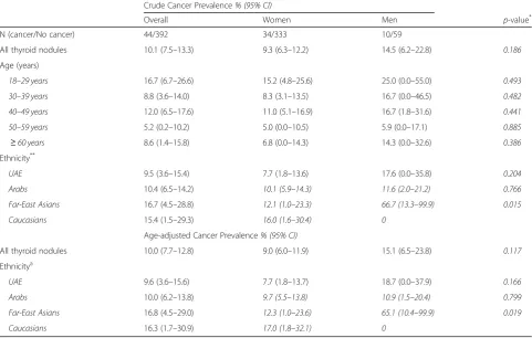
The overall crude prevalence of thyroid cancer in pa-tients with thyroid nodules was 10.1% (95% CI 7.5– 13.3). The crude prevalence was 9.3% (95% CI 6.3–12.2) in women, and 14.5% (95% CI 6.2–22.8) in men (Table2). Similar to the crude prevalence, the overall age-adjusted prevalence was 10.0% (7.7–12.8), and 9.0% (6.0–11.9) in women; whereas slightly higher in men (15.1% (6.5– 23.8)). Overall, there was no significant difference in age-adjusted thyroid cancer prevalence between women and men (p= 0.117).
The age-adjusted prevalence of thyroid cancer among UAE nationals, Arabs, Far East Asians, and Caucasians were 9.6% (3.6–15.6), 10.0% (6.2–13.8), 16.8% (4.5– 29.0) and 16.3% (1.7–30.9), respectively; with relatively higher cancer prevalence in men. Only for Far-East Asians patients, the cancer prevalence was significantly higher in men compared to women (p= 0.019). No can-cer was reported in thyroid nodules detected in South-East Asian or‘Other’ethnicities’patients.
Ultra-sonographic and cytological characteristics of the nodules
In total of 555 thyroid nodules underwent UG-FNA cytological examinations. As Table 3 shows, more than 60% of the nodules were sized 2 cm or more with no difference in the size distribution between the cancerous and noncancerous nodules (p= 0.176). Around 54% of the nodules were of complex echo-genicity. The echogenicity features were significantly different between the cancerous and noncancerous nodules (p= 0.025). Cancerous nodules were relatively more hyper- and hypo-echoic, while noncancerous nod-ules were mostly complex. Although 70% of the nodnod-ules had no calcifications, coarse calcification occurred more frequently with cancerous nodules compared to noncancerous nodules (p< 0.001). Around 79% of all
Fig. 1Numbers of patients and thyroid nodules included in the analyses
Table 1Characteristics of patients with thyroid nodules (N= 436) that underwent ultrasound guided fine needle aspiration (UG-FNA) cytology between January 2011 and December 2015 in a tertiary hospital in Abu Dhabi, United Arab Emirates
Number(%)
N
Gender
Women 367 (84.2)
Men 69 (15.8)
Age (years)mean (SD) 44.1 (12.7)
Age groups
18–29 years 54 (12.4)
30–39 years 114 (26.2)
40–49 years 133 (30.5)
50–59 years 77 (17.7)
≥60 years 58 (13.3)
Ethnicitya
UAE 95 (21.4)
Arabs 241 (55.4)
Far-East Asians 36 (8.3)
South-East Asians 29 (6.7)
Caucasians 26 (6.0)
Others 8 (1.8)
a
the nodules were classified as Bethesda II while cancer-ous nodules were more frequently classified as Bethesda IV, V, or VI compared to noncancerous nodules (p< 0.001).
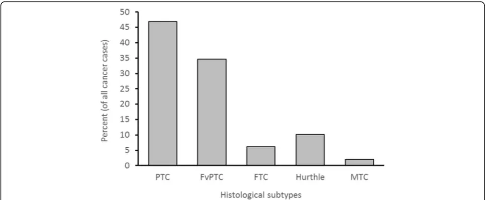
Among the cancerous thyroid nodules, 47% were PTC while 35% were FvPTC (Fig.3). No significant histological
differences were found between women and men in can-cerous nodules (p= 0.443).
Discussion
This study is the first to estimate the prevalence of thy-roid cancer among thythy-roid nodules in the UAE and
Fig. 2Age and gender distribution of patients with thyroid nodules (N= 555*) who underwent ultrasound guided fine needle aspiration (UG-FNA) cytology between January 2011 and December 2015 in a tertiary hospital in Abu Dhabi, United Arab Emirates (*471 nodules in women and 84 nodules in men)
Table 2Crude and age-adjusted overall and gender-specific thyroid cancer prevalence (95% CI) among patients with thyroid nodules who underwent ultrasound guided fine needle aspiration (UG-FNA) biopsy between January 2011 and December 2015 in a tertiary hospital in Abu Dhabi, United Arab Emirates
Crude Cancer Prevalence% (95% CI)
Overall Women Men p-value*
N (cancer/No cancer) 44/392 34/333 10/59
All thyroid nodules 10.1 (7.5–13.3) 9.3 (6.3–12.2) 14.5 (6.2–22.8) 0.186
Age (years)
18–29 years 16.7 (6.7–26.6) 15.2 (4.8–25.6) 25.0 (0.0–55.0) 0.493
30–39 years 8.8 (3.6–14.0) 8.3 (3.1–13.5) 16.7 (0.0–46.5) 0.482
40–49 years 12.0 (6.5–17.6) 11.0 (5.1–16.9) 16.7 (1.8–31.6) 0.441
50–59 years 5.2 (0.2–10.2) 5.0 (0.0–10.5) 5.9 (0.0–17.1) 0.885
≥60 years 8.6 (1.4–15.8) 6.8 (0.0–14.3) 14.3 (0.0–32.6) 0.386
Ethnicity**
UAE 9.5 (3.6–15.4) 7.7 (1.8–13.6) 17.6 (0.0–35.8) 0.204
Arabs 10.4 (6.5–14.2) 10.1 (5.9–14.3) 11.6 (2.0–21.2) 0.766
Far-East Asians 16.7 (4.5–28.8) 12.1 (1.0–23.3) 66.7 (13.3–99.9) 0.015
Caucasians 15.4 (1.5–29.3) 16.0 (1.6–30.4) 0
Age-adjusted Cancer Prevalence% (95% CI)
All thyroid nodules 10.0 (7.7–12.8) 9.0 (6.0–11.9) 15.1 (6.5–23.8) 0.117
Ethnicitya
UAE 9.6 (3.6–15.6) 7.7 (1.8–13.7) 18.7 (0.0–37.9) 0.166
Arabs 10.0 (6.2–13.8) 9.7 (5.5–13.8) 10.9 (1.5–20.4) 0.799
Far-East Asians 16.8 (4.5–29.0) 12.3 (1.0–23.6) 65.1 (10.4–99.9) 0.019
Caucasians 16.3 (1.7–30.9) 17.0 (1.8–32.1) 0
*
p-value comparing women vs men
a
covered the period from 2011 to 2015. Relatively high prevalence ranging between 9 to 15% were found in women and men. High malignancy rates were found in nodules with sizes between 2 and 4 cm.
The overall cancer crude prevalence of 10.1% which was found among thyroid nodules in our study is rela-tively high compared to the prevalence reported in some Western countries. The prevalence of thyroid malig-nancy in a single nodule in a study performed in the United States was 5% [6]. Finding of a similar study con-ducted in Italy supported the same lower prevalence (3.3%) of thyroid malignancy among thyroid nodules [6]. A study by Santos J. et al. found an association between increased risk of thyroid nodules and thyroid malignancy in iodine-deficient areas [5]. Another regional study re-ported the same association of increased malignant thy-roid nodules among iodine-deficient subjects [10]. This
could explain the relatively higher malignancy formation in thyroid nodules found in our study as most of the study population descend from iodine deficient parts of the world. However, the rate found in our study was more or less similar to other reported prevalence in Saudi Ara-bia, Cyprus, and Taiwan, where thyroid cancer rates among thyroid nodules ranged from 7 to 15% [11–13].
We found an 10.0% overall age-adjusted prevalence of thyroid cancer; with 9.0% in women and 15.1% in men. This gender differences with higher rate in men is con-sistent with findings from an Asian population study [8]. In this study, although the highest proportions of thyroid and cancerous nodules occurred among subjects aged 40–49 years, thyroid malignancy prevalence was highest among thyroid nodules in women aged 18–29 years and men aged 30–39 years. An increased unmet demand of iodine during pregnancy in the reproductive age group Table 3Characteristics of thyroid nodules (N= 555) that underwent ultrasound guided fine needle aspiration (UG-FNA) biopsy between January 2011 and December 2015 in a tertiary hospital in Abu Dhabi, United Arab Emirates
Total N (%*)
Cancer status p
-value** Cancer N (%a) No cancer N (%a)
N 555 (100) 49 (100) 506 (100)
Nodule size 535 (100) 48 (100.0) 487 (100.0) 0.176
< 1 cm 27 (5.1) 5 (10.4) 22 (4.5)
1–< 2 cm 174 (32.5) 17 (35.4) 157 (32.2)
2–4 cm 222 (41.5) 20 (41.7) 202 (41.5)
> 4 cm 112 (20.9) 6 (12.5) 106 (21.8)
Echogenicity 531 (100) 47 (100.0) 484 (100.0) 0.025
Cystic 15 (2.8) 1 (2.1) 14 (2.9)
Complex 287 (54.1) 17 (36.2) 270 (55.8)
Hyper-echoic 18 (3.4) 4 (8.5) 14 (2.9)
Hypo-echoic 124 (23.4) 17 (36.2) 107 (22.1)
Isoechoic 87 (16.4) 8 (17.0) 79 (16.3)
Calcification 547 (100) 48 (100.0) 499 (100.0) < 0.001
Coarse 25 (4.6) 14 (29.2) 11 (2.2)
Yes 125 (22.9) 10 (20.8) 115 (23.0)
No 397 (72.6) 24 (50.0) 373 (74.8)
Lymphadenopathy 554 (100) 49 (100) 505 (100) 0.999
No 530 (95.7) 47 (95.9) 483 (95.6)
Yes 24 (4.3) 2 (4.1) 22 (4.4)
FNA Cytology 551 (100) 49 (100) 502 (100) < 0.001
Bethesda, I 29 (5.3) 1 (2.0) 28 (5.6)
Bethesda II 435 (79.0) 4 (8.2) 431 (85.9)
Bethesda III 32 (5.8) 6 (12.2) 26 (5.2)
Bethesda IV 25 (4.5) 17 (34.7) 8 (1.6)
Bethesda V 16 (2.9) 12 (24.5) 4 (0.8)
Bethesda VI 14 (2.5) 9 (18.4) 5 (1.0)
a
Column percent.**p-value comparing cancerous vs noncancerous groups
may explain the increased frequency of thyroid nodules with a younger age presentation in women; and hence, the associated occurrence of malignancy in women com-pared to men [5,14, 15]. Another possible factor for the earlier presentation in women during the childbearing years is the associated level of oestrogen which is known to induce thyroid cancer in mice [16].
Interestingly, the prevalence of thyroid malignancy in this study varies between patients of different ethnic back-grounds. It was found to be highest among Far-East Asians and among Caucasian populations presenting with thyroid nodules. Higher rates of thyroid malignancy were reported previously among Far-East Asians in North America [17]. A similar study was performed in North America where it was found that thyroid cancers were proportionally higher in both foreign- and United States-born Arabs compared with other metropolitan eth-nicities [18]. Further research to identify risk factors influ-encing malignancy occurrence in certain ethnic groups is warranted. Ultrasound examination is readily available in the UAE in secondary care centres; therefore, the in-creased detection rate of thyroid nodules by ultrasound might have contributed to the increased prevalence of thy-roid malignancy in this study. Therefore, we speculate that ethnicity, iodine-deficiency (which leads to the formation of multinodular goiters), and the wide spread use of ultra-sound, led to the detection of relatively higher prevalence of thyroid cancer in our study.
Most nodules in this study were classified as Bethesda II cytology type (76.6%), in which thyroid malignancy is not common. Conventional PTC was found in 46.9% of all cancer types while FvPTC constituted 34.7% of all thyroid malignancies in our study. This result is consistent with
reports from different parts of the world that PTC consti-tutes 80–90% of all thyroid malignancy types [1, 19]. In terms of ultrasound characteristics, around 41.3% of nod-ules with a size of between 2 and 4 cm was associated with the occurrence of thyroid malignancy in our study. This result is consistence with other studies demonstrating similar association with size [20,21]. Even the most recent ATA guidelines (2015) showed that the potential of malig-nancy in nodules < 2 cm is low in the absence of other worrisome features [22]. Therefore, FNA of thyroid nod-ules is indicated for all thyroid nodnod-ules above 2 cm. An ex-ception to that is a nodule that is purely cystic or that has a spongiform pattern on ultrasound [22]. The presence of such ultra-sonographic worrisome features could indicate the need to decrease the threshold required for perform-ing thyroid nodule FNA, especially in certain ethnic groups or in all ethnicities when certain size criteria is met.
Limitations and strengths
This retrospective data analysis occurred in a single centre. The analysis was performed on nodules managed according to the ATA 2009 standard of practice guidelines for managing thyroid nodules. Referrals to our tertiary centre could have created a referral bias, however, FNA indication was based on certain clinical criteria. Since histological confirmation was not performed on all thyroid nodules, the rates of thyroid malignancy could have been underestimated. Among the strength of this study is the large sample size of patients and nodules included. More-over, all patients included in the study had normal TSH to ensure validity of the results because patients from iodine-deficient areas exhibit more autoimmune thyroid
conditions as well as increased risk of thyroid malignancy when presenting with high TSH [14].
Conclusion
The prevalence of thyroid malignancy in this study is higher relative to that reported in other parts of the world. Far-East Asian and Caucasian ethnic groups were found to have higher rates of thyroid malignancy.
Abbreviations
ATA:American Thyroid Association; CPTC: Conventional papillary thyroid cancer; FTC: Classical follicular thyroid cancer; FvPTC: Follicular variant of papillary thyroid cancer; MTC: Medullary thyroid cancer; SKMC: Sheikh Khalifa Medical City; TSH: Thyroid-stimulating hormone; UAE: United Arab Emirates; UG-FNA: Ultrasound guided fine needle aspiration
Acknowledgments Not Applicable
Funding
This research received no specific grant from any funding agency in the public, commercial or not-for-profit sectors.
Availability of data and materials No additional data are available.
Authors’contributions
EA, RB, AA, RM, JG, and LAA conceptualized and designed the study. EA, RB, AA, RM, and JG obtained the data. LAA analysed the data. EA and LAA drafted the manuscript. EA, RB, AA, RM, JG, and LAA critically revised the manuscript. EA, RB, AA, RM, JG, and LAA approved the final submission of the manuscript.
Ethics approval and consent to participate
An institutional research board approval was obtained from SKMC.
Consent for publication
No individual person’s data were included in the manuscript.
Competing interests
The authors declare that they have no competing interests.
Publisher’s Note
Springer Nature remains neutral with regard to jurisdictional claims in published maps and institutional affiliations.
Author details
1Division of Endocrinology, Sheikh Khalifa Medical City, P.O. Box 51900, Abu
Dhabi, UAE.2Division of Nuclear Medicine, Sheikh Khalifa Medical City, Abu Dhabi, UAE.3Division of Pathology, Sheikh Khalifa Medical City, Abu Dhabi, UAE.4Division of Internal Medicine, Sheikh Khalifa Medical City, Abu Dhabi, UAE.5Division of Radiology, Sheikh Khalifa Medical City, Abu Dhabi, UAE. 6Institute of Public Health, College of Medicine and Health Sciences, United Arab Emirates University, Al Ain, UAE.
Received: 18 June 2018 Accepted: 25 November 2018
References
1. Siegel RL, Miller KD, Jemal A. Cancer statistics, 2016. CA Cancer J Clin. 2016;66:7–30.
2. Alzahrani AS, Alomar H, Alzahrani N. Thyroid cancer in Saudi Arabia: a histopathological and outcome study. Int J Endocrinol. 2017;2017:8423147. https://doi.org/10.1155/2017/8423147. Epub 2017 Feb 27.
3. Guth S, Theune U, Aberle J, et al. Very high prevalence of thyroid nodules detected by high frequency (13 MHz) ultrasound examination. Eur J Clin Invest. 2009;39:699–706.
4. Al-Zaher N, Al-Salam S, El Teraifi H. Thyroid carcinoma in the United Arab Emirates: perspectives and experience of a tertiary care hospital. Hematol Oncol Stem Cell Ther. 2008;1:14–21.
5. Santos JE, Kalk WJ, Freitas M, et al. Iodine deficiency and thyroid nodular pathology--epidemiological and cancer characteristics in different populations: Portugal and South Africa. BMC Res Notes. 2015;8:284. 6. Brito JP, Yarur AJ, Prokop LJ, et al. Prevalence of thyroid cancer in
multinodular goiter versus single nodule: a systematic review and meta-analysis. Thyroid. 2013;23:449–55.
7. Mohammadi M, et al. Iodine deficiency status in the WHO eastern Mediterranean region: a systematic review. Environ Geochem Health. 2018; 40(1):87-97.https://doi.org/10.1007/s10653-017-9911-z. Epub 2017 Feb 21. 8. Cooper D, et al. Revised American Thyroid Association management
guidelines for adult patients with thyroid nodules and differentiated thyroid cancer: the American Thyroid Association guidelines task force on thyroid nodules and differentiated thyroid cancer. Thyroid. 2009;19(11):1167–214. 9. Cibas E, Ali S. The Bethesda system for reporting thyroid cytopathology.
Thyroid. 2009;19(11):1159-65.https://doi.org/10.1089/thy.2009.0274. 10. Al-Salamah SM, Khalid K, Bismar HA. Incidence of differentiated cancer in
nodular goiter. Saudi Med J. 2002;23:947–52.
11. Lin JD, Chao TC, Huang BY, et al. Thyroid cancer in the thyroid nodules evaluated by ultrasonography and fine-needle aspiration cytology. Thyroid. 2005;15:708.
12. Werk EE Jr, Vernon BM, Gonzalez JJ, et al. Cancer in thyroid nodules. A community hospital survey. Arch Intern Med. 1984;144:474–6.
13. Hadjisavva I, et al. Prevalence of Cancer in patients with thyroid nodules in the island of Cyprus: predictive value of ultrasound features and thyroid autoimmune status. Eur Thyroid J. 2015;4:123–8.
14. Belfiore A, Giuffrida D, La Rosa GL, et al. High frequency of cancer in cold thyroid nodules occurring at young age. Acta Endocrinol. 1989;121:97–202. 15. Harding KB, Peña-Rosas JP, Webster AC, et al. Iodine supplementation for
women during the preconception, pregnancy and postpartum period. Cochrane Database Syst Rev. 2017;3:CD011761.https://doi.org/10.1002/ 14651858.CD011761.pub2.
16. Hussain F, Iqbal S, Mehmood A, et al. Incidence of thyroid cancer in the Kingdom of Saudi Arabia, 2000-2010. Hematol Oncol Stem Cell Ther. 2013;6:58–64.
17. Laudico AV, Mirasol-Lumague MR, Mapua CA, et al. Cancer incidence and survival in metro Manila and Rizal province, Philippines. Jpn J Clin Oncol. 2010;40:603–12.
18. Khan F, Ruterbusch JJ, Gomez SL, et al. Differences in the cancer burden among foreign-born and US-born Arab Americans living in metropolitan Detroit. Cancer Causes Control. 2013;24:1955–61.
19. Vaccarella S, Franceschi S, Bray F, et al. Worldwide thyroid-cancer epidemic? The increasing impact of overdiagnosis. N Engl J Med. 2016;375:614–7. 20. Smith-Bindman R, Lebda P, Feldstein VA, et al. Risk of thyroid cancer based
on thyroid ultrasound imaging characteristics: results of a population-based study. JAMA Intern Med. 2013;173:1788–96.
21. Cappelli C, Castellano M, Pirola I, et al. The predictive value of ultrasound findings in the management of thyroid nodules. Q J Med. 2007;100:29–35. 22. Haugen BR, Alexander EK, Bible KC, et al. 2015 American Thyroid Association