R E V I E W
Open Access
Rabbit haemorrhagic disease (RHD) and rabbit
haemorrhagic disease virus (RHDV): a review
Joana Abrantes
1,2*, Wessel van der Loo
1, Jacques Le Pendu
2and Pedro J Esteves
1,3Abstract
Rabbit haemorrhagic disease virus (RHDV) is a calicivirus of the genusLagovirusthat causes rabbit haemorrhagic disease (RHD) in adult European rabbits (Oryctolagus cuniculus). First described in China in 1984, the virus rapidly spread worldwide and is nowadays considered as endemic in several countries. In Australia and New Zealand where rabbits are pests, RHDV was purposely introduced for rabbit biocontrol. Factors that may have precipitated RHD emergence remain unclear, but non-pathogenic strains seem to pre-date the appearance of the pathogenic strains suggesting a key role for the comprehension of the virus origins. All pathogenic strains are classified within one single serotype, but two subtypes are recognised, RHDV and RHDVa. RHD causes high mortality in both domestic and wild adult animals, with individuals succumbing between 48-72 h post-infection. No other species has been reported to be fatally susceptible to RHD. The disease is characterised by acute necrotising hepatitis, but haemorrhages may also be found in other organs, in particular the lungs, heart, and kidneys due to disseminated intravascular coagulation. Resistance to the disease might be explained in part by genetically determined absence or weak expression of attachment factors, but humoral immunity is also important. Disease control in rabbitries relies mainly on vaccination and biosecurity measures. Such measures are difficult to be implemented in wild populations. More recent research has indicated that RHDV might be used as a molecular tool for therapeutic applications. Although the study of RHDV and RHD has been hampered by the lack of an appropriate cell culture system for the virus, several aspects of the replication, epizootology, epidemiology and evolution have been disclosed. This review provides a broad coverage and description of the current knowledge on the disease and the virus.
Table of contents 1. Natural history 2. Aetiological agent 3. Clinical signs and lesions 4. Epidemiology
5. Virus life cycle
6. Mechanisms of resistance to RHD 7. Genetic diversity/RHDV evolution
7.1. Pathogenic RHDV
7.2. Non-pathogenic rabbit calicivirus 8. Host-virus co-evolution
9. Prevention, control and vaccination 10. Therapeutic applications of RHDV 11. Conclusions
12. List of abbreviations
13. Competing interests 14. Authors’contributions 15. Acknowledgements 16. References
1. Natural history
In the 1980s, the European rabbit populations were devastated by a new viral disease characterised by being extremely lethal and highly contagious in both domestic and wild rabbits (Oryctolagus cuniculus). The first out-break of this new disease, designated as rabbit haemor-rhagic disease (RHD), was noticed in 1984 in the Jiangsu Province of the People’s Republic of China within a group of commercially-bred Angora rabbits imported from Germany [1]. In less than a year, RHD killed 140 million domestic rabbits in China and spread over an area of 50 000 km2 [1,2]. Korea was the next country to report RHD outbreaks which were associated with rabbit fur importation from China [3]. The disease
* Correspondence: jabrantes@mail.icav.up.pt
1
CIBIO/UP, Centro de Investigacao em Biodiversidade e Recursos Geneticos/ Universidade do Porto, Campus Agrario de Vairao, 4485-661 Vairao, Portugal Full list of author information is available at the end of the article
then appeared in Europe and was first reported in Italy in 1986 [4] from where it spread to the rest of Europe, becoming endemic in several countries. In the Iberian Peninsula, where European rabbits originated and where they constitute a key species of the ecosystem [5], the first outbreaks date back to 1988 for Spain [6] and to 1989 for Portugal [7] and caused severe reduction of wild populations [8,9]. At the same time, domestic popula-tions from several countries in North Africa experienced RHD outbreaks [10]. In the Americas, the first outbreaks were recorded in 1988 in Mexico following the importa-tion of rabbit products from China [11]. Nevertheless, Mexico is currently the only country that has managed to successfully eradicate RHD with the last outbreak having occurred in 1992 [11]. This successful eradication of the disease might correlate with the absence of natural popu-lations of wild European rabbits. North America recorded the first outbreak only in 2000 and experienced a few additional outbreaks since then [12]. As the virus spread worldwide, naturally occurring RHD outbreaks were reported in geographically distant regions, such as Cuba, Uruguay and Reunion Island [13,14].
RHD causes important economic losses in the rabbit meat and fur industry and has a significant negative eco-logical impact among wild rabbit populations and indir-ectly on its dependant predators [2,11,15,16]. In Australia and New Zealand, where the rabbit is consid-ered an important agricultural pest, as well as a major threat to the endemic wildlife flora and fauna [17,18], rabbit haemorrhagic disease virus (RHDV) was soon considered as an agent for rabbit control [19]. In 1991, a scientific research program was initiated in laboratory under quarantine measures to assess the host specificity and efficacy of the RHDV Czech reference strain (Czech V351) as a biocontrol agent. After approval of the Aus-tralian authorities, RHDV was released in the Wardang Island in Spencer Gulf, South Australia. Despite the rig-orous quarantine measures, in 1995 RHDV escaped from the island, possibly transported by insects or air currents and reached the mainland [20]. In less than two years, it became established across southern Austra-lia. The initial spread was estimated to be 50 km per week. In some areas a reduction of more than 95% of the wild rabbit populations was observed, particularly in the more arid regions [21]. In New Zealand, after a careful investigation on the benefits and risks of intro-ducing RHDV, the government decided not to introduce the virus [19]. The virus was later illegally introduced by landholders [22]. Posterior characterisation of the New Zealand virus showed it to be similar to the Czech V351 strain introduced in Australia suggesting that it was imported from there [23].
Nowadays, RHDV outbreaks still occur on almost all continents and cause significant mortality rates, being
endemic in most parts of Europe, Asia, and parts of Africa, Australia and New Zealand. As a general trend, it seems that in areas where the European rabbit is his-torically present as wild populations, RHDV is also pre-sent and endemic. In contrast, in regions where the European rabbit is mainly present as a domestic or industrial animal, the occurrence of RHDV (epidemics or rare outbreaks) seems to be correlated with rabbit colony number and density.
2. Aetiological agent
Early efforts to classify RHDV were erratic, mostly due to its non-cultivable nature. Initially suspected to be a picornavirus [24], a parvovirus [25] and a parvo-like virus [2], it was finally assessed in the early 1990s as a member of the Caliciviridaefamily [26-30].
The International Committee on Taxonomy of Viruses (ICTV) recognises four genera in the Caliciviridae
family: Lagovirus, Vesivirus, Norovirus and Sapovirus. Three more genera were recently proposed as part of this family: Nabovirusor Becovirus[31], Recovirus[32] and Valovirus [33], but are not yet recognised by the ICTV. Caliciviruses infect a broad range of animals, including humans, and cause a variety of diseases, such as gastroenteritis byNorovirus andSapovirus, haemor-rhagic disease by Lagovirus, and vesicular lesions, respiratory infections and reproductive failure by Vesi-virus. TheLagovirus genus comprises both RHDV and European brown hare syndrome virus (EBHSV), a virus first detected in Sweden in the early 1980s prior to the first RHDV outbreak [34] which affects hare species (Lepus europaeus andLepus timidus). European brown hare syndrome (EBHS) is closely related to RHD with regards to clinical signs, pathological and histopathologi-cal alterations, mortality rates, virion morphology and antigenicity, but cross-species infection and cross-spe-cies protection could not be obtained in a reproducible way. Despite the similarities, RHDV and EBHSV repre-sent distinct agents, infecting different species although causing similar diseases [35-40].
subdomain P2, located at the most exposed region of the capsid, displays the greatest genetic variation. This variation is probably, at least in part, due to selection pressure because host antibodies recognise and target regions located in this subdomain [43,44,48]. In order to avoid this recognition and the inherent selective pres-sure, these regions tend to evolve faster [49,50] which results in an increase of the genetic variability and hence, of the antigenic variation. In addition, in noro-viruses, the P2 domain has been shown to contain the carbohydrate-binding domain, contributing to several more conserved amino acids [51-53]. By analogy, this should also apply to RHDV.
Sporadically, in rabbits affected by subacute or chronic forms of RHD with long clinical courses, it is possible to detect a second type of virus particle, the RHDV core-like particles (CLP), also referred to as smooth particles or s-RHDV [35]. These particles are found in large amounts in the liver and spleen [54] and present unique characteristics when compared to RHDV particles: a smooth surface due to the lack of the cup-shaped depressions; a smaller diameter of 25-29 nm; a molecu-lar weight of 28-30 kDa indicating that CLP correspond to the N-terminus (the buried shell domain) of the cap-sid; no haemagglutinating activity, most likely as the result of the absence of the C-terminus, but presenting reactivity with sera from RHDV convalescent rabbits and monoclonal antibodies directed towards the N-terminal part of the RHDV capsid [35,54-56]. CLP seem to be associated with the appearance of specific anti-RHDV IgM [54]. Indeed, these particles have been sug-gested to result from the degradation of the RHDV-IgM
immune-complexes formed during the humoral response [54]. Although defective gene expression has been suggested to be at the genesis of CLP [55], recent data indicate that CLP directly derive from intact virions with dissociated protrusion [45].
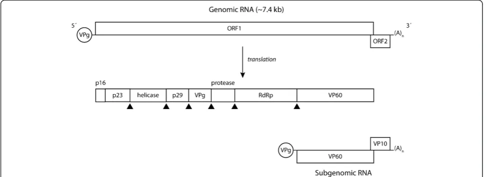
RHDV virions contain the genomic RNA (gRNA) and an additional RNA species with 2.2 kb designated subge-nomic RNA (sgRNA), which is collinear with the 3’end of the genomic RNA [26,57]. Subgenomic RNA usually contributes to the production of high levels of products required during the intermediate and late stages of infection (e.g. structural proteins) [58]. For RHDV, these comprise the capsid protein and VP10 [59-61]. Both the genomic and subgenomic RNA are polyadenylated at the 3’ end and at their 5’ region they are covalently linked through a Tyr-21 residue to the VPg (virus gen-ome-linked) protein [62]. The genomic RNA consists of a positive-sense single-stranded molecule of 7437 nucleotides consisting of two slightly overlapping open reading frames (ORF): ORF1, comprising nucleotides 10 to 7044 and ORF2, comprising nucleotides 7025 to 7378 [26]. ORF1 encodes a large polyprotein of ca. 257 kDa [26] which is cleaved into the mature non-structural proteins and a major structural protein, the capsid pro-tein, by post-translational proteolytic processing by a virus-encoded trypsin-like cysteine protease (Figure 1) [57,63-65]. Some of these proteins derive from larger precursors that result from further post-translational modifications of the precursor proteins [57,64]. The bio-logical role of some of the non-structural proteins encoded by the genome of caliciviruses has been eluci-dated by relying on previous knowledge gathered from
members of the closely related Picornaviridae family [26,64,65]. For RHDV, two proteins involved in the replication of the viral RNA, a helicase and an RNA-dependent RNA polymerase (RdRp), and a protease responsible for the proteolytic processing of the large polyprotein, have been characterised [63,66,67]. RdRp has been shown to also catalyse VPg uridylation [62,68] while a role in translation has been suggested for VPg [e.g. [69]]. The function of the RHDV non-structural proteins p16, p23 and p29 remains to be assessed. VP10, a minor structural protein encoded by the 3’end of gRNA and sgRNA in a different reading frame (ORF2), was recently shown to increase the levels of virus replication and to promote apoptosis [70]. In addi-tion, its ability to downregulate the expression of VP60 was demonstrated [71]. Together, this suggests that VP10 might regulate virus replication and virion release from infected host cells [71].
3. Clinical signs and histopathological lesions The incubation period of the disease ranges between 1 to 3 days and rabbits usually succumb within 12 h to 36 h after the onset of fever (> 40°C). Depending on the clinical evolution of the disease, three different clinical courses can occur [38,72]. In the peracute form, infected animals show no clinical signs and die suddenly. Acute infections are accompanied by anorexia, apathy and con-gestion of the palpebral conjunctiva and neurologic symptoms such as opisthotonos, excitement, paralysis and ataxia may also be observed. There are occasionally some respiratory signs (tracheitis, dyspnea and cyanosis) and a foamy and bloody nasal discharge; lacrimation, ocular haemorrhages and epistaxis can also occur. Suba-cute forms of the disease present similar, but milder clinical symptoms and most rabbits survive. Rabbits experiencing subacute infections develop antibodies against RHDV which confer protection upon re-infec-tion [73]. In addire-infec-tion, it has been reported that during an outbreak of RHD, a low percentage of rabbits may experience a chronic form of the disease with symptoms including a severe and generalised jaundice, anorexia and lethargy [35]. These animals tend to die 1-2 weeks later [54], but animals that overcome the disease present a potent seroconversion [35]. Interestingly, this form of the disease has been shown to be associated with the presence of RHDV core-like particles [35,55].
The liver, lung and spleen are the primary target tis-sues of RHDV. The major histopathological lesions found at necropsy are acute hepatitis due to liver cell loss as the result of RHDV-induced apoptosis, and sple-nomegaly [74,75]. Haemorrhages and congestions can be seen in several organs, particularly in the lungs, heart and kidneys, as a result of a massive disseminated intra-vascular coagulation (DIC) which is usually the cause of
death [76]. Depletion of both B and T lymphocytes in the liver and the spleen accompanies the disease and accounts for an impairment of the immune response [72,77] and a fatal progression of the disease within 2-3 days. In contrast, resistant rabbits develop high titres of IgM (and then of IgA and of IgG) already at day 3 pi, thus presenting an effective humoral immune response [54]. Table 1 presents a summary of the histopathologi-cal alterations that can be observed upon RHDV infection.
4. Epidemiology
The possible routes for transmission of the disease are the oral, nasal, conjunctival and parenteral, as blood-feeding insects have also been shown to be efficient mechanical vectors [72,78]. Transmission of RHDV may occur through direct contact with an infected animal, since infected rabbits may shed viral particles in their secretions and excretions [79], or indirectly by means of fomites-contaminated food, bedding, water, clothing, cages and equipment [19]-or vector-borne transmission
Table 1 Pathological and histopathological lesions [16,77,222,241,243]
Organ Lesions
Liver Enlarged with marked lobular pattern, yellow-grey colour, brittle, circumscribed infiltration with granulocytes, degenerative alterations of hepatocytes compatible with apoptosis (extensive vacuolization, severe alterations in the
mitochondrial structure, karyopyknosis and karyolysis) activation of Kupffer cells, leukopenia
Trachea Hyperaemia of mucous membrane, petechial or diffuse haemorrhages, may be filled with bloody foam
Lung Hyperaemia, pulmonary oedema, intra-alveolar and perivascular haemorrhages, sometimes slight catarrhal bronchiolitis, proliferation of lymphocytes
Kidneys Enlargement with spotted dark red coloration, hyperaemia, haemorrhages within glomerular loops and renal medulla, hyaline thrombi, dilated tubuli, lymphocytic infiltration, degeneration of tubular epithelium
Spleen Enlargement (splenomegaly), spotted dark red colorations, hyperaemia, occasionally karyorrhexis within follicles, hemosiderosis, leukopenia
Digestive tract Contents usually normal, occasional enteritis, subserous haemorrhages
Chest and abdominal cavity
Small amounts of serous, occasionally bloody exudate, sometimes subserous haemorrhages
Muscles Anaemia in the area of the thighs, petechiae in the heart muscle, focal necrosis in myocardium, degenerative alterations, hemosiderosis
Central Nervous System
Congestion of cortical vessels, dilated vessels in the area of the pia mater of the cortex and cerebellum, hyperaemia, small haemorrhages in the cortex, occasionally non-purulent
by scavenging mammals, birds and insects [e.g. [78,80,81]]. The natural doors for viral entry have been suggested to be located in the upper respiratory and digestive tract [16,82]. In natural infections, the faecal-oral route is considered the preferential mode of trans-mission [10,11].
In the field, carcasses of RHDV-infected rabbits may be a major source for viral spreading since the virus seems to be highly resistant and stable when exposed to harsh environmental conditions. Indeed, carcasses of RHDV-infected rabbits exposed to environmental condi-tions have been found to contain viable viral particles for up to three months [83,84]. This ability is para-mount for the epidemiology of RHD and supports the importance of indirect routes in transmission. Environ-mental factors have also been suggested to impact on the effectiveness of RHD in rabbit populations [reviewed in [20]]. Temperature and humidity seem to be the most important climate variables. Indeed, in Australia, mortality rates due to RHD are higher in arid and semi-arid inland areas than in moist coastal regions experien-cing milder temperatures and the disease becomes active during the breeding season, peaks in early spring and is absent in the summer [85]. Climate variables might con-tribute to the geographic and seasonality observed for the RHDV outbreaks by affecting the abundance and activity of the vectors involved in RHDV transmission [reviewed in [20]]. Other non-climatic factors have also been suggested to contribute to the variable pattern of the impact of RHD in rabbit populations such as the timing of the breeding season, the presence of a related and protective RHDV-like calicivirus in rabbit popula-tions or the negative interaction of the myxomatosis outbreaks in the populations [reviewed in [86]]. In addi-tion, modelling studies indicated that population dynamics and spatial structure may greatly influence disease impact and host-virus co-evolution [87,88].
Caliciviruses occur in a wide range of animals apart from rabbits, which include mustelids (minks and skunks), reptiles, cattle, felids (cats and cheetahs), dogs, humans, chimpanzees, pigs and sea mammals (sea lions, seals, walrus, whales, and dolphins), but they are usually restricted to their primary host and closely related spe-cies [89]. Indeed, rabbits and hares are the only hosts for the RHDV and EBHSV lagoviruses, respectively. Other leporid species have been shown not to be sus-ceptible to RHDV [11]. Additionally, several non-host species from the Australian fauna, including domestic, feral animals and wildlife, were assessed for susceptibil-ity to RHDV. No viral replication could be detected with an extensive panel of tests which included clinical observations, pathology, electron microscopy, virology and serology, reinforcing the idea that susceptibility to RHDV is restricted to the European rabbit (Oryctolagus
cuniculus) [90]. Both subspecies of the European rabbit,
O. c. cuniculusandO. c. algirus, seem equally suscepti-ble to RHDV [91]. Interestingly, antibodies against RHDV had been found in animals that live in sympatry with rabbit populations infected with RHDV [81,92,93] and, more recently, RHDV RNA was isolated from sym-patric wild micromammals opening the possibility of other species being involved in the epidemiology of the disease [94].
5. Virus life cycle
In adult rabbits, the targets of the initial stages of the virus life cycle have been determined. Indeed, viral anti-gens are detected in the liver within the first hours fol-lowing infection with RHDV with viral replication occurring in the cytoplasm of hepatocytes located mostly in centriacinar areas [30,74,95-98]. The number of infected hepatocytes clearly increases in the course of the disease, reaching a maximum between 36-48 h [95-97]. Detection of viral antigens in Kupfer cells has also been reported [74,98] associated with viral replica-tion [98]. Extrahepatic presence of the virus has also been observed, but some discrepancies exist between the different studies since different techniques had been employed. Nevertheless, viral antigens have been detected in the spleen, in particular in the macrophages located in the red pulp [96-98], kidney [96], and alveolar macrophages in the lungs. It has been suggested that the presence of replicating virus in alveolar macro-phages, which are in contact with the bloodstream, might be important for initial virus dissemination, and later when the virus reaches the liver, Kupfer cells may be important for spreading the infection into other organs [98].
In contrast, viral dissemination in young resistant rab-bits is far unclear. Viral antigens have been detected in hepatocytes from experimentally-infected 2-week old rabbits [99], but most studies were only able to detect them in rabbits older than 4-weeks [96,100]. Viral anti-gens were found to be scattered and present in only a small percentage of cells. Nevertheless, this suggests that some hepatocytes in young (resistant) rabbits are able to support viral replication, but that major changes must occur in the liver to support a full infectious process. In addition, clearance of the virus seems to be extremely rapid as no viral antigens were detected after day 4 pi [100]. The presence of the virus in other organs has not been fully assessed.
expressed in insect cells. These particles have the advan-tage of being morphologically and antigenically indistin-guishable from native virions, despite being devoid of viral RNA [59,101-105]. RHDV VLP strongly aggluti-nated human adult erythrocytes as the result of binding to glycolipid ligands present on the erythrocyte surfaces [72]. These ligands must develop with age since VLP did not agglutinate erythrocytes from human umbilical cords or foetuses presenting no agglutination [106]. Sub-sequent studies revealed that several caliciviruses use the carbohydrate moiety of host-cell histo-blood group anti-gens (HBGA) for attachment (e.g. ABH/O and Lewis antigens) initiating their replication cycle (Figure 2) [82,107-115]. Histo-blood group antigens are complex glycans either attached to proteins or lipids present on the surface of epithelial cells and erythrocytes, either as
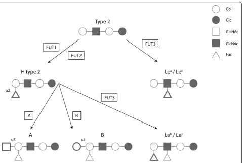
free oligosaccharides in biological fluids (milk, saliva, blood and intestinal contents). HBGA are formed by the sequential addition of monosaccharides to an oligosac-charide precursor chain attached to the cell glycans. This process, designated glycosylation, is catalysed by glycosyltransferase enzymes with specific substrate affi-nity and by a defined linkage [116]. Several genes encode the glycosyltransferases resulting in ABO, Lewis and secretor polymorphic phenotypes (Figure 3).
RHDV was shown to bind to the HBGA H type 2, A type 2 and B type 2 oligosaccharides [82,117]. These structures were shown to be present on the surface of the epithelial cells of the upper respiratory and digestive tracts that the virus first encounters when infecting the host and therefore where doors for virus entry are most likely located [16,82]. Synthesis of H type 2 requires the
addition of a fucose residue, the minimal structural epi-tope [118], in a1,2 linkage to a precursor. This reaction is catalysed by ana1,2-fucosyltransferase which in rab-bits is encoded by three functional genes, Fut1, Fut2
and Sec1 that have undergone multiple events of gene conversion during evolution [119]. Synthesis of the A and B type 2 antigens involves the addition of either a N-acetylgalactosamine or a galactose residue in a1,3 linkage to the H type 2 trisaccharide in a reaction cata-lysed by ana1,3-N-acetylgalactosaminyltransferase or an
a1,3-galactosyltransferase (A or B transferases), respec-tively. In rabbits, theABOblood group locus is largely unresolved, but preliminary data suggest that at least 6
Abogenes exist in the genome, arranged in tandem (K Nyström and J Le Pendu, personal communication).
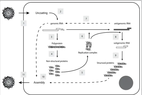
Following the attachment of RHDV to the cell surface, internalisation, by an unknown mechanism, and desen-capsidation occur, leading to the release of the viral gen-ome into the cytoplasm. The virus life cycle then proceeds to the translation of the polyprotein precursor encoded by the ORF1 of the viral genome through
interaction with the host cellular machinery. The gRNA and the sgRNA covalently-linked VPg uses the cellular translation machinery, positioning the ribosome at the initiation codon AUG without ribosome scanning and initiating translation [69,120]. Post-translational proteoly-tic processing by the viral gRNA encoded protease cleaves the polyprotein precursor into the mature non-structural proteins and, in RHDV, into the capsid protein VP60 [57]. The non-structural proteins, helicase and RdRp, then form a replication complex synthesising a complementary negative-sense RNA from the genomic RNA which is used as a template for the synthesis of gRNA and the sgRNA [reviewed in [121]]. The resulting RNA can either be de novo translated or packaged into viral particles that will be released from the infected cell. The mechanism used by RHDV for dissemination of the viral progeny is still unclear, but the ability of VP10 to induce apoptosis may suggest a role in programmed cell death in virion release and dissemination [70,95].
Translation of the RHDV ORF2 produces VP10 through a unique mechanism of reinitiation after
Figure 3Schematic biosynthesis of the HBGA ABH and Lewis. Several transferase enzymes (boxed) are involved in the addition of relevant monosaccharides (in bold) to synthesise the ABH and Lewis ligands in a variety of tissues. Gal, Galactose; Glc, Glucose; GalNAc,
termination of translation of the preceding major capsid protein [122]. This mechanism, although not fully understood, is dependent on the RNA sequence located upstream of the start/stop site, designated TURBS (ter-mination upstream ribosomal binding site), but it is independent on the presence of the AUG initiation codon. Two critical motifs for VP10 expression have been identified within TURBS: motif 1, which is highly conserved among caliciviruses and shows complemen-tarity with a short sequence in the 18S rRNA thus sug-gesting an interaction between the viral RNA and the ribosomal 18S rRNA, and motif 2, which is believed to be involved in the correct positioning of the ribosome at the translational start site [122,123].
6. Mechanisms of resistance to RHD
The lack of a cell culture system has been hampering the study of RHDV pathogenesis and, as a consequence, the mechanisms of resistance to the disease. The indir-ect strategies that have been employed for the study of the pathogenesis of RHDV allowed the identification of the HBGA H type 2 as an attachment factor for RHDV [82]. In humans, identification of alleles at the ABO, FUT2 and FUT3 loci that generate failure to express antigens recognised by different human Norovirus
strains and that confer resistance to infection led to the search of such alleles in the a1,2-fucosyltransferase genes in adult rabbits [124]. This represented the first study on the genetic mechanisms underlying resistance to RHDV. A link between a rabbit allele at the a 1,2-fucosyltransferase geneSec1, that also intervenes in the H type 2 synthesis, and survival to a devastating RHDV outbreak was demonstrated [124]. This Sec1 allele encoded a weakly functionala1,2-fucosyltransferase, but was always found associated withFut2alleles coding for active enzymes that could compensate for the inability of Sec1 to synthesise H type 2. The authors hypothe-sised that thisSec1allele was probably associated with a mutation located in the regulatory region ofFut2which had compromised the Fut2 enzymes and, therefore, the synthesis of the virus’ligand. This result suggests that allelic variation in the a1,2-fucosyltransferase gene appears to have a significant role in resistance to RHDV. More recently, experimental challenge experi-ments indicated that at low virus titres, adult rabbits expressing low amounts of the HBGA ligands were less susceptible to the disease than animals expressing high amounts, although all animals were infected [117].
One striking characteristic of the pathogenesis is that of resistance of young rabbits less than 2 months of age to RHD [72]. Indeed, kittens less than 3 weeks old are fully resistant, but when infected at an age of 4 weeks or older the mortality rates increase to reach, at about 9 weeks old, the rates observed for adult individuals [10].
Thus, the mechanisms of resistance to RHDV have also been studied in light of the differences observed between adult and young rabbits. Interestingly, in young-resistant rabbits the attachment factor H type 2 has been shown to be weakly expressed on the epithelial cells of the upper respiratory and digestive tracts, where primary infection by the virus is believed to occur [82,106], which could explain their resistance to infec-tion. Nevertheless, the reasons behind this differential expression have not yet been disclosed and the picture seems to be far more complicated. Indeed, and despite compelling evidence that supports a role of carbohy-drates in facilitating infection by RHDV in epithelial cells of the upper respiratory and digestive tracts, other attachment factors or receptors must be playing a role at the epithelial level since low expression of the carbo-hydrate receptor at the doors of entry confers only par-tial protection against infection [117]. In addition, hepatocytes, the main cellular target for viral replication, have been shown not to express HBGA [82] and, in young rabbits, infection is accompanied by hepatic lesions due to virus replication as in adult individuals, although they tend to be more severe in 4 week old than in 2 week old rabbits [96,99,100,125,126]. This indicates the existence of at least additional hepatic cel-lular receptor(s) and that the genetic basis for the resis-tance mechanisms goes beyond the attachment of the virus to host cells through histo-blood group antigens. Immune response related-genes, either of the innate or the adaptive responses, are obvious candidate genes to be involved in the resistance mechanisms to RHDV and should deserve attention in future studies.
Differences in the innate immune response between RHDV-infected adult and young rabbits have also been observed [96,99,125-127]. Heterophils seem to be the predominant type of leukocyte in the liver inflammatory infiltrates in adult rabbits and are in close proximity with damaged hepatocytes probably being involved in the clearance of the dead cells. At variance, in young rabbits, this infiltrate is composed mostly by lympho-cytes associated with undamaged and possibly antigen-presenting hepatocytes [127] and which are likely to mount a more effective and specific immune response than heterophils. In addition, in young rabbits only a small fraction of hepatocytes supports viral replication indicating that structural and functional changes have to occur in the liver to support RHDV replication [10,96,126].
to RHDV where shown to present high levels of sero-conversion [35,72,128,129]. A correlation between cELISA titres and protection has been established which is important for determination of the applicability of vac-cination and to assess the current status of the disease [54,128,130,131]. Recovering rabbits present IgM titres that quickly reach a maximum within 2 weeks and then sharply decrease. IgA titres are more prolonged in time, but they also face a decrease. In contrast, IgG slowly increase and are able to persist for months. With regards to IgA, this suggests a mucosal response [128]. Passive immunization (serotherapy) was also shown to be effec-tive in stopping RHD in a rabbit farm hit by an outbreak [132], demonstrating the importance of humoral immu-nity in protection against RHD. In young rabbits, resis-tance has also been associated with the presence of maternal antibodies which are maintained during the period of life when they are considered RHD-resistant [128]. These are exclusively IgG acquired through the placenta in the last days of pregnancy and show a decline with age and body weight [19]. Additionally, if young rab-bits are infected in their early life, they will become resis-tant when adult, suggesting that their immune system is capable of recognising the virus and producing an effec-tive immune response that will confer long-term protec-tion [133]. Therefore, humoral immunity clearly provides protection against RHDV when present [28,103].
7. Genetic diversity/RHDV evolution
7.1. Pathogenic RHDV
The origin and evolution of RHDV are not well under-stood. Although first reported in China in Angora rab-bits imported from Germany, it was not clear if rabrab-bits were already infected with RHDV when they arrived in China, since the disease might have been previously observed in Germany [73], or if they became infected later in China. The idea of RHDV being of Chinese ori-gin has been challenged by several studies [50,134-136]. Indeed, these studies have shown that the pathogenic form of RHDV originated before 1984 [50,134,136] and that the Chinese strain isolated in 1984 had its origin in European isolates [135]. In light of these results, it seems that RHDV had its origins in Europe and that it had been circulating for some time, but that mortalities went unnoticed. Some hypotheses have been put for-ward regarding how RHDV originated. One of them, the transmission of the European brown hare syndrome virus to the European rabbit [137] has been discarded since EBHSV does not infect European rabbits. Other hypotheses propose that a virus from another species jumped to the rabbit where it became pathogenic [138], but the presently favoured hypothesis would be the change of a non-pathogenic virus closely related to RHDV and that rendered it pathogenic (see below).
Identifying novel features in the genome of RHDV might give some indications on the origin of the virus and its virulence. The first complete genome sequence of RHDV was obtained in 1991 by Meyers et al. [26]. The characterisation of the genetic diversity was initiated by sequencing and comparing partial sequences of a few European isolates [139-141]. The isolates were found to be highly similar and closely related.
Later, in an attempt to characterise the relation between EBHSV and RHDV, Wirblich et al. presented evidence that the N terminal portion of the capsid was highly conserved while the highest degree of variability was located in the C terminal half [39]. Indeed, while for the N-terminus homology between caliciviruses is ~80%, no strict conservation was observed for the C-ter-minus. In RHDV, this highly variable portion seems to correspond to the C and E domains as defined by Neill [47], where the majority of the differences between cali-civirus isolates have been detected and where the main antigenic determinants have been found to be located [43,44,47,142-144]. These domains were predicted to be located at the capsid surface [46] and therefore more variable as a result of the strong selective pressure due to exposure to the host immune system.
early as 1985 in China, where they might have emerged [13,136,146-150]. In some areas, these variants seem to be replacing the original strains [148].
In the Iberian Peninsula, only G1 strains have been found, even among contemporary strains [91,134]. This differs with the general pattern of RHDV evolution observed in other European countries, but, overall, G1 strains seem to evolve following a temporal rather than a geographic pattern as observed in the other gen-ogroups [91]. This temporal structure is common in RNA viruses and likely results from the strong selection imposed by adaptive immune recognition by the host [151]. This was confirmed for RHDV, where a few posi-tively selected codons were detected located in the major antigenic determinants of the capsid [49,50,134]. Interestingly, all the positively selected codons were associated with potential N-glycosylation sites. Glycosy-lation is known to play a role in the infectious process in other non-enveloped viruses such as rotavirus and Hepatitis E virus [152-155]. Positive selection recorded at N-glycosylation sites in the capsid protein of RHDV indicates that glycans might influence viral pathogeni-city. This is further supported by the finding of non-pathogenic RHDV-like strains which do lack some of
the positively selected N-glycosylation sites [49] although it is not known at present if the capsid protein from authentic virions is actually glycosylated.
Overall, with the exception of the RHDVa isolates, the evolution of RHDV is associated with a high degree of genetic homogeneity, with maximum nucleotide and amino acid differences of 10% and 6%, respectively [14,90,91,137,139-141,145,147,150,156-163], and mostly located in the regions C and E. These differences are much lower than those observed for other caliciviruses (e.g. forNorovirus, amino acid differences can reach a maximum of 61.4% while forSapovirus they can reach 55%) [164,165]. This high homology may have resulted from the rapid spread of a new virus, expanding into a susceptible host population [139], but also from the fact that RHDV is a newly emerging pathogen whose evolu-tion started recently, at variance from that ofNorovirus
andSapovirus.
7.2. Non-pathogenic rabbit calicivirus
The emergence of RHDV from a pre-existing non-pathogenic rabbit calicivirus that has mutated and become pathogenic to rabbits has been hypothesised [136,139,160,166-168]. The detection of antibodies spe-cific to RHDV in rabbit sera collected before the first RHDV outbreak, the identification of RHDV-seroposi-tive rabbits where RHD was never recorded and the pre-sence or persistence of viral RNA in populations where no overt signs of disease could be observed, provided compelling evidence for the pre-existence of a non-pathogenic RHVD-like virus in European rabbit popula-tions [29,128,160,169-179]. This non-pathogenic virus would share antigenic properties with RHDV and circu-lated asymptomatically amongst rabbit populations before the first RHDV outbreak in China. Isolation of several non-pathogenic rabbit caliciviruses related to RHDV but with a tropism limited to the gut and no obvious pathogenicity further substantiated this hypoth-esis [160,166,168,176,180,181] and showed that the non-pathogenic strains are still circulating. Protection to RHD had been shown to be conferred by some of these non-pathogenic strains which might provide an explana-tion for the low level of RHD incidence in some regions of Australia and Britain [166,168,169,171,173,179, 182-184]. More recently this hypothesis was confirmed experimentally when Strive et al. showed that the non-pathogenic Australian strain RCV-A1 is able to generate an antibody response that cross-reacts to RHDV and further protects animals from RHD, but not completely and not from infection [184]. Other strains, however, do not confer any kind of protection [181,185].
Understanding the evolutionary history and the origin of RHDV will benefit from further studies of the non-pathogenic strains. The isolation of non-non-pathogenic
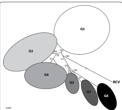
Figure 4 Phylogenetic relationships between the RHDV genogroups G1-G6 and the Italian non-pathogenic strain RCV. The tree was obtained using the neighbour-joining method and using nucleotide sequences from RHDV strains isolated worldwide. Bootstrap values greater than 50% are presented at the nodes. RCV was used as outgroup to root the tree. With kind permission from Springer Science + Business Media: Archives of Virology,
strains has so far already revealed striking differences between these strains and the pathogenic RHDV strains, with the most important being, beside pathogenicity, tis-sue tropism and capsid variability [160,166,168,176,180]. Interestingly, MRCV, a new variant of the non-patho-genic rabbit calicivirus (RCV)-like group that was recently characterised [180,186], displayed a pathogeni-city of approximately 30% which is significant and in contrast with reports from other non-pathogenic strains. The viral RNA of this strain was detected in the liver rather than in the intestine which seems to indicate that the ability of a strain to cross the epithelia barrier and reach other organs such as the liver is an important fea-ture for the emergence of the pathogenic forms and should be further explored. However, these results should be considered with caution since experimental reproduction of the disease failed, with only 2 out of the 14 inoculated rabbits presenting very mild symptoms.
Despite the accumulation of data, no mutations have yet been attributed that would explain the switch from a non-pathogenic to a pathogenic form. Recombination within the RHDV genome is not uncommon [187,188] and might have played a role in the origin of RHDV. Indeed, the recombination event reported by Forrester et al. [188] in the strain isolated in 1984 in China may suggest that recombination was a common mechanism at the time of emergence of the pathogenic forms. Nevertheless, not all pathogenic forms are direct descen-dants of this strain [135] and despite the relatively high frequency at which recombination occurs (4 out of 10), it does not seem to be widespread in all RHDV lineages [136]. This might be the result of an incomplete and non-systematic sampling or of the scarcity of complete genomic sequences.
Recently, novel phylogenetic analysis approaches were used to assess the emergence of RHDV [50,134,136]. Although the results could have brought an insight into the timing of the appearance of the non-pathogenic and the pathogenic forms of the virus, the studies led to incongruent results most likely as the result of the dif-ferent capsid fragment lengths used in each study. This poses the question if the capsid is indeed the best gene for inferring RHDV history. While Kerr et al. could establish the Time to Most Recent Common Ancestor (TMRCA) between RHDV and the non-pathogenic forms of < 550 years and of < 150 years for the patho-genic forms [136], Kinnear and Linde set the existence of the ancestor of RHDV-RCV later in the 1930s and of the ancestor of the pathogenic RHDV strains between 1957-1976 [50]. Consistent with the findings of Kerr et al., Alda et al. set the TMRCA for all the RHDV to ~1884 [134]. Nevertheless, and considering these esti-mates, it is surprising that the disease had not been reported earlier than 1984. This might suggest an
alternative scenario for the virus emergence where the virus would have come from another species through a species jump, that would have acted as a reservoir and was not affected by RHDV [136]. This scenario, how-ever, implies the existence of such a reservoir host in which the virus was able to replicate. Viral RNA has been recently detected and isolated in micromammals living in sympatry with European rabbit populations that could represent the unidentified reservoir [94], but viral replication within these species could not be con-firmed. Since these species might be important for virus transmission and spread and perhaps represent the
“unknown” reservoir for when the virus seems to be inactive, i.e. between outbreaks, this hypothesis should be further explored.
8. Host-virus co-evolution
The virus-host dynamics result in a co-evolutionary pro-cess between the host resistance mechanisms and the virus escape mechanisms with attenuation of the virus and/or increase in resistance of the host. Therefore, the study of the host-virus co-evolutionary processes requires the analysis of each element of this dynamical pair simultaneously. Regarding the host, and in order to identify signatures of selection due to infectious agents, the natural history of this species should be considered. The fossil record suggests that the European rabbit ori-ginated in the Iberian Peninsula during the medium Pleistocene [189-192] and two morphologically differen-tiated subspecies have been distinguished:O. cuniculus algirus and O. cuniculus cuniculus[193]. O. c. algirus
inhabits the southwestern Iberian Peninsula, whileO. c. cuniculus is present in the northeastern Iberian Penin-sula. These two subspecies diverged ~1.8 Mya [reviewed in [194]] and then, by a post-glaciation expansion from the southwestern refugium to North and from the Northeastern to South or West, a contact zone was established. While the natural populations of O. c. algirus remained confined to the southwest of the Peninsula, the natural populations of O. c. cuniculus
later expanded its range north towards France, likely after the last glacial peak [195], where they still remain present. The expansion of these populations with suc-cessive bottleneck events caused a significantly lower genetic diversity of the wild French O. c. cuniculus
populations compared to the Iberian populations [195-201]. The European rabbit gene pool has been manipulated by man through a recent single domestica-tion event of French origin, and therefore, all domestic rabbits belong to the subspecies O. c. cuniculus
populations worldwide have been shaped differently by these events and this might have also interfered with the resistance mechanisms. Therefore, when studying the co-evolution between the European rabbits and RHDV, these events should be taken into account.
The introduction of myxoma virus in Australia was soon followed by an increase in genetic resistance in rabbits and the appearance of less virulent strains [203]. Recent field studies conducted in Australia suggest that RHDV also has become less effective in keeping wild rabbit numbers low and that in some populations rabbit numbers are returning to the pre-RHDV levels [204]. Laboratory challenges have confirmed this scenario when inoculation of rabbits from different Australian populations with the original introduced strain (Czech strain V351) failed to induce mortality or induced mor-tality rates lower than that which was expected [205]. In addition, in an Australian population, the frequency of host resistant phenotypes, i.e., phenotypes that confer a weak binding of the virus to the host HBGA that facili-tate infection and thus provide protection to the host, has significantly increased [117]. A similar co-adaptation process seems to be occurring both in New Zealand [92] and in rabbit populations from Europe [117]. Indeed, Nyström et al. found that in a French wild rab-bit population recovering from a major RHDV outbreak the frequency of resistant phenotypes increased among the survivors [117]. As with the Australian population, the resistant phenotypes are associated with weak viral binding. This indicates that the virus has contributed to select resistant hosts in accordance to the binding speci-ficities of the circulating RHDV strains and gives further support for a role of the HBGA in the virus epidemiol-ogy and suggests that the virus is shaping the hosts’ HBGA diversity.
The virus also seems to be evolving to overcome the host resistance mechanisms since significant mortalities are still observed in the field, at least in Australia. Evi-dence for this comes from the fact that in comparative trials in rabbits known to be resistant to infection with Czech 351, modern field strains appeared more virulent than the original released strain, suggesting they had evolved to keep pace with changes in rabbit resistance [205]. In addition, HBGA specificities of the strains that evolved in France from 1988 to 2009 progressively shifted, allowing preferential recognition of subgroups of animals that express distinct HBGA motifs, which sug-gests an adaptation to the host genetic diversity [117].
9. Prevention, control and vaccination
In animals presenting subclinical or no clinical signs, passively acquired immunity has been shown to act suc-cessfully in emergency situations [132]. Indeed, this therapy, which is achieved by inoculation with a
hyperimmune antiserum, confers short-term protection, preventing death. Nevertheless, passive immunization is ineffective on animals presenting clinical signs. Thus, as yet, no cure is available for RHDV-dying rabbits. Pre-vention and control of the disease through biosecurity and immunoprophylactic measures such as vaccination are, therefore, of utmost importance. Due to the lack of a cell culture system for efficient virus propagation, commercially available vaccines against RHDV are pro-duced from tissue suspensions of experimentally infected rabbits, followed by chemical inactivation of the virus [132,177,206]. However, and to obviate the risks inherent to the manufacturing and use of this kind of vaccines (the use of infectious particles, the need for a safe disposal of contaminant residues, social concerns on animal welfare) the RHDV capsid protein has been tested in various studies as a subunit vaccine against RHD. Several heterologous expression systems or recombinant animal viruses have been developed to pro-duce recombinant versions of the VP60 protein. The VP60 recombinant protein has been produced in Escher-ichia coli[140]; insect cultured cells [59,101-104]; yeast [207,208]; plants [209-213]; insect larvae [214] and recombinant animal-derived viruses [215-219]. Most of these systems were shown to be immunogenic and to confer protection against lethal doses of RHDV by elicit-ing a humoral response indicatelicit-ing that they are good substitutes for the tissue vaccines. Features such as low cost, high yields and ease of scaling up are amongst the most important factors for their commercial viability.
Although commercially available vaccines have proven effective in rabbitries, in wild rabbit populations vaccina-tion campaigns are economically and logistically imprac-ticable and their effects are considered insignificant [220]. Indeed, administration to wild rabbits implies capturing and handling of rabbits which by being a stress factor might increase the mortality rates [221]. In addition, this would need to be performed systematically since induced-immunity lasts no longer than 1 year [222] and efficacy has been shown to be dependent on several physiological parameters of the individuals [223]. Therefore, alternative approaches are being explored to overcome these limitations such as the development of vaccines with the capacity for horizontal transmission to ensure appropriate immunization of a relevant portion of the population [219], vaccines that may be adminis-tered by the oral or nasal routes [105,208,213,217,219, 224,225] or the construction of bivalent vaccines [216,226]. Nevertheless, as yet, none of these vaccines has been registered or is commercially available.
the rabbit industry. In countries where RHDV circulates in wild rabbits and where eradication is not achievable, these measures might prevent large-scale infection in rabbitries. Thus, a careful and correct management of the RHDV outbreaks is always dependent on the epide-miological situation of the regions where they occur. In addition, a continuous monitoring of the viral evolution in the field is fundamental for the quick detection of new genetic and antigenic variants which might be determinant for the application of the most appropriate measures.
10. Therapeutic applications of RHDV
More recently, RHDV VLP have been considered as a mean for cancer and pathogen immunotherapies [227-232]. The capsid protein of RHDV spontaneously assembles into VLP which are morphologically and anti-genically indistinguishable from native virions, but devoid of the viral RNA [101]. By genetic engineering, RHDV VLP have been shown to efficiently incorporate antigens that might be presented to immune cells and to elicit an adequate cell-mediated and humoral immune response [228-232]. In addition, RHDV VLP have the advantage of being easily produced and at low cost, and, by deriving from a non-human virus, they are not sus-ceptible to pre-existing neutralising antibodies [228], thus providing a reliable molecular tool for therapeutic applications.
RHDV has also been investigated for the study of virally-induced acute liver failure (ALF) in humans [233-239] as it fulfils several of the requirements to be a good animal model [233]. ALF is a condition charac-terised by severe liver injury, hepatic encephalopathy, coagulopathy and multiorgan failure, with viral infec-tions (e.g. hepatitis A, B and E) and drug use (e.g. para-cetamol overdose) amongst the commonest causes [240]. The hepatic lesions observed following infection by RHDV [99] resemble those caused by ALF in humans, but other physiologic, histological and bio-chemical alterations are also shared. Indeed, the hemo-dynamic changes, alterations in the intracranial pressure and histological alterations such as apoptosis observed in RHDV are common to ALF [74,233,241,242]. In addi-tion, clinical symptoms such as prostration or convul-sions observed on RHD are also observed in ALF [38]. RHDV has also been used for the study of therapeutic approaches for ALF [236,238,239].
11. Conclusions
Despite the lack of an appropriate cell culture system, some light has been shed on several aspects of RHDV and RHD. Nevertheless, the host-parasite interactions established between RHDV and the European rabbit are still unclear. Indeed, the role of some of the proteins
encoded by RHDV is still unknown and the emergence of RHDV as a pathogenic form has not yet been resolved. In order to clarify these gaps, an effort should be made in obtaining full genomic sequences, including for non-pathogenic strains as these might contribute to understand the pathogenesis of RHDV. As for the host, and in particular as for key factors of susceptibility and resistance, the rabbit genome project should be consid-ered and used for the study of candidate genes. By using temporal samples, i.e., samples of rabbits collected before and after RHDV outbreaks, one might determine those candidate genes. Further studies on immunity to RHDV and on the related non-pathogenic viruses are also warranted for a better understanding of the host-pathogen relationships. In addition, the study of the clo-sely related EBHSV and its host, might contribute to the understanding of the interplay between lagoviruses and leporid species. The possibility that RHDV might be used as a model for the study of other calicivirus infec-tions, in particular in view of its non-pathogenicity for humans, as well as for the development of anti-cancer and pathogen therapies transforms it into a valuable research molecular tool.
12. List of abbreviations
RHDV: rabbit haemorrhagic disease virus; RHD: rabbit haemorrhagic disease; EBHSV: European brown hare syndrome virus; EBHS: European brown hare syndrome; gRNA: genomic RNA; sgRNA: subgenomic RNA; VPg: virus genome-linked protein; ORF: open reading frame; RdRp: RNA-dependent RNA polymerase; DIC: dissemi-nated intravascular coagulation; pi: post-infection; VLP: virus-like particles; HBGA: histo-blood group antigens; TURBS: termination upstream ribosomal binding site; RCV: rabbit calicivirus; TMRCA: Time to Most Recent Common Ancestor; ALF: acute liver failure.
Acknowledgements
The Portuguese Foundation for Science and Technology supported the post-doctoral fellowships of JA (SFRH/BPD/73512/2010) and PJE (SPRH/BPD/ 27021/2006). JLP was supported by a grant from the Région des Pays de la Loire (Calilago).
Author details
1CIBIO/UP, Centro de Investigacao em Biodiversidade e Recursos Geneticos/
Universidade do Porto, Campus Agrario de Vairao, 4485-661 Vairao, Portugal.
2
INSERM, U892, Université de Nantes, 44007 Nantes, France.3CITS, Centro de Investigacao em Tecnologias de Saude, CESPU, Gandra, Portugal.
Authors’contributions
JA performed a study on the available literature on the subject, analysed the retrieved information and wrote the manuscript. WvdL, JLP and PJE revised the manuscript critically according to their own area of expertise. All authors read and approved the final manuscript.
Competing interests
Received: 20 July 2011 Accepted: 10 February 2012 Published: 10 February 2012
References
1. Liu SJ, Xue HP, Pu BQ, Qian NH:A new viral disease in rabbit.Anim Husb Vet Med1984,16:253-255.
2. Xu WY:Viral haemorrhagic disease of rabbits in the People’s Republic of China: epidemiology and virus characterisation.Rev Sci Tech1991,
10:393-408.
3. Park NY, Chong CY, Kim JH, Cho SM, Cha YH, Jung BT, Kim DS, Yoon JB:An outbreak of viral haemorrhagic pneumonia (tentative name) of rabbits in Korea.J Korean Vet Med Assoc1987,23:603-610.
4. Cancellotti FM, Renzi M:Epidemiology and current situation of viral haemorrhagic disease of rabbits and the European brown hare syndrome in Italy.Rev Sci Tech1991,10:409-422.
5. Delibes-Mateos M, Delibes M, Ferreras P, Villafuerte R:Key role of European rabbits in the conservation of the Western Mediterranean basin hotspot.
Conserv Biol2008,22:1106-1117.
6. Argüello JL, Llanos A, Pérez LI:Enfermedad hemorrágica del conejo en España.Med Vet1988,5:645-650, (in Spanish).
7. Anonymous:Doença hemorrágica a vírus do Coelho em Portugal.Rev Port Ciênc Vet1989,84:57-58, (in Portuguese).
8. Villafuerte R, Calvete C, Blanco JC, Lucientes J:Incidence of viral hemorrhagic disease in wild rabbit populations in Spain.Mammalia
1995,59:651-660.
9. Delibes-Mateos M, Ferreras P, Villafuerte R:Rabbit populations and game management: the situation after 15 years of rabbit haemorrhagic disease in central southern Spain.Biodivers Conserv2008,17:559-574. 10. Morisse JP, Le Gall G, Boilletot E:Hepatitis of viral origin in Leporidae:
introduction and aetiological hypotheses.Rev Sci Tech1991,10:283-295. 11. Gregg DA, House C, Meyer R, Berninger M:Viral haemorrhagic disease of
rabbits in Mexico: epidemiology and viral characterization.Rev Sci Tech
1991,10:435-451.
12. Rabbit calicivirus infection confirmed in Iowa rabbitry. J Am Vet Med Assoc2000,216:1537.
13. Farnos O, Rodriguez D, Valdes O, Chiong M, Parra F, Toledo JR, Fernandez E, Lleonart R, Suarez M:Molecular and antigenic characterization of rabbit hemorrhagic disease virus isolated in Cuba indicates a distinct antigenic subtype.Arch Virol2007,152:1215-1221.
14. Le Gall-Recule G, Zwingelstein F, Laurent S, de Boisseson C, Portejoie Y, Rasschaert D:Phylogenetic analysis of rabbit haemorrhagic disease virus in France between 1993 and 2000, and the characterisation of RHDV antigenic variants.Arch Virol2003,148:65-81.
15. Delibes-Mateos M, Redpath SM, Angulo E, Ferreras P, Villafuerte R:Rabbits as a keystone species in southern Europe.Biol Conserv2007,137:149-156. 16. Mitro S, Krauss H:Rabbit hemorrhagic disease: a review with special
reference to its epizootiology.Eur J Epidemiol1993,9:70-78.
17. Gibb JA, Williams JM:The rabbit in New Zealand.InThe European rabbit: the history and biology of a successful colonizer.Edited by: Corbet GB, Flux JEC, Rogers PM, Arthur CP, Soriguer RC, Myers K, Parer I, Wood D, Cooke BD, Gibb JA Williams JM, Fenner F, Ris J, Thompson HV, King CM. Oxford: Oxford University Press; 1994:158-200.
18. Fenner F:Deliberate introduction of the European rabbit, Oryctolagus cuniculus, into Australia.Rev Sci Tech2010,29:103-111.
19. Cooke BD:Rabbit haemorrhagic disease: field epidemiology and the management of wild rabbit populations.Rev Sci Tech2002,21:347-358. 20. Cooke BD, Fenner F:Rabbit haemorrhagic disease and the biological
control of wild rabbits, Oryctolagus cuniculus, in Australia and New Zealand.Wildl Res2002,29:689-706.
21. Mutze G, Cooke B, Alexander P:The initial impact of rabbit hemorrhagic disease on European rabbit populations in South Australia.J Wildl Dis
1998,34:221-227.
22. Thompson J, Clark G:Rabbit calicivirus disease now established in New Zealand.Surveillance1997,24:5-6.
23. O’Keefe JS, Tempero J, Atkinson PH, Pacciarini L, Fallacara F, Horner GW, Motha J:Typing of rabbit haemorrhagic disease virus from New Zealand wild rabbits.N Z Vet J1998,46:42-43.
24. An S-H, Kim B-H, Lee JB, Song JU, Park BK, Kwon YB, Jung JS, Lee YS:
Studies on Picornavirus hemorrhagic fever (tentative name) in rabbit. 1. Physico-chemical properties of the casuative virus.Res Rep Rural Dev Adm
1988,30:55-61.
25. Gregg DA, House C:Necrotic hepatitis of rabbits in Mexico: a parvovirus.
Vet Rec1989,125:603-604.
26. Meyers G, Wirblich C, Thiel HJ:Rabbit hemorrhagic disease virus– molecular cloning and nucleotide sequencing of a calicivirus genome.
Virology1991,184:664-676.
27. Ohlinger VF, Haas B, Meyers G, Weiland F, Thiel HJ:Identification and characterization of the virus causing rabbit hemorrhagic disease.J Virol
1990,64:3331-3336.
28. Parra F, Prieto M:Purification and characterization of a calicivirus as the causative agent of a lethal hemorrhagic disease in rabbits.J Virol1990,
64:4013-4015.
29. Rodak L, Smid B, Valicek L, Vesely T, Stepanek J, Hampl J, Jurak E: Enzyme-linked immunosorbent assay of antibodies to rabbit haemorrhagic disease virus and determination of its major structural proteins.J Gen Virol1990,71:1075-1080.
30. Moussa A, Chasey D, Lavazza A, Capucci L, Smid B, Meyers G, Rossi C, Thiel HJ, Vlasak R, Ronsholt L, Nowotny N, McCullough K, Gavier-Widen D:
Haemorrhagic disease of lagomorphs: evidence for a calicivirus.Vet Microbiol1992,33:375-381.
31. Oliver SL, Asobayire E, Dastjerdi AM, Bridger JC:Genomic characterization of the unclassified bovine enteric virus Newbury agent-1 (Newbury1) endorses a new genus in the family Caliciviridae.Virology2006,
350:240-250.
32. Farkas T, Sestak K, Wei C, Jiang X:Characterization of a rhesus monkey calicivirus representing a new genus of Caliciviridae.J Virol2008,
82:5408-5416.
33. L’Homme Y, Sansregret R, Plante-Fortier E, Lamontagne AM, Ouardani M, Lacroix G, Simard C:Genomic characterization of swine caliciviruses representing a new genus of Caliciviridae.Virus Genes2009,39:66-75. 34. Gavier-Widen D, Morner T:Descriptive epizootiological study of European
brown hare syndrome in Sweden.J Wildl Dis1993,29:15-20. 35. Capucci L, Scicluna MT, Lavazza A:Diagnosis of viral haemorrhagic
disease of rabbits and the European brown hare syndrome.Rev Sci Tech
1991,10:347-370.
36. Chasey D, Lucas M, Westcott D, Williams M:European brown hare syndrome in the U.K.; a calicivirus related to but distinct from that of viral haemorrhagic disease in rabbits.Arch Virol1992,124:363-370. 37. Fuchs A, Weissenbock H:Comparative histopathological study of rabbit
haemorrhagic disease (RHD) and European brown hare syndrome (EBHS).J Comp Pathol1992,107:103-113.
38. Marcato PS, Benazzi C, Vecchi G, Galeotti M, Della Salda L, Sarli G, Lucidi P:
Clinical and pathological features of viral haemorrhagic disease of rabbits and the European brown hare syndrome.Rev Sci Tech1991,
10:371-392.
39. Wirblich C, Meyers G, Ohlinger VF, Capucci L, Eskens U, Haas B, Thiel HJ:
European brown hare syndrome virus: relationship to rabbit hemorrhagic disease virus and other caliciviruses.J Virol1994,
68:5164-5173.
40. Lavazza A, Scicluna MT, Capucci L:Susceptibility of hares and rabbits to the European brown hare syndrome virus (EBHSV) and rabbit haemorrhagic disease virus (RHDV) under experimental conditions.
Zentralbl Veterinarmed B1996,43:401-410.
41. Thouvenin E, Laurent S, Madelaine MF, Rasschaert D, Vautherot JF, Hewat EA:Bivalent binding of a neutralising antibody to a calicivirus involves the torsional flexibility of the antibody hinge.J Mol Biol1997,
270:238-246.
42. Valicek L, Smid B, Rodak L, Kudrna J:Electron and immunoelectron microscopy of rabbit haemorrhagic disease virus (RHDV).Arch Virol1990,
112:271-275.
43. Barcena J, Verdaguer N, Roca R, Morales M, Angulo I, Risco C, Carrascosa JL, Torres JM, Caston JR:The coat protein of Rabbit hemorrhagic disease virus contains a molecular switch at the N-terminal region facing the inner surface of the capsid.Virology2004,322:118-134.
44. Capucci L, Frigoli G, Ronshold L, Lavazza A, Brocchi E, Rossi C:Antigenicity of the rabbit hemorrhagic disease virus studied by its reactivity with monoclonal antibodies.Virus Res1995,37:221-238.
45. Hu Z, Tian X, Zhai Y, Xu W, Zheng D, Sun F:Cryo-electron microscopy reconstructions of two types of wild rabbit hemorrhagic disease viruses characterized the structural features of Lagovirus.Protein Cell2010,
46. Prasad BV, Rothnagel R, Jiang X, Estes MK:Three-dimensional structure of baculovirus-expressed Norwalk virus capsids.J Virol1994,68:5117-5125. 47. Neill JD:Nucleotide sequence of the capsid protein gene of two
serotypes of San Miguel sea lion virus: identification of conserved and non-conserved amino acid sequences among calicivirus capsid proteins.
Virus Res1992,24:211-222.
48. Martinez-Torrecuadrada JL, Cortes E, Vela C, Langeveld JP, Meloen RH, Dalsgaard K, Hamilton WD, Casal JI:Antigenic structure of the capsid protein of rabbit haemorrhagic disease virus.J Gen Virol1998,
79:1901-1909.
49. Esteves PJ, Abrantes J, Carneiro M, Muller A, Thompson G, van der Loo W:
Detection of positive selection in the major capsid protein VP60 of the rabbit haemorrhagic disease virus (RHDV).Virus Res2008,137:253-256. 50. Kinnear M, Linde CC:Capsid gene divergence in rabbit hemorrhagic
disease virus.J Gen Virol2010,91:174-181.
51. Bu W, Mamedova A, Tan M, Xia M, Jiang X, Hegde RS:Structural basis for the receptor binding specificity of Norwalk virus.J Virol2008,
82:5340-5347.
52. Cao S, Lou Z, Tan M, Chen Y, Liu Y, Zhang Z, Zhang XC, Jiang X, Li X, Rao Z:Structural basis for the recognition of blood group trisaccharides by norovirus.J Virol2007,81:5949-5957.
53. Choi JM, Hutson AM, Estes MK, Prasad BV:Atomic resolution structural characterization of recognition of histo-blood group antigens by Norwalk virus.Proc Natl Acad Sci USA2008,105:9175-9180. 54. Lavazza A, Capucci L:How Many Caliciviruses are there in Rabbits? A
Review on RHDV and Correlated Viruses Lagomorph Biology.Edited by: Alves PC, Ferrand N, Hackländer K. Springer Berlin Heidelberg; 2008:263-278. 55. Granzow H, Weiland F, Strebelow HG, Liu CM, Schirrmeier H:Rabbit
hemorrhagic disease virus (RHDV): ultrastructure and biochemical studies of typical and core-like particles present in liver homogenates.
Virus Res1996,41:163-172.
56. Alexandrov M, Peshev R, Bozhkov S, Yanchev I, Doumanova L: Electron-and immunoelectron-microscopic investigation on the rabbit haemorrhagic disease virus.Comp Immunol Microbiol Infect Dis1993,
16:21-27.
57. Meyers G, Wirblich C, Thiel HJ, Thumfart JO:Rabbit hemorrhagic disease virus: genome organization and polyprotein processing of a calicivirus studied after transient expression of cDNA constructs.Virology2000,
276:349-363.
58. Miller WA, Koev G:Synthesis of subgenomic RNAs by positive-strand RNA viruses.Virology2000,273:1-8.
59. Sibilia M, Boniotti MB, Angoscini P, Capucci L, Rossi C:Two independent pathways of expression lead to self-assembly of the rabbit hemorrhagic disease virus capsid protein.J Virol1995,69:5812-5815.
60. Boga JA, Marin MS, Casais R, Prieto M, Parra F:In vitro translation of a subgenomic mRNA from purified virions of the Spanish field isolate AST/89 of rabbit hemorrhagic disease virus (RHDV).Virus Res1992,
26:33-40.
61. Neill JD, Reardon IM, Heinrikson RL:Nucleotide sequence and expression of the capsid protein gene of feline calicivirus.J Virol1991,65:5440-5447. 62. Machin A, Martin Alonso JM, Parra F:Identification of the amino acid
residue involved in rabbit hemorrhagic disease virus VPg uridylylation.J Biol Chem2001,276:27787-27792.
63. Boniotti B, Wirblich C, Sibilia M, Meyers G, Thiel HJ, Rossi C:Identification and characterization of a 3C-like protease from rabbit hemorrhagic disease virus, a calicivirus.J Virol1994,68:6487-6495.
64. Konig M, Thiel HJ, Meyers G:Detection of viral proteins after infection of cultured hepatocytes with rabbit hemorrhagic disease virus.J Virol1998,
72:4492-4497.
65. Wirblich C, Thiel HJ, Meyers G:Genetic map of the calicivirus rabbit hemorrhagic disease virus as deduced from in vitro translation studies.J Virol1996,70:7974-7983.
66. López Vázquez A, Martin Alonso JM, Casais R, Boga JA, Parra F:Expression of enzymatically active rabbit hemorrhagic disease virus RNA-dependent RNA polymerase in Escherichia coli.J Virol1998,72:2999-3004.
67. Marin MS, Casais R, Alonso JM, Parra F:ATP binding and ATPase activities associated with recombinant rabbit hemorrhagic disease virus 2C-like polypeptide.J Virol2000,74:10846-10851.
68. Machin A, Martin Alonso JM, Dalton KP, Parra F:Functional differences between precursor and mature forms of the RNA-dependent RNA
polymerase from rabbit hemorrhagic disease virus.J Gen Virol2009,
90:2114-2118.
69. Goodfellow I, Chaudhry Y, Gioldasi I, Gerondopoulos A, Natoni A, Labrie L, Laliberte JF, Roberts L:Calicivirus translation initiation requires an interaction between VPg and eIF 4 E.EMBO Rep2005,6:968-972. 70. Liu G, Ni Z, Yun T, Yu B, Chen L, Zhao W, Hua J, Chen J:A DNA-launched
reverse genetics system for rabbit hemorrhagic disease virus reveals that the VP2 protein is not essential for virus infectivity.J Gen Virol2008,
89:3080-3085.
71. Chen L, Liu G, Ni Z, Yu B, Yun T, Song Y, Hua J, Li S, Chen J:Minor structural protein VP2 in rabbit hemorrhagic disease virus downregulates the expression of the viral capsid protein VP60.J Gen Virol2009,90:2952-2955.
72. Xu ZJ, Chen WX:Viral haemorrhagic disease in rabbits: a review.Vet Res Commun1989,13:205-212.
73. Patton NM:Viral hemorrhagic disease. A major new disease problem of rabbits.Rabbit Res1989,12:64-67.
74. Alonso C, Oviedo JM, Martin-Alonso JM, Diaz E, Boga JA, Parra F:
Programmed cell death in the pathogenesis of rabbit hemorrhagic disease.Arch Virol1998,143:321-332.
75. Park JH, Lee YS, Itakura C:Pathogenesis of acute necrotic hepatitis in rabbit hemorrhagic disease.Lab Anim Sci1995,45:445-449. 76. Ueda K, Park JH, Ochiai K, Itakura C:Disseminated intravascular
coagulation (DIC) in rabbit haemorrhagic disease.Jpn J Vet Res1992,
40:133-141.
77. Marques RM, Costa ESA, Aguas AP, Teixeira L, Ferreira PG:Early acute depletion of lymphocytes in calicivirus-infected adult rabbits.Vet Res Commun2010,34:659-668.
78. Asgari S, Hardy JR, Sinclair RG, Cooke BD:Field evidence for mechanical transmission of rabbit haemorrhagic disease virus (RHDV) by flies (Diptera:Calliphoridae) among wild rabbits in Australia.Virus Res1998,
54:123-132.
79. Ohlinger VF, Haas B, Thiel HJ:Rabbit hemorrhagic disease (RHD): characterization of the causative calicivirus.Vet Res1993,24:103-116. 80. Chasey D:Possible origin of rabbit haemorrhagic disease in the United
Kingdom.Vet Rec1994,135:496-499.
81. Frolich K, Klima F, Dedek J:Antibodies against rabbit hemorrhagic disease virus in free-ranging red foxes from Germany.J Wildl Dis1998,
34:436-442.
82. Ruvoen-Clouet N, Ganiere JP, Andre-Fontaine G, Blanchard D, Le Pendu J:
Binding of rabbit hemorrhagic disease virus to antigens of the ABH histo-blood group family.J Virol2000,74:11950-11954.
83. McColl KA, Morrissy CJ, Collins BJ, Westbury HA:Persistence of rabbit haemorrhagic disease virus in decomposing rabbit carcases.Aust Vet J
2002,80:298-299.
84. Henning J, Meers J, Davies PR, Morris RS:Survival of rabbit haemorrhagic disease virus (RHDV) in the environment.Epidemiol Infect2005,
133:719-730.
85. Mutze G, Bird P, Kovaliski J, Peacock D, Jennings S, Cooke B:Emerging epidemiological patterns in rabbit haemorrhagic disease, its interaction with myxomatosis, and their effects on rabbit populations in South Australia.Wildl Res2002,29:577-590.
86. Mutze G, Bird P, Cooke B, Henzell R:Geographic and Seasonal Variation in the Impact of Rabbit Haemorrhagic Disease on European Rabbits, Oryctolagus cuniculus, and Rabbit Damage in Australia.Lagomorph BiologySpringer Berlin Heidelberg; 2008, 279-293.
87. Calvete C:Modeling the effect of population dynamics on the impact of rabbit hemorrhagic disease.Conserv Biol2006,20:1232-1241.
88. Fouchet D, Le Pendu J, Guitton JS, Guiserix M, Marchandeau S, Pontier D:
Evolution of microparasites in spatially and genetically structured host populations: the example of RHDV infecting rabbits.J Theor Biol2009,
257:212-227.
![Table 1 Pathological and histopathological lesions[16,77,222,241,243]](https://thumb-us.123doks.com/thumbv2/123dok_us/827509.1580292/4.595.304.539.378.730/table-pathological-and-histopathological-lesions.webp)