2 8 0 6 0 9 7 2 3 5 U ) k 1 1 0
j >Ü\ '
CELL SURFACE MOLECULES
INVOLVED IN THE SURVIVAL AND GROWTH
OF PURKINJE CELLS
Thesis submitted by
Valérie M-C DUMON
to
The University of London
For the degree of Doctor of Philosophy
Department of Physiology Royal Free Hospital School of Medicine
All rights reserved
INFORMATION TO ALL USERS
The quality of this reproduction is dependent upon the quality of the copy submitted.
In the unlikely event that the author did not send a complete manuscript
and there are missing pages, th ese will be noted. Also, if material had to be removed, a note will indicate the deletion.
uest.
ProQuest 10106711
Published by ProQuest LLC(2016). Copyright of the Dissertation is held by the Author.
All rights reserved.
This work is protected against unauthorized copying under Title 17, United States Code. Microform Edition © ProQuest LLC.
ProQuest LLC
789 East Eisenhower Parkway P.O. Box 1346
H
gH t \ iTi>
r^-ifrrf. I [1 ’I' I
S/J
rr^fm ',
mrjly,?/ • t 'r
j jl+aP, J B i l
A ma soeur et à mon frère.
MEDICAL UBRARY
ROYAL FREE HOSPITAL
During the development o f the central nervous system (CNS), cell surface molecules play an important role in determining the fate o f neurones. The expression o f these cell surface molecules changes throughout development. The changes are indicative of their involvement in events such as proliferation, differentiation, synaptic plasticity and neurite outgrowth. As a neurone matures and gains a differentiated character, synaptic plasticity remains but neurite outgrowth in the mature CNS is limited. The reasons for this limitation are unclear.
This study investigates some o f the events which determine the survival and the development o f mature cerebellar Purkinje cells in vitro. It was found that the yield of live Purkinje cells is greatly increased (6-fold) when sucrose and pyruvate are present in the buffer used for cell isolation. The enzyme pronase is also found to be o f benefit in preserving the Purkinje cell’s dendritic tree. The increase o f cell adhesion to the culture plate via Thy-1 or membrane-bound glutamate acid decarboxylase (GAD) support the survival of mature neurones in vitro. Purkinje cells’ morphological and molecular characteristics were preserved and these cells survived up to one month. Soluble antibodies to Thy-1, added to the medium of the culture were found to promote significant neurite outgrowth from mature Purkinje cells and their growth is greatly enhanced when NGF is also added. The neurite outgrowth pattern induced by anti-Thy-1 antibodies treatment differs from those induced by NGF. Anti-Thy-1 antibodies promote a single-branched type of elongation while NGF enhances mainly arborisation as well as a neurite elongation.
This study concludes that modulation o f cell surface molecules can cause neurones to elongate neurites de novo and further differentiate even after they have reached maturity.
TABLE OF CONTENTS
ABSTRACT... 3
TABLE OF CONTENTS... 4
FIGURES AND TABLES... 8
ACKNOWLEDGEMENTS... 10
PUBLICATIONS AND ABSTRACTS... 11
ABBREVIATIONS... 12
CHAPTER ONE INTRODUCTION 14 1.1 Introduction... 15
1.1.1 Background... 15
1.1.2 Scope o f the project ... 15
1.1.3 Overview... 16
1.2 Thecerebellum... 17
1.2.1 Organisation o f the cerebellum ... 17
Anatomical organisation... 17
Functional organisation... 18
1.2.2 Cell types and connections... 19
The molecular layer... 20
The Purkinje cell layer... 21
The granular layer... 21
1.3 The Purkinjec ells... 22
1.3.1 Purkinje cell ontogenesis... 23
1.3.2 Developmental phases... 23
Phase 1. The presynaptogenic phase... 23
Phase 2. Formation o f transient structures... 25
Phase 3. Synaptic maturation o f the soma... 25
Phase 4. Maturation o f the lower synaptic domain o f Purkinje cell dendrites 25 Phase 5. Maturation o f the upper synaptic domain ofPurkinje cell dendrites 26 1.3.3 Markers for Purkinje cells... 27
Calcium-binding proteins expression... 28
Glutamic acid decarboxylase expression... 28
Thy-1 expression... 29
1.4. The In v it r oC u lt iv a t io n o f P u rk in je c e l l s ... 32
1.4.1 Explant or organotypic cultures 32
Integrins 40
Cadherins... 41
Cell surface receptors... 41
1.6 Ai m s o f t h e s t u d y... 43
CHAPTER TWO ISOLATION AND CULTURE 45 2.1 Introduction... 46
2.2 Methods... 48
2.2.1 Reagents and m aterials... 48
2.2.3 Buffers and solutions used for cell isolation... 49
2.2.4 Isolation of Purkinje cells... 49
2.2.5 Assessment of Cell Viability... 50
Trypan blue exclusion test,... 50
Esterase activity assessment... 50
22.6 In vitro Cultivation o f Purkinje cells 51 Culture plate substrates... 51
Culturing procedure... 52
2.3 Results... 53
2.3.1 Optimisation o f Cell Isolation... 53
Yield.... 53
Cell viability... 53
Statistical tests... 60
Optimisation o f cell isolation... 60
2.3.2 Cultures... 66
Cells plated on peptide or protein substrates... 66
Cells plated on glial cell cultures... 71
Cells plated on granule cell cultures... 71
Cells plated on antibody-coated chamber plates... 71
2.4 Discussion... 75
2.4.1 Purkinje cell isolation... 75
Methodology... 75
Parameters examined fo r an optimal cell isolation... 76
2.4.2 vitro Cultivation o f Purkinje cells... 78
Cell adherence to various substrates... 78
Growth factors.... 79
Cell-cell interactions... 79
CHAPTER THREE MOLECULAR CHARACTERISATION 81 3.1 In t r o d u c t i o n... 82
3.2 Me t h o d s... 83
3.2.1 Materials, reagents and buffers... 83
3.2.2 Protocol for immunocytochemistry... 83
Immunological reagents... 83
Immunocyîochemical reaction... 86
3.2.3 Protocols for Western blotting... 88
Preparation o f samples... 88
Protein estimation... 89
Western blotting... 89
3.2.4 Reverse Transcriptase Polymerase Chain Reaction (RT-PCR)... 90
Precautions... 90
Schematic representation o f the protocol... 91
Sample preparation... 92
Total RNA extraction (GITC method)... 92
Messenger RNA preparation... 94
Primer design... 94
RT-PCR... 99
3.3 Results... 101
3.3.1 Western blotting... 101
Detection o f calbindin and Thy-1 proteins... 101
“Before and after dissociation ” studies... 102
3.3.2 Sections... 104
3.3.3 Isolated cells... 112
Embedded cells.... 112
Cultured cells... 116
3.3.4 RNA expression study... 130
RNA expression in cerebellar slices... 130
RNA expression in individual Purkinje cells... 136
3.4. Discussion 139 3.4.1 Protein studies by Western Blotting... 139
3.4.2 General cerebellar organisation: section studies... 139
3.4.3 Isolated Purkinje cells: freshly dissociated/ cultured... 140
3.4.4 RNA expression in Purkinje cells... 141
Optimisation o f RT-PCR... 141
Single cell observation... 143
CHAPTER FOUR INVOLVEMENT OF THY-1 IN THE MODULATION OF NEURITE OUTGROWTH 145 4.1. In t r o d u c t i o n... 146
4.2. Ma t e r i a l s a n d Me t h o d s... 147
4.2.1 Materials and buffers... 147
4.2.2 Cultures... 147
4.2.3 Antibody and NGF treatments... 147
4.2.4 Assessment of neurite outgrowth... 148
4.2.5 Statistical tests... 149
4.3. Re s u l t s... 150
4.4.2 Evidence o f Neurite Outgrowth... 162
CHAPTER FIVE GENERAL DISCUSSION 165 5.1 INTRODUCTION... 166
5.2 Is o l a t i o n o f m a t u r e Pu r k i n j e c e l l s... 168
5.3 S u r v i v a l o f m a t u r e n e u r o n e s i n v it r o 170 5.4 MOLECULAR EXPRESSION OF CALBINDIN, GAD AND THY-1... 173
5.4.1. Calcium-binding proteins... 173
5.4.2 GAD expression... 174
5.4.3 Thy-1 expression... 175
5.5 Ce l l s u r f a c e m o l e c u l e s a n d n e u r i t e o u t g r o w t h... 176
5.6 Pe r s p e c t i v e s... 183
APPENDIX... 184
A1. Reagents and materials used for the isolation and the culture of Purkinje 185 cells... A2 Buffers used for the isolation and the culture procedures... 186
A3. Materials and reagents used for immunocytochemistry and Western 187 blotting A4. Buffers and solutions used for immunodetection and Western blotting 189 A5. Reagents used for RT-PCR... 190
A6. Primer sequences used for RT-PCR... 190
A7. Solutions used for RT-PCR... 191
FIGURES AND TABLES
Figure I-l Figure 1-2 Figure 1-3. Figure 1-4 Figure 1-5 Figure 1-6 Figure II-l Figure II-2A Figure II-2B Figure II-3 Figure II-4 Figure U-S Figure U-6 Figure II-7 Figure II-8 Figure II-9 Figure 11-10 Figure 11-11 Figure IIl-l Figure III-2 Figure III-3 Figure III-4 Figure III-5 Figure m -6 Figure III-7 Figure III-8 Figure III-9 Figure HI-IO Figure UI-11 Figure n i-12 Figure n i-13 Figure HI-14 Figure DI-15 Figure n i-16Anatomical organisation of the cerebellum ... 18
Neuronal organisation of the adult mammalian cerebellar cortex 20 Purkinje cell labelled with anti-calbindin antibodies... 22
Purkinje cell developmental phases ... 24
Schematic drawing of Thy-1 m olecule... 31
Schematic drawing of some o f the glycoproteins with Ig domains 39 Schematic representation o f the apparatus used for cell isolation 48 Cell viability assessed by Trypan blue exclusion test ... 56
Esterase activity in cultured Purkinje c e lls... 58
Influence o f oxygenation on cell viability ... 60
Influence o f sucrose on cell viability... 61
Influence o f glucose and pyruvate on cell viability... 62
Effect o f different enzymatic treatments on cell viability 63 Effect o f ascorbic acid on cell viability... 65
Photographs o f Purkinje cells cultured on laminin ... 69
Photographs o f cultured Purkinje cells isolated from 12 day old ra ts... 70
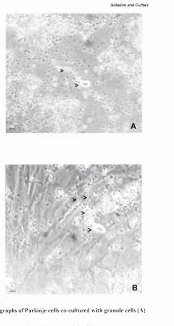
Photographs o f Purkinje cells co-cultured with granule cells (A) or glial cells ( B ) ... 73
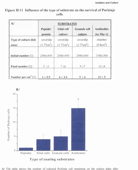
Influence o f the type o f substrate on the survival of Purkinje cells... 74
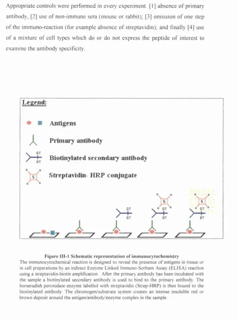
Schematic representation of immunocytochemistry ... 87
Schematic representation o f RT-PCR ... 91
Single cell collection for R T-PC R ... 93
GAD65 DNA sequence... 96
Sample o f ‘primer design’ program o u tp u t... 97
Sample o f ‘blast’ program o u tp u t... 98
Detection o f GAD, Thy-1 and calbindin ... 101
Immunoblots on samples taken before and after cell isolation .... 102
12% SDS-PAGE o f proteins from samples taken before and after cell isolation... 103
Photographs o f a cerebellar section... 105
Photographs o f anti-calbindin staining in cerebellar sections 106 Photographs o f anti-parvalbumin staining in cerebellar sections .. 108
Photographs o f anti-GAD staining in cerebellar sections 110 Influence o f Triton in fixed and unfixed samples ... 112
Figure 111-19 Figure DI-20 Figure ni-21 Figure DI-22 Figure I I I 23 Figure n i-2 4 Figure HI-25 Figure n i-26 Figure III-27 Figure IV-1 Figure IV-2 Figure IV-3 Figure IV-4 Figure FV-5 Figure IV-6 Figure IV-7 Figure FV-8 Figure V-1 Table I-l Table H-1 Table H-2 Table H-3 Table H-4 Table HI-l Table m -2 Table-IV-1
Photographs o f various GABAergic neurones co-cultured with
granule c e lls ... 123
Photographs o f Purkinje cells plated on glial cells, cultured for 10 days and stained with anti-Thy-1 antibodies... 126
Photograph o f Purkinje cells displaying various morphologies and labelled with anti-GAD antibodies... 128
PCR products obtained with GAD 65, GAD67 and actin primers. 130 PCR products obtained from mRNA and total RNA preparations 132 PCR products obtained with different amount o f Tth and after different number of cy cles... 133
Influence o f Magnesium concentration and pH in RT-PCR 134 Detection of Thy-1 mRNA from single c e l l... 137
Detection o f actin mRNA from single c e ll... 138
Effect o f soluble 0X 7 antibodies in presence and absence of N G F ... 151
Effect o f soluble TY antibodies in presence and absence o f NGF 152 Effect o f NGF in presence and absence of soluble TY antibodies 153 Distribution o f neurite lengths obtained with different treatments 154 Pictures o f typical Purkinje cells observed in the different treatments ... 156
Number o f primary and secondary neurites per cell in the different treatm ents... 158
Influence o f NGF on neurite outgrowth from Purkinje cells plated on anti-GAD antibodies... 159
Soluble antibodies-treated cell on anti-GAD antibodies coated chamber p la te s... 160
Schematic representation o f possible mechanisms o f action involved in neurite outgrow th... 177
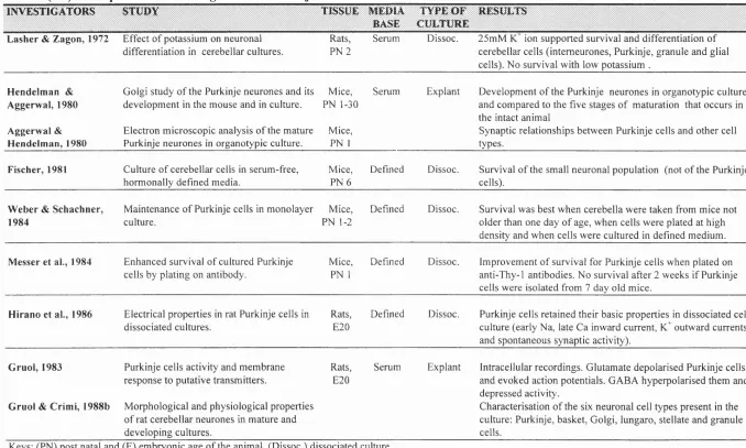
Principal Purkinje cell investigators and a summary o f their stu d ies... 33
Variations in the procedure used for Purkinje cells isolation ... 55
Morphology o f Purkinje cells depending o f the enzymatic treatm ent... 64
Summary o f Purkinje cell c u ltu re s ... 67
Adherence o f Purkinje cells on different substrates... 72
Percentage o f positively stained embedded cells... 113
Antibody-staining o f the cell types in culture... 116
ACKNOWLEDGEMENTS
I would like to thank Professor Annette Dolphin and Professor Mike Spyer for their support. I am particularly grateful to Nandita Ray, Anastasia Gratsa, Pierre Izzo, Liz Bradbury and Alex Yule for helping me as well as for being my friends.
Dumon V. and Docherty M. (1995) Cell-surface molecules important to the maintenance o f rat cerebellar Purkinje cells in culture. Abstract presented at the 25th Annual meeting o f the American Society for Neuroscience, 519.2.
In preparation:
Dumon V. Isolation and maintenance of fully differentiated cerebellar Purkinje cells. J. Neuroscience Methods.
ABBREVIATIONS
cAMP cyclic adenosine 5’-monophosphate
ATP Adenosine Tri Phosphate
BSA Bovine serum albumin
CAM Cell Adhesion Molecule
cDNA clone DNA
CNS Central nervous system
CSF Cerebro-spinal fluid
C(x) Culture number x
DEPC Diethyl pyrocarbonate
DMEM Dulbeco Minimum Eagle’s medium
DNA Deoxyribonucleic acid
D IT Dithiothreitol
E(x) Embryonic day x
ECM Extracellular matrix
Fig. Figure
FNin Fibronectin domain HI
GABA gamma-aminobutyric acid
GAD Glutamate acid decarboxylase
GMP Guanosine monophosphate
GPI Glycosyl-phosphatidylinositol
HGMP Human genome mapping project
HRP Horseraddish peroxydase
IDDM Insulin-dependent diabetes mellitus
Ig Immunoglobulin
IGF Insulin-like growth factor
kDa Kilo Daltons
LTP Long-term potentiation
MAP kinase mitogen-activated protein kinase
MEM Minimum Eagle’s medium
MuLV-RT Murine leukeamia virus reverse transcriptase
N-CAM Neural-cell adhesion molecule
NGF Nerve growth factor
PBS Phosphate buffered saline
PCR Polymerase chain reaction
PDGF Platelet derived-growth factor
PKC Protein kinase C
PL Poly-D-lysine
PLC Phospholipase C
PN(x) Postnatal day x
SDS-PAGE SDS-polyacrylamide gel electrophoresis
SEM Standard error of the mean
SMS Stiff man’s syndrome
Tth Thermus thermophilus (DNA polymerase)
U Unit
V/V Volume per volume
Introduction
C
h a p t e r
O
n e
1.1 In t r o d u c t i o n
1.1.1 Ba c k g r o u n d
The ability of cells to respond to signals from their micro-environment is fundamental to their development. In the developing nervous system, neurones migrate and extend neurites to establish an intricate network o f synaptic connections. During migration and neurite outgrowth, cells are guided by both attractive and repulsive signals (Hynes and Lander, 1992; Keynes and Cook, 1992). The ability of neurones to respond to these signals is dependent upon cell surface molecules which receive signals and transmit them to the cell’s interior. This results in specific biological responses such as promoting morphological plasticity (Goodman and Shatz, 1993) and maintaining stable contact between cells (Bailey et al., 1992). This stability, however, may also contribute to the poor regenerative capacity of the adult central nervous system (CNS), (Doherty et al., 1995). In fact, as a neurone reaches maturity, synaptic plasticity remains but neurite outgrowth remains limited. It has been shown that by blocking certain cell surface molecules of a neurone, mainly using specific antibodies directed against them, the ability to extend neurite can be modulated (Leifer et al., 1984; Mahanppatha et al., 1992(a); Doherty et al., 1993, Shea and Benowitz, 1995).
Investigation of this latter phenomenon might provide some valuable insight into practical means of promoting neuronal repair after injury or disease-induced neurodegeneration. However, none o f the published studies have been confined to mature CNS neurones. In such a model, their ability to extend neurites would be naturally limited.
1.1.2 Sc o p eo f t h e p r o j e c t
Introduction
vitro is more difficult as the age o f the animal from which they are isolated increases (Messer et a l, 1984; Hockerberger et a l, 1989; Cohen-Cory et a l, 1991). It was therefore important to study a cell type for which differentiation is easily recognisable in vitro, well described in the literature and occurs early in development.
Use o f a cerebellar cell type was considered to hold possible advantages. The cellular organisation o f the cerebellum is well characterised and composed of only a limited number of cell types, thereby simplifying the task of identification. O f these cell types, Purkinje cells have a unique and highly distinctive morphology. Their developmental maturation has been well studied in animal models and a variety of biochemical and immunological markers have been described. Post natal day twenty, in the rat, corresponds to the start of the final stage in the maturation o f the Purkinje cells. They already present the typical morphology of a highly differentiated neurone and from now on are considered as mature neurones. They therefore provided an excellent model for this study.
However, no successful studies on the culture of mature Purkinje cells has been yet reported. The optimal conditions for isolating and maintaining mature Purkinje cells with preserved dendritic trees in vitro are largely undefined. A detailed investigation o f isolation and culture conditions was required before progressing to functional studies o f neurite outgrowth.
1.1.3 Ov e r v i e w
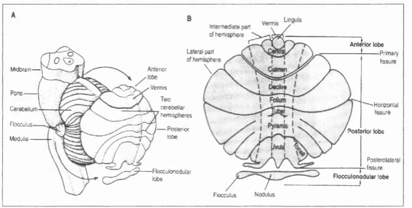
1.2 Th e c e r e b e l l u m
The cerebellum constitutes only 10% of the total volume o f the brain but contains more than half of all its neurones. However the apparent anatomical organisation is relatively simple; five types of neurones (stellate, basket, Purkinje, Golgi and granule cells) which are organised in three distinct layers, two afferent pathways (the mossy fibres and the climbing fibres) and only one output pathway (the axon of the Purkinje cells). These components are highly organised in a regular repetition of simple circuits of neuronal connections (Leiner et al., 1989). These circuits mediate different functions according to the information they receive and relay it outwards.
1.2.1 Or g a n i s a t i o n o f t h e c e r e b e l l u m
Anatomical organisation
Introduction
Functional organisation
The cerebellum can be divided into three regions running from the anterior to the
posterior lobe: the central part known as vermis, the intennediate and the lateral
zones o f each hemispheres (Fig. I-l.).
Flocculus Nodukis
Anljrk* lobe
*— — Primary fissure verm is Intermediate part of fiemispftere Lateral part of ftemisphere
\
Anterior Midbram-^— Vermis Horizontal cerebellar Cerebellum fissure hem ispheres Flocculus Posteriorlobe Posterior lobe Medulla
Posterolateral
tissure
Flocculonodular
lobe Flocculonodulsr lobe
Figure I-l Anatom ical organisation the cerebellum A. the cerebellum is unfolded to reveal the lobes normally hidden from view.
B. the main body o f the cerebellum is divided by the primary fissure into anterior and posterior lobes. The posterolateral fissure separates the flocculonodular lobe. Shallower fissures divide the anterior and posterior lobes into nine lobules. The cerebellum has three functional regions: the central vermis and the lateral and interm ediate zones in each hemisphere, (adapted from Kandel et al., 1991)
A second functional division is based on the connections made to the different
regions of the CNS, into the vestibulocerebellum, the spinocerebellum and the
cerebrocerebellum (Ito et al., 1984). This division relates to the phylogenetical
evolution of cerebellar regions. The oldest region is the vestibulocerebellum
(anatomically the flocculonodular lobe) which is vital for movements. The second
region, the spinocerebellum, receives proprioceptive information from the cortex
accomplished by controlling the firing patterns of the alpha and gamma motomeurones. This region therefore mediates the detailed execution of movements and helps to minimise disturbances that might throw them out of course (Stein et al., 1986).
The most recent phylogenetical region, the cerebrocerebellum, has more complex functions. It makes reciprocal connections with the cerebral hemispheres but receives no proprioceptive information from the spinal cord. This allows the cerebrocerebellum to rapidly control and plan learned, skilled movements without the delay of sensory feedback pathways needed for the actual execution and adjustment of movement. Lesion studies suggest that it also functions as a timing device as patients with cerebellar dysfunction are unable to reproduce precisely- timed tapping sequences (Ivry et al., 1988).
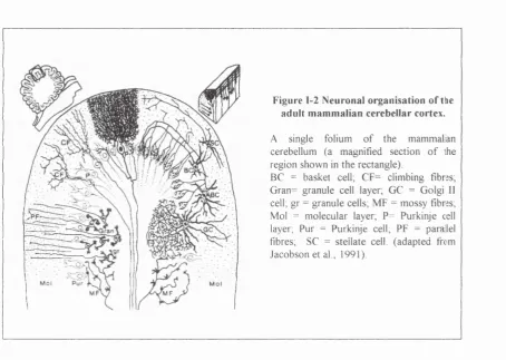
1.2.2 Ce l l t y p e s a n d c o n n e c t i o n s
Introduction
Figure 1-2 Neuronal organisation o f thie adult m am m alian cerebellar cortex.
A single folium o f the m am m alan cerebellum (a magnified section o f ihe region shown in the rectangle).
BC = basket cell, CF= climbing fibres; Gran= granule cell layer, GC = Golgi II
cell; gr = granule cells; M F = m ossy fibres; Mol = m olecular layer, P= Purkinje cell layer, Pur = Purkinje cell, PF = parallel fibres, SC = stellate cell, (adapted from Jacobson et al., 1991).
The molecular layer
Nearest to the surface of the cerebellum, the molecular layer is built up of the
dendritic arborisation o f Purkinje cells and densely packed thin axons of the
granule cells also called parallel fibres. They run parallel to the longitudinal axis
o f the folium. These fibres form synaptic connections on the dendritic spines of
the Purkinje cells and also on the dendrites of the stellate, basket and Golgi cells.
The cell bodies o f the basket and the stellate cells, both interneurones, are also
located in this layer. The stellate neurones form synapses on Purkinje cell
dendritic shafts while the Golgi neurones form a local circuit with the granule
neurones and do not interact directly with the Purkinje cells. The afferent
climbing fibres which originate in the inferior olive, innervate the spine of the
The Purkinje cell layer
The Purkinje cell layer is located beneath the molecular layer and contains the characteristic, large, pear-shape like cell bodies o f the Purkinje cells which are arranged uniformly side by side in a single sheet. Their extensive dendritic trees, confined to the transversal plane of the folium, rise up to the molecular layer. The Purkinje cell bodies receive inputs essentially from the basket cells. The soma of specialised astroglial cells (the processes o f which are known as the Bergmann glial fibres) are also present in this layer.
The granular layer
Innermost of the three, the granular layer is o f uneven thickness. A vast number of densely packed small neurones, mostly granule cells, are confined to this layer. A few Golgi cells are found on the outer border. The important structural element of this layer is the cerebellar glomeruli where dendrites of the granule cells form complex synaptic contacts with the afferent mossy fibres.
Introduction
1.3 Th e Pu r k i n j e c e l l s
The Purkinje cells have been described as one o f the "most characteristic example
o f highly differentiated neurones with respect to the specific beauty o f their
dendritic arborisations as well as the many different kinds o f synaptic relations
they have" (Eccles, 1967). Purkinje cells are GABAergic, easily recognisable,
abundant, and have the potency to elaborate extended dendritic trees. At postnatal
day 20 (PN20), in the rat, these cells are fully differentiated and are considered as
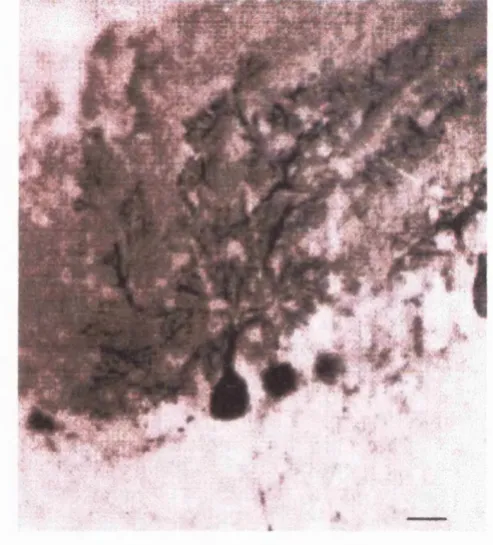
mature (Fig. 1-3).
Figure 1-3 Purkinje cells labelled w ith anti-calbindin antibodies
1.3.1 PURKINJE CELL ONTOGENESIS
In general, the stage o f development of the cerebellum at birth can be correlated with the development o f an animal's power of locomotion and motor co ordination. Time and sites of origin of the Purkinje cell and of the four types of cerebellar local-circuit neurones, as well as their migration routes to specific positions in the cortex, their distinctive pattern of differentiation and growth, and their synaptogenesis, have been described in several species (reviewed by Jacobson, 1991).
Well defined germinal zones can be demarcated which give origin to different types of neurones in a regular timetable. Histogenesis o f large principal neurones occurs first, followed by genesis of local-circuit neurones. Cells originate from two separate germinal zones. A zone in the roof of the fourth ventricle (the rhombic lip) gives origin to deep cerebellar neurones in addition to the Purkinje, Golgi 11 cells, and the Golgi epithelial glial cells. Later another germinal zone, called the external granule layer, is formed immediately beneath the pia covering the cerebellar plate, and this gives origin to granule, stellate and basket cells and some glial cells, all o f which migrate deeper into the cerebellar cortex.
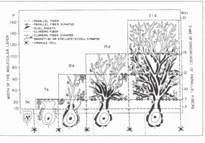
1.3.2 De v e l o p m e n t a lp h a s e s
The development of Purkinje cells can be divided into five phases. The first is embryonic (E) and all the others are postnatal (PN), (Fig. 1-4).
Phase 1. The presynaptogenic phase.
Introduction
Purkinje cells are the most numerous cellular elements. Purkinje cells are
scattered in 6 to 12 rows and have dendrites which are budding from all sides
(Addison et al., 1911; Altman et al., 1972 a, b). Ramon y Cajal (1911) pointed out
that in the same region, the Purkinje cells closer to the surface appear to be more
mature than the others. Synapses are absent on the soma o f newborn animals.
More often, desmosome-like attachment membranes are visible between the
perikarya of the Purkinje cells and unidentified processes and sometimes with
adjoining Purkinje cells. The Golgi II glial cells start to appear during this phase.
PARALLEL FIB E R - -P A R A L L E L F IB E R S Y N A P S E
-G L IA L S H EA TH -C L IM B IN G F IB E R ■ ^ . - C L I M B I N G F IB E R S Y N A P SE
» Y ^ - B A S K E T ( B ) o r S T E L L A T E ( S ) C E L L SYNAPSE
tC - g r a n u l e c e l l
Figure 1-4 Purkinje cell developm ental phase
Diagrammatic illustration o f some m ajor events in the m aturation o f a Purkinje cell. The w idth o f the m olecular layer is a function o f the animal’s age. The five columns represent the five developmental phases. At day 3, the Purkinje cell is still imm ature and resembles to a neuroblast with a visible axon. D uring phase 2 (7 days), the Purkinje cell acquires transient structures (perisom atic processes) and the main dendrite start to grow. The climbing fibres and the granule cell axons develop their first synaptic contacts with the Purkinje cells. The m aturation o f the soma occurs in phase 3 .(1 2 days). The upw ard ‘m arch’ o f glial sheeting o f the Purkinje cells commences. Basket cells (B) and stellate cells (S) form synaptic contacts respectively with the cell body and the newly elaborated dendrites o f the Purkinje cell. Phase 4 and 5 represent the m aturation o f the dendritic tree o f the cell. Permanent synaptic contacts are made as the cell elaborates secondary and tertiary dendritic branches, as well as spiny branchelets, and expands to the upper part o f the layer.
Phase 2. Formation o f transient structures
Maturation of the Purkinje cells begins with the permanent dispersion of Purkinje cells into a monolayer, five days after birth in the rat (Addison et al., 1911) and ten days after birth in the mouse (Miale et al., 1961). Two transient cytoplasmic formations appear; an apical cone composed of a large concentration of mitochondria and agranular vesicles, and many perisomatic processes o f the soma. The appearance of the latter is associated with the establishment of complex asymmetrical synapses, presumably those of climbing fibres. In the seven days old rat (or postnatal day 7), “early synapses” are formed between parallel fibres and the outgrowing dendrites o f the Purkinje cells.
Phase 3. Synaptic maturation o f the soma.
Purkinje cells begin to differentiate rapidly after the granule cells migrate past them from the external granule layer to the internal granule layer. During that phase, the synaptic maturation of the soma o f the Purkinje cell is indicated by the gradual disappearance of perisomatic processes and of temporary synapses with climbing fibres and by the acquisition of permanent symmetrical basket cell contacts (Altman et al., 1972b). The axon o f the Purkinje cell which can be seen at birth, spreads small collaterals which contact neighbouring Purkinje cells. Glial cell processes start spreading around the soma o f the Purkinje cells. The neurones o f the deep cerebellar nuclei make their first synaptic contacts. The nuclei o f the Purkinje cell continues to increase in size until PNIO in the mouse and PN14 in the rat.
Phase 4. Maturation o f the lower synaptic domain o f Purkinje cell dendrites
Introduction
and Aggerwall, 1980) and in the first 21 postnatal days in the rat (Addison et al., 1911; Altman et al., 1972b). Dendritic growth is stimulated by contact with the parallel fibres and by the stellate cells.
Phase 5. Maturation o f the upper synaptic domain o f Purkinje cell dendrites.
The development of permanent synaptic connections with climbing fibres and granule cell axons (the parallel fibres) continues over an extended time (up to 30 days), during which elimination of synapses also occurs. Growth is now shifted to the rapidly arborising Purkinje cell dendrites, to secondary and tertiary branches and to terminal branchelets with spines in the upper molecular layer. The progressive upward march of synaptogenesis continues until the glial insulation over the Purkinje cell and its extended dendritic tree is completed.
1.3.3 M a r k e r s f o r P u r k i n j e c e l l s
An array of antibodies directed against a number of Purkinje cell proteins have been applied in developmental studies. Markers include vitamin D-dependent calcium binding protein also named calbindin (Jande et al., 1981; Baimbridge et al., 1982; Rogers et al., 1989; Celio et al., 1990), parvalbumin (Legrand et al.,
1983), motilin (Nilaver et al., 1982), cerebellin (Slemmon et al., 1984, 1985) and cGMP-dependent protein kinase (De camilli et al., 1984). Other immunochemical makers used include Glutamate decarboxylase (GAD; Chan-Palay et al., 1981), UCHT and Leu-4 (Garson et al., 1982), PEP-19 (Mugnaini et al., 1987; Sangameswaran et al., 1989), mabQl 13 (Ha^vkes et al., 1985), and zebrin I and II (Brochut et al., 1990; Wassef et al., 1990).
Expression o f several of these markers appears to be related to different stages of Purkinje cell development. For example, calbindin appears at E l 6, cGMP at E l 7 and PEP-19 at E19 (Jacobson, 1991). Markers are present on both migrating and static Purkinje cells.
Introduction
The present study focused on three Purkinje cell markers: calcium binding proteins, GAD and Thy-1.
Calcium-binding proteins expression
Calcium binding proteins were first described in skeletal muscles where troponin-C was shown to bind calcium in order to induce muscle contraction (Ebashi,
1960). Then the number of others calcium-binding proteins was soon added to; a vitamin D-dependent calcium binding protein (more commonly designated calbindin) was added to the list by Wassermann and Taylor (1966), followed by S- 100 by Calissano et al., (1969), and parvalbumin by Heizmann (1984). The discovery of the ubiquitous calcium binding protein calmodulin by Cheung (1980), prompted the search for related proteins. They are involved in calcium transport and act as intracellular calcium buffers (Celio, 1990). All o f them, except troponin-C, have been isolated from the brain o f various species. Calmodulin (Lin et al., 1980; Biber et al., 1984), parvalbumin (Celio and Heizmann, 1981; Celio, 1986) and calbindin (Baimbridge and Miller, 1982; Garcia-Segura et al., 1984; ) are present in neurones while S-100 is mainly found in astrocytes (Cocchia et al., 1981). Calmodulin is ubiquitous and occurs in all neurones while calbindin and parvalbumin are present in certain subsets of neurones. The majority of cerebellar Purkinje cells display at least three different calcium-binding proteins in co-existence: calmodulin, calbindin and parvalbumin. Antibodies directed against calbindin or parvalbumin have been extensively used in culturing studies to identify Purkinje cells (Gruol and Crimi, 1988 a, b; Mariani et al., 1991; Cohen-Cory et al., 1991; Torres-Aleman et al., 1992; de Talamoni et al., 1993; Baptista et al., 1994; Mount et al., 1993; 1994a, b).
Glutamic acid decarboxylase expression
terminal amino acid compositions (Erlander et al., 1991, Solimena and De Camilli, 1993). Both forms require the cofactor pyridoxal phosphate (PLP or vitamin B6) for activity. However, GAD67 possesses a higher affinity for PLP and is therefore constitutively active (Denner and Wu, 1985). The lower affinity of GAD 65 for PLP means that its activity is more dependent on PLP availability. GAD65 cycles between PLP-bound (active, apo form) and PLP-unbound (inactive, holo form) forms (Martin et al., 1991). Differential tissue expression and sub-cellular distributions of these forms of GAD have been reported (Erdo and Wolff, 1990; Reetz et al., 1991; Kaufmann et al., 1991) as well as developmental expression (Bond et al., 1988, 1990). It has been suggested that GAD67 is predominantly located in cell bodies whereas the lower molecular weight form is primarily situated in the neurites and terminals (Kaufmann et al., 1991). Membrane forms of GAD have also been identified (Covarrubias and Tapia, 1978; Christgau et al., 1992, Nathan et al., 1993). Purkinje cells, as well as the other inhibitory neuronal population o f the cerebellum, express GAD (McLaughlin et al., 1975; Oertel et al., 1981; Chan-Palay et al., 1981).
GAD has been implicated in the aetiology of autoimmune diseases including insulin-dependent diabetes mellitus (IDDM) and stiff man syndrome (SMS, a rare neurological disorder). Autoantibodies to GAD have been identified in the CSF of patients with these diseases (De Aizpuria et al., 1992). These pathological conditions result from the destruction of GABAergic cells: the pancreatic beta cells in IDDM and the cerebellar Purkinje cells in SMS (reviewed by Ellis et al., 1996). The membrane form of GAD has been implicated in the abnormal cellular recognition by the immune system.
Thy-1 expression
Introduction
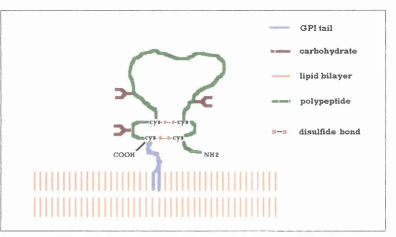
c y t - t - t - c y i
G PI tail "Il ■■ carb oh yd rate ——• lip id b ila y e r
Il p oly p ep tid e S“ 5 d isu lfîd e bond COOH ^ " N H 2
F ig u re 1-5 S chem atic d ra w in g o f Thy-1 m olecule (adapted from Williams and Gagnon, 1982)
It has been suggested that Thy-1 mediates some intercellular adhesive function
involved in regulating development (Messer et al., 1984; Morris, 1986, 1992a).
Many groups have shown that antibodies to Thy-1 promote neurite outgrowth.
These studies were undertaken on chromaffin cells (Mahanthappa et al., 1992a),
on PC12 cell line (Mahanthappa et al., 1992a; Doherty et al., 1993) and on retinal
ganglion cells (Lipton et al., 1992) but never in mature Purkinje cells maintained
in vitro. Thy-1 produces inhibition of neurite outgrowth in the presence of
astrocytes (Tiveron et al., 1992). Recently, studies in transgenic mice lacking Thy-
1 have reported some alteration in LTP and learning processes (Nosten-Bertrand
et al, 1996; Michalovich et al., 1996). The full picture o f the function of Thy-1 is
still unclear and its mode o f action undefined.
Introduction
1 .4 . T h e In v it r o C u l t i v a t i o n o f P u r k i n j e c e l l s _________________
The in vivo cellular events that lead to the morphological and functional differentiation of the Purkinje cell (Altman et al., 1972b; Berry et a l, 1976) and the role that environmental events such as contacts with afferent pathways or glial cells play in cerebellar ontogeny has been extensively studied (see Ito et a l, 1984 for review). The importance of afferent innervations (Mariani et a l, 1991; Baptista et a l, 1994), the migration of granule cells (Rakic and Sidman, 1973; Sotelo et a l, 1976) and the role o f growth factors (Cohen-Cory et a l, 1991; Torres-Aleman et a l, 1992; Lârkfors et a l, 1994) or neurotransmitters (Hockberger et a l, 1989; Mount et a l, 1993, 1994a) have been investigated during Purkinje cell development in vitro using embryonic tissue from rats or mice (see table I-l). The in vitro cultivation of Purkinje cells has been undertaken using three types of cerebellar cultures: explant or organotypic cultures, dissociated cultures prepared from embryonic tissue and dissociated cultures prepared from postnatal tissue (see table I - l. for summary).
1.4.1 Ex p l a n t o ro r g a n o t y p ic c u l t u r e s
IN V E ST IC A T O R S ST U D Y TISSUE MEDIA BASE
TY PE O F C U L T U R E
R ESU L TS
L a sh e r & Z ag o n , 1972 E ffect o f potassium on neuronal differentiation in cerebellar cultures.
Rats, P N 2
Serum Dissoc. 25m M ion supported survival and differentiation o f cerebellar cells (intem eurones, Purkinje, granule and glial cells). No survival with low potassium .
H en d e lm a n & A ggerw al, 1980
A g gerw al & H en d e lm a n , 1980
G olgi study o f the Purkinje neurones and its developm ent in the m ouse and in culture.
Electron m icroscopic analysis o f the m ature Purkinje neurones in organotypic culture.
Mice, PN 1-30
Mice, PN 1
Serum Explant Developm ent o f the Purkinje neurones in organotypic culture and com pared to the five stages o f m aturation that occurs in the intact anim al
Synaptic relationships between Purkinje cells and other cell types.
F isch e r, 1981 Culture o f cerebellar cells in serum -free, horm onally defined media.
Mice, P N 6
Defined Dissoc. Survival o f the small neuronal population (not o f the Purkinje cells).
W e b e r & S ch a c h n e r, 1984
M aintenance o f Purkinje cells in m onolayer culture.
Mice, PN 1-2
Defined Dissoc. Survival was best w hen cerebella w ere taken from m ice not older than one day o f age, w hen cells w ere plated at high density and w hen cells w ere cultured in defined m edium .
M esser et aL, 1984 Enhanced survival o f cultured Purkinje cells by plating on antibody.
Mice, PN 1
Defined Dissoc. Im provem ent o f survival for Purkinje cells when plated on anti-Thy-1 antibodies. N o survival after 2 w eeks if Purkinje cells w ere isolated from 7 day old mice.
H ira n o et al., 1986 Electrical properties in rat Purkinje cells in dissociated cultures.
Rats, E20
Defined Dissoc. Purkinje cells retained their basic properties in dissociated cell culture (early Na, late Ca inward current, K^ outw ard currents and spontaneous synaptic activity).
G ru o l, 1983
G ru o l & C rim i, 1988b
Purkinje cells activity and m em brane response to putative transm itters.
M orphological and physiological properties o f rat cerebellar neurones in m ature and developing cultures.
Rats, E20
Serum Explant Intracellular recordings. G lutam ate depolarised Purkinje cells and evoked action potentials. GABA hyperpolarised them and depressed activity.
Characterisation o f the six neuronal cell types present in the culture: Purkinje, basket, G olgi, lungaro, stellate and granule cells.
Keys: (PN) post natal and (E) em bryonic age o f the anim al. (Dissoc.) dissociated culture.
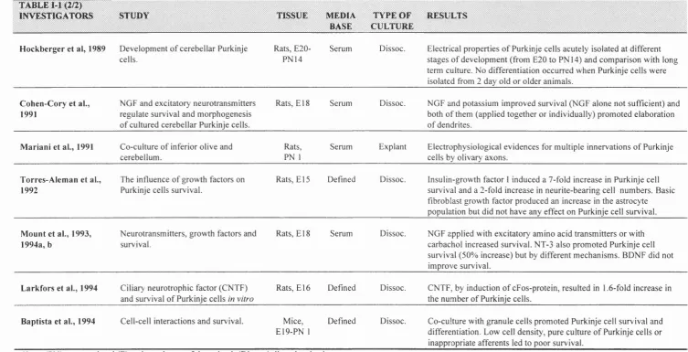
T A B L E M (2/2)
IN V ESTIG A T O R S S T U D Y TISSU E M EDIA
BASE
T Y PE O F C U LTURE
R ESU L TS
H ockberger et al, 1989 D evelopm ent o f cerebellar Purkinje cells.
Rats, E20- PN14
Serum Dissoc. Electrical properties o f Purkinje cells acutely isolated at different stages o f developm ent (from E20 to PN14) and com parison with long term culture. No differentiation occurred w hen Purkinje cells were isolated from 2 day old or older anim als.
C ohen-C ory et al., 1991
NGF and excitatory neurotransm itters regulate survival and m orphogenesis o f cultured cerebellar Purkinje cells.
Rats, E l 8 Serum Dissoc. NGF and potassium im proved survival (NG F alone not sufficient) and both o f them (applied together or individually) prom oted elaboration o f dendrites.
M ariani et al., 1991 C o-culture o f inferior olive and cerebellum .
Rats, PN 1
Serum Explant Electrophysiological evidences for m ultiple innervations o f Purkinje cells by olivary axons.
Torres-A lem an et al., 1992
The influence o f grow th factors on Purkinje cells survival.
Rats, E l 5 Defined Dissoc. Insulin-grow th factor 1 induced a 7-fold increase in Purkinje cell survival and a 2-fold increase in neurite-bearing cell num bers. Basic fibroblast growth factor produced an increase in the astrocyte population but did not have any effect on Purkinje cell survival.
Mount et al., 1993, 1994a, b
N eurotransm itters, grow th factors and survival.
Rats, E l 8 Serum Dissoc. N G F applied with excitatory am ino acid transm itters or with carbachol increased survival. NT-3 also prom oted Purkinje cell survival (50% increase) but by different m echanism s. BDNF did not im prove survival.
Lârkfors et al., 1994 Ciliary neurotrophic factor (CNTF) and survival o f Purkinje cells in vitro
Rats, E16 Defined Dissoc. CNTF, by induction o f cFos-protein, resulted in 1.6-fold increase in the num ber o f Purkinje cells.
Baptista et al., 1994 Cell-cell interactions and survival. Mice, E19-PN 1
Defined Dissoc. C o-culture with granule cells prom oted Purkinje cell survival and differentiation. Low cell density, pure culture o f Purkinje cells or inappropriate afferents led to poor survival.
1.4.2 Dis s o c ia t e d c u l t u r e sf r o m e m b r y o n ic t is s u e
Several investigators have attempted to culture Purkinje cells following their dissociation from cerebellar tissue (Woodmans et al., 1980; Hirano et al., 1986; Hockberger et al., 1989; Cohen-Cory et al., 1991; Torres-Aleman et al., 1992; Mount et al., 1993; Lârkfors et al., 1994; Baptista et al., 1994). Some approaches have employed defined serum-ffee medium, while others have opted for the more traditional approach o f a serum-containing media (often horse serum, see Table I- 1). Fischer (1981) was the first to define a hormonally-based medium which replaced the traditional serum-supplemented Eagle medium. The defined medium was composed of BME-Earle's supplemented with BSA, insulin, 1-thyroxine, transferrin, aprotinin, selenium, glucose and glutamine and it was found to greatly enhance the survival of cerebellar cells (Fischer, 1981; Weber et al., 1984; Torres- Aleman et al., 1992). Three vital conditions were required to obtain good survival and differentiation of Purkinje cells: (1) the early age of cerebella (E14-PN1), (2) sufficient plating densities (4xl0^cells/ coverslip of 1.5cm in diameter), and (3) a chemically defined culture medium (Weber et al., 1984).
Introduction
1.4.3 Dis s o c ia t e d c u l t u r e s f r o mp o s t n a t a l t is s u e
Purkinje cell cultures obtained from postnatal tissues have never attained the survival rates obtained with Purkinje cells derived from embryonic tissues (Weber et al., 1984). Hockberger et al. (1989) in parallel to his embryonic study, attempted to grow Purkinje cells from different postnatal ages. With cerebella originating from postnatal day 2 or older animals Purkinje cells survived poorly and did not differentiate properly even in the presence o f high potassium, glutamate or aspartate. The reason for the addition of these neurotransmitters was to mimic the in vivo synaptic innervations (the parallel and climbing fibres).
1 .5 Ce l l Su r f a c e Mo l e c u l e s a n d t h e i r In v o l v e m e n t in
Ad h e s i o n a n d Ne u r it e Ou t g r o w t h_______________________________
The intrinsic capacity o f a mature neurone to grow and /or regenerate is not lost but rather inhibited or down regulated by specific post-natal expression of particular molecules. Cells express an assortment of surface molecules that enable them to respond specifically to soluble signals (such as hormones, growth factors or neurotransmitters) or to bind in a characteristic way to other cells or to the extracellular matrix (ECM) in order to modify their behaviour. The expression of such molecules is not rigid, and it is plasticity that allows the cell to behave differently. (Krushel et al., 1993).
Cell adhesion molecules have been shown to play an important role in a variety of processes, including the migration o f neuroblasts, neurite outgrowth and fasciculation, axonal guidance and establishment of functional synapses (Edelman and Crossin, 1986; Walsh and Doherty, 1991, [reviews]). Glycoproteins with immunoglobulin (Ig)-like domains have been implicated in adhesion and neurite outgrowth.
1.5.1 Gl y c o p r o t e i n s W ITH IG -L IK E D O M A IN S
glycosyl-Introduction
phosphatidylinositol (GPI) linkage and a fourth form is secreted. N-CAMs form homophilic or heterophilic interactions with other cells (Fig.I-6). Twenty forms of N-CAM exist which are generated from alternative splicing o f RNA transcript produced from a single gene. When N-CAM is highly glycosylated, sialic acid is formed and the charge of the surface becomes very negative, preventing cell adhesion and therefore promoting nerve outgrowth.
Other members of the Ig superfamily group have restricted distribution and participate in more selective interactions. They usually do not span the membrane but are attached to the plasma membrane by a GPI anchor. These include cell- surface molecules such as LI (Persohn and Schachner, 1987), TAG-1 (Dodd et a l, 1988), F3/ FI I (Gennarini et a l, 1989; Brummendorf et a l, 1989) and Thy-1 (Morris et a l, 1985). Some molecules can also be secreted and/ or incorporated into the ECM. The cellular distribution can be restricted to subsets of neurones or subcellular compartments within a given neurone. For instance, the mouse F3 protein (FI 1 counterpart of the chicken molecule) is confined to a subset of neurones in the cerebellum. Developing granule cell axons and growing parallel fibres strongly express F3. By contrast, Purkinje cell bodies and dendritic arborisations as well as the stellate cells do not express F3 (Faivre-Sarrailh et a l, 1992).
1992a). It is possible that an imbalance in favour of stability may contribute to the
poor regenerative capacity o f the adult CNS (Doherty et al., 1995). Studies on the
modulation o f the expression of cell surface molecules in mature neurones would
provide invaluable clues on the mechanisms of action o f these molecules. In
addition, the second messenger pathways involved in these events need to be
investigated.
H.CiLM s
se c re ted GPI an ch o r w ith tran sm e m b ra n e ^ dom ains
Ig d o m a in
D
S - S b o n d
3
FN d o m ain
Thv-1 O O H
CYTOSOL OOH
10 |im
Figure 1-6 Schem atic draw ing o f som e o f the glycoproteins w ith Ig dom ains
The extracellular part o f the polypeptide chain in each case is folded into one to five Ig-like domains and one or tw o fibronectin type III repeats (case o f N -CAM s). The disulfide bonds connect the ends o f each loop forming each Ig-like domain. (A dapted from Alberts et al.,
1994).
Introduction
1.5.2 Ot h e r t y p e s o f c e l l s u r f a c e m o l e c u l e s
How cell surface molecules activate intracellular pathways in order to alter the cell behaviour is not well understood. In the case o f immunoglobulins, which do not possess an intracellular domain, the problem is even more cloudy. One supposition is that cell surface molecules activate intracellular pathways by interacting with other types o f cell surface molecules which have full intracellular pathways. In addition, by binding to each other (in an homophilic or heterophilic manner), they promote cell to cell and cell to substrate adhesion; a process which is also related to neurite outgrowth. Three other families o f cell surface molecules have been found to be involved in adhesion and/or neurite outgrowth: the integrins, the cadherins and the cell surface receptors.
Integrins
Cadherins
The cadherins are calcium-dependent cell surface molecules which keep cells attached to each other (Geiger and ayalon, 1992). In vitro, the removal of calcium is followed by tissue disruption and the isolation of individual cells. Three types of binding are possible, homophilic binding (same type o f molecule), heterophilic binding (different type o f molecules bind to each other) or binding to a cell surface receptor by a linker molecule (Geiger and Ayalon, 1992; Takeichi, 1990). There are at least 15 types of cadherins depending on where they are expressed. Another member o f this group, the selectins, are cell surface carbohydrate-binding proteins also known as lectin.
The majority function as transmembrane linker proteins which mediate interactions between the actin cytoskeleton o f the cells which are joined together. Developmental phases exist during which the loss of these molecules allows the cell to migrate. Later, the cell re-expresses them, aggregation occurs, and a ganglion is formed.
Cell surface receptors
These are transmembrane receptors to either channel-linked receptors, enzyme-linked receptors or G-protein-enzyme-linked receptors (Kandel et al., 1991). Many of them have already been implicated in morphological changes o f a cell such as growth.
Introduction
protein kinases or any elements of the subsequent cascade (e.g.. the ras proteins) can lead to excessive cell division and proliferation which are no longer under the normal 'social' control of adjacent cells. Other enzyme-linked receptors function as receptor- tyrosine phosphatases which remove phosphate groups from tyrosine residues. Finally, some cell surface receptors belong to the guanylyl cyclases family and catalyse the production of cyclic GMP in the cytosol (Yuen and Garbers, 1992).
The channel-linked cell surface receptors are transmitter-gated ion channels involved in rapid synaptic signalling between electrically excitable cells. Small neurotransmitters transiently open or close the ion channel to which they bind, briefly changing the permeability of the plasma membrane and thereby the excitability of the postsynaptic cell.
1 .6 Ai m s o f t h e s t u d y
The structure and pattern o f connectivity o f the mature nervous system is a result of complex developmental processes which include cell proliferation, migration, neurite extension, and synaptogenesis (Mendell et al., 1991). Cell surface molecules such as Thy-1 have been implicated in cell survival (Messer et al.,
1984) and neurite outgrowth (Mahanppatha et al., 1992a; Doherty et al., 1993). The studies on neurite outgrowth have been undertaken in several cell types (chromaffin cells, cell lines and sympathetic neurones) but never on mature CNS neurones. The aim o f my project was to investigate the role of such molecules in mature neurones of the CNS. The maintenance of differentiated neurones in vitro
has proven to be extremely difficult. However, as these molecules are likely to play an important role in the survival and growth of mature cells, it is essential to carry out such a study on primary mature neurones in order to understand their involvement. The present study investigated the ability o f differentiated neurones to survive and grow in culture. The isolation and maintenance in vitro of fully differentiated Purkinje cells would provide an excellent model to study the role of these molecules.
Introduction
effect on Purkinje cells isolated from older animals (age superior to 7 days). The conditions for in vitro survival and growth of more mature Purkinje cells (isolated from animals older than 7 days) probably rely on the supply of different types of additives and other parameters such as adhesiveness and contact with other cell types. The role of synaptic innervations, cell surface molecules and soluble signals from the environment present later in development still need to be further identified in order to determine the conditions which will influence a mature cell to die or to survive and grow when its fate has been altered. Cerebellar mixed cultures (neuronal and non-neuronal cells) were chosen for this study because this type o f culture allows the Purkinje cells to evolve in a relatively rich environment, as they would do in vivo. Culturing conditions for mature neurones were determined and survival conditions are detailed in Chapter two.
Purkinje cells isolated from cerebella o f twenty day old rats were identified and characterised in four separate conditions: [1] cerebellar sections, [2] individual cells embedded into agarose gel, [3] long-term cultures and [4] at the mRNA expression level (Chapter Three). Several antibodies directed against Purkinje cells were used for immunocytochemical detection: anti-calbindin, anti-GAD and anti-Thy-1 antibodies. Total and messenger RNA expressions in Purkinje cells were analysed by reverse-transcriptase polymerase chain reaction (RT-PCR) This technique allowed the study o f mRNAs expression in cerebellar slices and in single cells.
C
h a p t e r
T
w o
Isolation and Culture
2 .1 In t r o d u c t i o n
The in vivo survival and differentiation of the Purkinje cells are affected by multiple epigenetic factors which include the excitatory innervation of the parallel fibres (axons of cerebellar granule cells) and of the climbing fibres projecting from the inferior olive. Loss of these inputs, through olivary or granule cell ablation (Berry and Bradley 1976; Sotelo and Arsenio-Nunes, 1976; Crepel et al., 1981), pharmacological blockade (Vogel et al., 1990) or mutation (Rakic and Sidman, 1973) can result in degeneration or aberrant development of Purkinje cells. Neurotrophic factors and neurotransmitters, such as NGF, IGF-1, NT-3 and glutamate, also play an important role in development and maturation of neuronal cells (Cohen-Cory et al., 1991; Wanaka and Jonhson, 1990; Mount et al., 1993, 1994a, b; Lârkfors et al., 1994). The majority of studies on Purkinje cells have been undertaken in explant cultures (Hendelman and Aggerwall, 1980; Gruol, 1983; Fields, 1984; Mariani et al., 1991; Hauser et al., 1994) and in dissociated cultures (Weber and Schachner, 1984; Messer et al., 1984; Hirano and Ohmori, 1986; Hockberger et al., 1989; Cohen-Cory et al., 1991; Schilling et al., 1991; Mount et al., 1993). The starting material for these culture systems was embryonic or early post-natal cerebellar tissue.
This chapter describes experiments performed to establish a method for the isolation of viable mature cerebellar Purkinje cells from 20 day old rats and the determination of the optimal conditions for their long term maintenance in vitro.
Isolation and Culture
2 .2 Me t h o d s
2.2.1 Re a g e n t s AND MATERIALS
A total of 300 twenty day old Wistar rats and 10 twelve days old Wistar rats,
either male or female, were used for this study. The materials and reagents used
for the experiments are listed in Appendix A l.
Û 2
Glass vessel
Bufifer
W ater bath
Brain slices
2.2.3 Bu f f e r s a n d s o l u t i o n s u s e d f o r c e l l i s o l a t i o n
The composition of the buffer used for cell isolation is detailed in table II-1 and the composition of all the solutions are described in Appendix A2.
2.2.4 Is o l a t i o n o f Pu r k i n j e c e l l s
Animals were either anaesthetised with isoflurane by inhalation and then decapitated, or directly decapitated with a guillotine. The brain was carefully removed and the cerebellum placed in 1ml of basic isolation buffer composed of 240mM sucrose, 20mM PIPES di-potassium, lOmM KCl, 25mM glucose, 6mM MgCl], ImM CaCl], pH 7 and pre-warmed to 37°C. Sagital slices were cut manually using a razor blade. From each cerebellum, 10 slices (thickness ranged from 0.5 to 1 mm) were obtained and placed in a glass vessel containing the buffer used for the dissociation (4 slices in 4ml of buffer per vessel).
Tissue slices were pre-incubated for one hour at 37°C and equilibrated with 100% O2 (see apparatus. Fig. II-1). This step was followed by enzymatic digestion o f the
slices using a solution containing either pronase E (0.1-0.2%), trypsin (0.06%), collagenase (0.1%), thermolysin (0.2%), (weight/volume in buffer) or combinations of these enzymes. The enzymatic treatment (4ml o f enzyme solution/vessel) was applied to the slices for 15-30 min at 37°C with oxygenation. Tissue slices were then washed four times with 4 ml of basic isolation buffer. Sections were held at 37°C for one hour with oxygenation in the fourth buffer change.
isolation and Culture
times ranging between 5- 120 min). For culturing purposes, isolated cells were used within 15 min of isolation.
2.2.5 As s e s s m e n t o f Ce l l Vi a b i l i t y
Trypan blue exclusion test
Isolated cells were centrifuged at 1500 rpm for 2 min and the resulting pellet was resuspended in 4ml volume of 300mM PBS. Coverslips bearing cultured cells were washed once with 0.4ml volume o f 300mM PBS per coverslip. A 1ml volume of Trypan blue stain (0.08%, w/v) was added. After 5 min at room temperature, the cells were repelleted and once again suspended in a 4ml volume of PBS. Aliquots of cells were removed and placed in the chambers of an haemocytometer slide. A minimum o f 100 isolated Purkinje cells were contained in each chamber. Purkinje cells were observed under the microscope noting whether or not Trypan blue was inside the cell. Two different aliquots per condition were assessed. For cells in culture, the Purkinje cells on two coverslips were observed. The percentage score of live cells (colourless and bright) over total number o f Purkinje cells was calculated.
Esterase activity assessment
Cell viability was also determined using the fluorescent dyes calcein and ethidium homodimer. These dyes were applied on freshly isolated cells or on cultured cells. A solution of 2pM ethidium homodimer and 4|liM calcein-AM made up in
2 .2 .6 I n v i t r o Cu l t iv a t io no f Pu r k in j ec e l l s
Culture plate substrates
a) peptide or protein substrates
Poly-D-lysine was coated on the coyerslips or on the 16-well-chamber plates at 1 pg/cm^ oyemight at 37°C. After four washes with sterile water (0.5ml per coyerslip or 0.2ml per well of chamber plate), culture dishes were air-dried before use.
Laminin or fibronectin was coated at 2.5pg/cm^on poly-D-lysine pre-coated plates for two hours at 37°C, and then washed and air-dried as for poly-D-lysine procedure.
ProNectin was applied at 2pg/cm^ for two hours at room temperature or oyemight at 4°C, and then washed 4 times and air dried as for poly-D-lysine procedure. Matrigel was used at 12.8mg/ml for thick coating or diluted (1:1) in culture medium for thin coating and applied while cool (+4°C) onto coyerslips or 8-well chambers.
b) on cerebellar glial cells pre-cultured fo r 10 days.
Cerebellar glial cells were obtained from either 2-4 day or 7-8 day old rats. The cerebella were sliced carefully using a razor blade and incubated with trypsin (0.05% in BBSS) for 15 min at 37°C. Slices were washed twice in BBSS and mechanically dissociated by four passages through a Pasteur pipette of 500pm diameter. The cell suspension was centrifuged (1500 rpm for 5 min), resuspended in culture medium A (see Appendix A2) and plated at a density of 5x10^ cells/cm^ on 13mm-coyerslips pre-coated with poly-D-lysine. The cells were kept at 37°C with 5% CO2, and the medium changed eyery fiye days.
c) on granule cells pre-cultured fo r 4 days
Isolation and Culture
d) on antibody-coated 16-well chamber plates.
A mouse monoclonal antibody to Thy-1 (TY, 50|ig/ml) diluted in a solution o f
lOjig/ml Poly-D-lysine was coated on the 16-well chamber plates (80pl/well)
overnight at 4®C. A rabbit polyclonal anti-GAD (SOpg/ml) and a normal mouse
serum IgG fraction (Zymed, 50 pg/ml) were coated onto 16-well chamber plates
in a similar manner. The plates were rinsed four times with sterile water and air
dried before use.
Culturing procedure
Cerebellar cell suspensions (containing Purkinje cells) were centrifuged for 2 min
at 1500 rpm and resuspended in various culture media. The cells were plated at a
density o f 5x10^ cells/ml on the protein substrates or on the monolayer cultures o f
glial or granule cells or on antibody-coated chamber plates. Cultures were
maintained at 37°C, in a humid atmosphere supplemented with 5% CO2. The medium was changed after two days in vitro and thereafter at weekly intervals.
Growth factors were added in some culture media; lO-lOOng/ml NGF, 20-40ng/ml
IGF or O.l-lOpg/ml Insulin. The growth factors were added at the beginning o f
the culture and maintained in the media as long as the culture was kept. Table II-3