Radical Reduction in the Rate of Extensive Corrective Surgery for
Clubfoot Using the Ponseti Method
Jose A. Morcuende, MD, PhD; Lori A. Dolan, PhD(c); Frederick R. Dietz, MD; and Ignacio V. Ponseti, MD
ABSTRACT. Objectives. The purpose of this study was to evaluate the efficacy of the Ponseti method in reducing extensive corrective surgery rates for congenital idiopathic clubfoot.
Methods. Consecutive case series were conducted from January 1991 through December 2001. A total of 157 patients (256 clubfeet) were evaluated. All patients were treated by serial manipulation and casting as described by Ponseti. Main outcome measures included initial cor-rection of the deformity, extensive corrective surgery rate, and relapses.
Results. Clubfoot correction was obtained in all but 3 patients (98%). Ninety percent of patients required <5 casts for correction. Average time for full correction of the deformity was 20 days (range: 14 –24 days). Only 4 (2.5%) patients required extensive corrective surgery. There were 17 (11%) relapses. Relapses were unrelated to age at presentation, previous unsuccessful treatment, or severity of the deformity (as measured by the number of Ponseti casts needed for correction). Relapses were re-lated to noncompliance with the foot-abduction brace. Four patients (2.5%) underwent an anterior tibial tendon transfer to prevent further relapses.
Conclusions. The Ponseti method is a safe and effec-tive treatment for congenital idiopathic clubfoot and radically decreases the need for extensive corrective sur-gery. This technique can be used in children up to 2 years of age even after previous unsuccessful nonsurgical treatment.Pediatrics 2004;113:376 –380;clubfoot, Ponseti method.
C
ongenital idiopathic clubfoot is a complex foot deformity that occurs in an otherwise normal child. In 1996, 2224 children were born with clubfoot in the United States, an incidence of ⬃0.6 cases per 1000 live births.1The goal of treatment is tocorrect all components of the deformity so that the patient has a pain-free, plantigrade foot with good mobility, without calluses, and without the need to wear special or modified shoes.
Most orthopedists agree that the initial treatment should be nonsurgical and started soon after birth. Many different methods of correction are used, most of them involving manipulation and casting. In many institutions, manipulation and serial casting require many months of treatment and frequently result in incomplete or defective corrections.2– 4As a
result, extensive corrective surgery is indicated in 50% to 90% of the cases, often with disturbing fail-ures and complications.5–11 In addition, depending
on the technique followed and the residual defor-mity, up to 47% of clubfeet undergo 1 or more revi-sion surgeries.12–17
The results at our institution differ radically from these reports. Since the late 1940s, we have followed the method of correction developed by the senior author.18This method involves weekly stretching of
the deformity followed by application of a long-leg cast. All components of the deformity usually correct within 4 to 5 weeks with the exception of the equi-nus. A simple percutaneous tendoachilles tenotomy often is necessary to correct completely the equinus. The first report of 67 patients who were younger than 6 months and treated by the Ponseti method demonstrated satisfactory and rapid initial correc-tion in the majority of cases (83%) with minimal complications.19 However, there was a relatively
high incidence clubfoot relapses (56%) in this patient population. Most relapses were treated successfully with repeat manipulation and castings and/or ante-rior tibial tendon transfers. More important, the long-term functional and clinical results at a 30-year follow-up were excellent or good using pain and functional limitation as the outcome criteria in the majority of these patients (78% compared with 85% of a matched control population born with normal feet).20
The technique has been refined over the years, and we have come to realize the necessity of hyperabduc-tion of the foot in the last cast and long-term use of the foot abduction brace. In addition, our referral base has radically changed as a result of patient referral from the Internet.21 This has resulted in an
increase in the number of children who present at an age older than 6 months and many who have had previous unsuccessful nonsurgical treatment else-where. This change in patient population has led us to expand the age range of our traditional indications for nonsurgical treatment rather than to default to extensive corrective surgery based solely on older age or previous treatment.
Because of this more recent experience, we are reevaluating the efficacy of the Ponseti method for the correction of congenital idiopathic clubfoot. This study also examined the effect of our changing pop-ulation in the context of the age of the patients at the onset of treatment and previous treatment.
From the Department of Orthopaedic Surgery, University of Iowa, Iowa City, Iowa.
Received for publication Dec 6, 2002; accepted Jul 7, 2003.
METHODS
Patient Population and Treatment Protocol
We reviewed the records of 157 patients with congenital idio-pathic clubfoot (256 clubfeet) who were treated consecutively under the supervision of the senior author from January 1991 to December 2001. Positional clubfeet were not included. Institu-tional review board approval was obtained. The average age at last follow-up was 26 months (range: 6 months to 8 years). No patient was lost to follow-up.
The guidelines for the treatment developed by the senior au-thor and followed at our institution are as follows18: 1) all
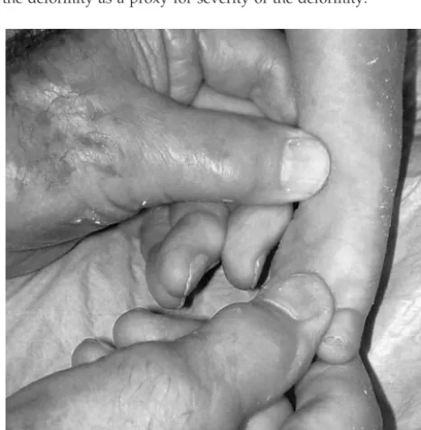
com-ponents of the deformity are corrected simultaneously, not in sequence, except for the equinus, which should be corrected last; 2) the cavus, which results from pronation of the forefoot in relation to the hind foot, is corrected together with the adduction by supinating and abducting the forefoot in proper alignment with the hind foot; 3) with the longitudinal arch of the foot well molded and the forefoot in some supination, the entire foot can be abducted gently and gradually under the talus, which is secured against rotation in the ankle mortise by applying counterpressure with the thumb against the lateral aspect of the head of the talus; 4) heel varus will correct when the entire foot is fully abducted under the talus; the heel is never touched (Fig 1); 5) finally, the equinus is corrected by dorsiflexing the foot; this is generally facilitated by a simple percutaneous tendoachilles tenotomy under local anesthesia.
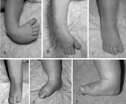
For maintaining the correction obtained by gentle manipula-tion, a plaster cast is applied in 2 sections. The first section extends from the toes to just below the knee, and the second covers the knee and the thigh. The knee is immobilized at a right angle. The plaster cast is molded to fit the anatomy precisely. Abduction of the foot is increased progressively with each manipulation and plaster cast application until hypercorrection to⬃70° of foot ab-duction is obtained. All of the casts in all of the patients included in this study were applied under the supervision of the senior author (I.V.P.; Fig 2). Radiographs are taken in the rare case of a deformity that resists correction to rule out any bony abnormality. If full, initial correction is not achievable, then surgical treatment is indicated. The surgical procedure is tailored to the residual deformity. No severity classification is used because the deformity is not necessarily related to the resistance to correction. However, we used the number of casts required to obtain a full correction of the deformity as a proxy for severity of the deformity.
After correction, a foot-abduction brace is used to maintain the correction. This brace consists of a bar with shoes attached at the ends at 70° of outward rotation on the affected side and 40° on the normal side. The length of the bar should be equal to the width of the child’s shoulders (Fig 3). The brace is used on a full-time basis for 2 to 3 months and at night and during naptime for 3 to 4 years. Parent self-report on brace wear was used to assess compliance. Noncompliance was defined when the foot abduction brace was not used for at least 10 hours a day.
A relapse is defined as the appearance of any of the compo-nents of the deformity, including cavus, adductus, varus, and/or equinus. Relapses after initial correction are treated with addi-tional manipulation and serial casting in marked foot abduction. A tendoachilles tenotomy is indicated when dorsiflexion of the ankle is⬍15°. When the anterior tibial muscle tends to strongly supinate the foot during gait, its transfer to the third cuneiform will prevent additional relapses in most patients.
Analysis
We evaluated the following variables: age of the patient at first visit to our institution, previous treatment and type of treatment before referral, number of casts, previous tendoachilles tenotomy, number of casts required at our institution, need for percutaneous tendoachilles tenotomy, degree of ankle dorsiflexion after tenot-omy, and compliance with the foot-abduction brace. These vari-ables were in turn related to the need for extensive corrective surgery and the incidence of relapses. Fisher exact tests,ttests, and odds ratios were used as appropriate.
RESULTS
A total of 107 (68%) patients were male. Seventy-nine (60%) patients were first-born. Thirty-three (22%) patients had a positive family history of club-foot deformity. Most children (92%) were full term, without complications during gestation or delivery. At initial Ponseti casting, 128 (81%) patients were younger than 6 months and 29 (19%) were older than 6 months.
A total of 113 (73%) patients had some form of treatment before their initial visit to our institution. Seven (4.5%) had physical therapy, and 111 (71%) had serial manipulation and casting. The number of casts ranged from 1 to 20, with a median of 10. Patients had primarily below-the-knee casts (49%) or a combination of below-the-knee and above-the-knee casts (24%). Fourteen (9%) patients had a percutane-ous tendoachilles tenotomy. Ninety-five percent of the patients came to our clinic with all of the com-ponents of the deformity uncorrected. Extensive cor-rective surgery was recommended to 75 (48%) pa-tients by their treating physicians.
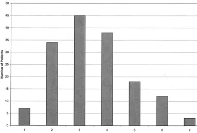
Clubfoot correction was obtained in all but 3 (98%) patients. These 3 patients were previously treated in other institutions and presented with very severe deformities. Correction was obtained with 1 to 7 casts; 90% of the patients requiredⱕ5 casts for cor-rection (Fig 4). The average time from the first cast to the tendoachilles tenotomy (full correction of the deformity) was 20 days (range: 14 –24 days). Percu-taneous tendoachilles tenotomy was performed in 86% of the cases. Average ankle dorsiflexion post-tenotomy was 20° (range: 0 –35°). The majority of patients started walking at an age of 13 months (range: 9 –17 months). Twelve (8%) patients had a cast complication, including erythema, slight swell-ing of the toes, or downward slippage of the cast. All these complications were attributed to a deficient casting technique. No infections, skin necrosis, neu-Fig 1. Manipulation as described by Ponseti. The thumb is
rovascular compromise, or profuse bleeding post-tenotomy were observed.
Four (2.5%) patients required extensive corrective surgery after treatment by the Ponseti method. The surgical procedures performed included 1 postero-medial release and 3 posterior releases with ten-doachilles tenotomy. It is interesting that of the 75 patients whose deformities were previously indi-cated for corrective surgery by the local physician, only 1 went on to surgery after treatment by the Ponseti method.
There were 17 (10%) relapses after initial success-ful treatment. Relapse of the deformity was not sig-nificantly related to age at presentation, previous unsuccessful treatment at other institution, or the number of casts required for correction (used as a measure of severity; allP ⬎ .05). Relapses were as-sociated with noncompliance with the foot-abduc-tion brace (P ⫽ .001; Table 1). Noncompliance was associated with a 17 times greater odds of relapse (15 Fig 2. Serial photographs at weekly intervals of the correction of a severe clubfoot deformity in a 3-week-old infant. A, At initial visit. B, After first cast. C, After second cast. D, After third cast. E, After fourth cast. F, Treatment result after percutaneous tendoachilles tenotomy.
of 17) compared with compliance (6 of 140; P ⫽
.0001). Relapses were treated with a second series of manipulation and casting, followed by the use of the foot-abduction brace. Three patients required a sec-ond tendoachilles tenotomy. For preventing addi-tional relapses in 4 (2.5%) patients who were non-compliant with the foot-abduction brace, an anterior tibial tendon transfer to the third cuneiform and a tendoachilles lengthening were performed.
DISCUSSION
This study demonstrates that with the use of the Ponseti method, ⬎95% of patients’ idiopathic club-foot can be corrected without the need for extensive corrective surgery. In addition, we found no in-creased difficulty correcting the deformity in chil-dren up to the age of 2 years or in patients who previously had a nonsurgical corrective attempt.
The results of this study are in contrast with most published series.2–17We believe that this discrepancy
in the results of treatment is attributable primarily to a deficient understanding of the nature of the defor-mity and the normal functional anatomy of the foot. This lack of understanding has led to poor corrective manipulation and casting techniques. It is not attrib-utable, on the basis of the current study, to the com-plexity or the severity of the deformity.
The main reason for failures is the application of
counterpressure on the calcaneocuboid joint during the manipulation. By so doing, the normal move-ment of the calcaneus under the talus, a motion that is fundamental for the correction of the deformity, is prevented. Because the 3 tarsal joints move simulta-neously, blocking the calcaneocuboid joint in turn prevents the movement of the talonavicular joint and therefore makes it impossible to correct the clubfoot. In addition, many physicians perform these maneu-vers somewhat forcefully, often causing the child great distress.10,22 The correction should be
per-formed very gently to avoid these problems because crying and pain are associated with increased muscle tension in the lower extremity, making the manipu-lation and casting more difficult.
An important finding in this study was the de-creased number of patients who presented with a relapse after initial full correction when compared with our previous publication.19We believe that this
is attributable to the hyperabduction now obtained in the last cast and to the education of parents about the need to use the foot-abduction brace at night and naptime until the age of 3 years.
Our study has a number of important implications for parents, children, and physicians. From the pub-lic health standpoint, our findings can be used to reassure the public that the congenital idiopathic clubfoot deformity, although complex in nature, is easily corrected and, more important, that our results can be replicated in other institutions and clinical practices and even in different health care systems. Herzenberg et al23 recently reported similar good
results in their institutions. They evaluated 27 pa-tients (34 clubfeet) after serial manipulation and cast-ing followcast-ing the Ponseti method. Control subjects included patients who were treated with initial serial short-leg casts for 3 months (range: 2– 6 months). Fig 4. Total number of casts required for full correction of the deformity.
TABLE 1. Factors Associated With Relapse After Full Correc-tion of the Deformity
PValue
Age of patient .98
Previous treatment .5
Only 1 (3%) clubfoot that was treated by the Ponseti method required extensive corrective surgery com-pared with 32 (94%) in the control group. In addition, a significant decrease in the range of motion in pa-tients who were treated surgically was found com-pared with those who were treated by the Ponseti method.
Pirani et al24,25 introduced the Ponseti method in
Uganda in November 1999 and developed a national program for the treatment of clubfoot. As in many other developing countries, many, if not most, chil-dren who are born with clubfoot do not receive any treatment of the deformity. Through training of phy-sicians and other health care personnel and public awareness campaigns, ⬎80% of the patients have their deformity completely corrected within 2 months of onset of treatment. The numbers of infants who are younger than 1 year and being referred for extensive corrective surgery has reduced remark-ably. The success of this program is such that it has already been expanded to Malawi, Tanzania, and Ethiopia (S. Pirani, J. N. Penny, and M. Steenbeeck, personal communication, 2002).
In conclusion, the Ponseti method is a very safe, efficient treatment for the correction of clubfoot that radically decreases the need for extensive corrective surgery. Furthermore, it can be used successfully in children up to ⬃2 years of age when no previous surgical treatment has been attempted. The decline in extensive clubfoot surgery should encourage na-tional efforts to make this method the gold standard in the treatment of congenital idiopathic clubfoot. Educational programs should be targeted to primary care physicians to increase awareness of the Ponseti method and its excellent results so that they can advise families accordingly. Physicians who adopt the Ponseti method will feel rewarded by the satis-faction of successfully correcting what traditionally has been a very frustrating deformity to treat.
REFERENCES
1. Ventura SJ, Martin, JA, Curtin, SC, Mathews TJ. Report of final natality statistics, 1996.Mon Vital Stat Rep. 1998;46(suppl):1–99
2. Kite JH. Nonoperative treatment of congenital clubfoot.Clin Orthop. 1972;84:29 –38
3. Dimeglio A, Bonnet F, Mazeau PH, De Rosa V. Orthopaedic treatment
and passive motion machine: consequences for the surgical treatment of clubfoot.J Pediatr Orthop B.1996;5:173–180
4. Dewaele J, Zachee B, De Vleeschauwer P, Fabry G. Treatment of idio-pathic clubfoot: critical evaluation of different types of treatment pro-grams.J Pediatr Orthop. 1994;3:89 –95
5. Turco VJ. Resistant congenital clubfoot: one-stage posteromedial release with internal fixation: a follow-up report of a fifteen-year experience. J Bone Joint Surg Am. 1979;61:805– 814
6. Crawford AH, Marxen JL, Osterfeld Dl. The Cincinnati incision: a comprehensive approach for surgical procedures of the foot and ankle in childhood.J Bone Joint Surg Am. 1982;64:1355–1358
7. Simons GW. Complete subtalar release in clubfeet. I: a preliminary report.J Bone Joint Surg Am. 1985;67:1044 –1055
8. Green ADL, Lloyd-Roberts GC. The results of early posterior release in resistant club feet. A long-term review.J Bone Joint Surg Br. 1985;67:588 9. Aronson J, Puskarich CL. Deformity and disability from treated
club-foot.J Pediatr Orthop. 1990;10:109 –112
10. Crawford AH, Gupta AK. Clubfoot controversies: complications and causes for failure.Instr Course Lect. 1991;45:339 –346
11. Atar D, Lehman WB, Grant AD. Complications in clubfoot surgery. Orthop Rev. 1991;20:233–239
12. Vizkelety T, Szepesi K. Reoperation in treatment of clubfoot.J Pediatr Orthop. 1989;9:144 –147
13. Tarraf YN, Carroll NC. Analysis of the components of residual defor-mity in clubfeet presenting for reoperation.J Pediatr Orthop. 1992;12: 207–216
14. Krauspe R, Vispo Seara JL, Lohr JF. Long-term results after surgery for congenital clubfoot.Foot Ankle Surg. 1996;2:77– 82
15. Sobel E, Giorgini RJ, Michel R, Cohen SL. The natural history and longitudinal study of the surgically corrected clubfoot.J Foot Ankle Surg. 2000;5:305–320
16. Uglow MG, Clarke NM. Relapse in staged surgery for congenital talipes equinovarus.J Bone Joint Surg Br. 2000;82:739 –743
17. Bradish CF, Noor S. The Ilizarov method in the management of relapsed club feet.J Bone Joint Surg Br. 2000;82:387–391
18. Ponseti IV. Congenital Clubfoot: Fundamentals of Treatment. Oxford, England: Oxford University Press; 1996
19. Ponseti IV, Smoley EN. Congenital clubfoot: the results of treatment. J Bone Joint Surg Am. 1963;45:261–275
20. Cooper DM, Dietz FR. Treatment of idiopathic clubfoot. A thirty-year follow- up.J Bone Joint Surg Am. 1995;77:1477–1489
21. Morcuende JA, Egbert M, Ponseti IV. The effect of the Internet in the treatment of congenital idiopathic clubfoot.Iowa Orthop J. 2003;23:83– 86 22. Grayev AM, Boal DK, Wallach DM, Segal LS. Metaphyseal fractures mimicking abuse during treatment for clubfoot.Pediatr Radiol. 2001;31: 559 –563
23. Herzenberg JE, Radler C, Bor N. Ponseti versus traditional methods of casting for idiopathic clubfoot.J Pediatr Orthop. 2002;22:517–521 24. Pirani S. The Ponseti method and results in developing nations.
Pre-sented at the Symposium on Conservative Methods on the Treatment of Clubfeet. SICOT. August 30, 2002; San Diego, CA
DOI: 10.1542/peds.113.2.376
2004;113;376
Pediatrics
Jose A. Morcuende, Lori A. Dolan, Frederick R. Dietz and Ignacio V. Ponseti
Using the Ponseti Method
Radical Reduction in the Rate of Extensive Corrective Surgery for Clubfoot
Services
Updated Information &
http://pediatrics.aappublications.org/content/113/2/376
including high resolution figures, can be found at:
References
http://pediatrics.aappublications.org/content/113/2/376#BIBL
This article cites 22 articles, 0 of which you can access for free at:
Subspecialty Collections
http://www.aappublications.org/cgi/collection/surgery_sub
Surgery
following collection(s):
This article, along with others on similar topics, appears in the
Permissions & Licensing
http://www.aappublications.org/site/misc/Permissions.xhtml
in its entirety can be found online at:
Information about reproducing this article in parts (figures, tables) or
Reprints
http://www.aappublications.org/site/misc/reprints.xhtml
DOI: 10.1542/peds.113.2.376
2004;113;376
Pediatrics
Jose A. Morcuende, Lori A. Dolan, Frederick R. Dietz and Ignacio V. Ponseti
Using the Ponseti Method
Radical Reduction in the Rate of Extensive Corrective Surgery for Clubfoot
http://pediatrics.aappublications.org/content/113/2/376
located on the World Wide Web at:
The online version of this article, along with updated information and services, is
by the American Academy of Pediatrics. All rights reserved. Print ISSN: 1073-0397.