Article
Sarcocystis neurona Protein Kinases: Computational
Identification, Evolutionary Analysis and Putative
Inhibitor Docking
Edwin K. Murungi 1 & Henry M. Kariithi 2
1 Department of Biochemistry and Molecular Biology, Egerton University, P.O. Box 536, 20115, Njoro, Kenya; eddkimm@gmail.com
2 Biotechnology Research Institute, Kenya Agricultural and Livestock Research Organization, P.O Box 57811, 00200, Kaptagat Rd, Loresho, Nairobi, Kenya; henry.kariithi@kalro.org
* Correspondence: eddkimm@gmail.com; Tel: (+254)-789-716059
Abstract: The apicomplexan parasite, Sarcocystis neurona causes the degenerative neurological equine protozoal myeloencephalitis (EPM) disease of horses. Due to its host range expansion, S. neurona is an emerging threat that requires close monitoring. In apicomplexans, protein kinases (PKs) have been implicated in a myriad of critical functions such as host cell invasion, cell cycle progression and host immune responses evasion. Here, we used various bioinformatics methods to define the kinome of S. neurona and phylogenetic relatedness of its PKs to other apicomplexans. Further, three-dimensional (3D) homology models for selected S. neurona putative PKs were constructed and evaluated for inhibitor docking. We identified 92 putative PKs clustering within the AGC, CAMK, CK1, CMGC, STE, TKL, aPK and OPK groups. Although containing the universally conserved PKA (AGC group), S. neurona kinome was devoid of PKB and PKC, but contained the six apicomplexan conserved CDPKs (CAMK group). The OPK group was represented by ROPKs 19A, 27, 30, 33, 35 and 37, but was devoid of the virulence-associated ROPKs 5, 6, 18 and 38. Two out of the three S. neurona CK1 enzymes had high sequence similarities to T. gondii TgCK1-α and TgCK1-β and the Plasmodium PfCK1. Docking of four inhibitors onto homology models of putative ROP27 and PKA indicated that inhibition of S. neurona PKs is feasible, but needs to be experimentally tested. The essentiality of apicomplexan PKs makes the elucidation of S. neurona kinome a key milestone for development of novel therapeutics for EPM.
Keywords: Sarcocystis neurona; EPM; Docking; Apicomplexans; Phylogeny; Homology modeling.
1. Introduction
Equine protozoal myeloencephalitis (EPM) is an infectious, progressive, degenerative neurological disease of horses caused by the apicomplexan parasite, Sarcocystis neurona [1]. To complete its life cycle, this heteroxenous parasite requires a reservoir host (i.e. opossums; Didelphis virginiana, D. albiventris) and an aberrant (horses) or intermediate host (cats, skunks, raccoons and sea otters) [2]. Opossums become infected upon ingestion of sarcocysts containing hundreds of bradyzoites, which undergo gametogony that sporulate into mature oocysts, which are then shed in the faeces. Upon ingestion by the intermediate or aberrant hosts, the oocysts release the environmentally resistant sporozoites, which chronically parasitize the neural and inflammatory cells of the host’s central nervous system (CNS). Clinical EPM symptoms depend on which part of the CNS is parasitized; parasitization generally results in abnormal gait, dysphagia and muscle atrophy in affected horses [3].
The intracellular nature of S. neurona and its ability to evade the host's immune surveillance [4] makes EPM treatment expensive, lengthy and challenging. Traditionally, clinical treatment of EPM
2 of 34
involved inhibitors of folate synthesis and metabolism (sulphonamides/pyrimethamine combination) over a prolonged period [5]. More recently, triazines derivatives (diclazuril, ponazuril) that target the parasite’s apicoplast [6], nitazoxanide, a pyruvate:ferridoxin oxidoreductase analogue that inhibits the parasite’s anaerobic metabolism [7], and anti-inflammatory agents and immune stimulants [8] have been used with variable success in eliminating clinical signs. Despite the availability of these drugs, EPM treatment is complicated by the emergence of drug-resistance (due to intermittent or periodic treatments), cost of therapies and drug toxicity (e.g. diarrhea and/or anemia) and infection relapses due to re-growth of residual parasites after the treatment regimes [2]. As such, discovery and development of novel therapeutics for EPM is imperative.
To successfully invade the host cells, apicomplexans utilize three specialized exocytic organelles (micronemes, rhoptries and dense-granules) [9]. The microneme is used for host cell recognition, binding, penetration and gliding along the cytoskeletal structures. Rhoptry proteins are discharged into the host cell during parasite internalization and are crucial in the formation of the parasitophorous vacuoles (PVs). Developing zoites contain non-pedunculated condensing vesicles that synthesize and package inactive rhoptry proteins, which are proteolytically activated when the rhoptry contents are condensed [10]. The PVs facilitate parasite development by allowing nutrient transport from the host cell and by blocking lysosomal fusion, which would otherwise kill the parasites [11]. Upon internalization, zoites use the dense-granules to remodel the PVs into functionally active organelles.
The virulence of the apicomplexans is highly dependent on protein kinases (PKs) that are involved in the invasion and modification of host cell structure and function. PKs are classified into various groups, comprising of many families and sub-families [12-14]. The major apicomplexans PK groups are the PKs A, G and C (AGCs), which are regulated by cyclic nucleotides and other second messengers; the calmodulin/calcium-dependent PKs (CAMKs); the casein kinase 1 (CK1) and close relatives, the PKs involved in cell-cycle control and signal transduction (containing cyclin-dependent, mitogen-activated and cell division cycle [CDC]-like PKs; or CMGCs); the receptor guanylate cyclase (RGC); the “sterile” serine/threonine kinases (STEs, including several kinases functioning in the MAPK cascades); the tyrosine kinases (TKs); and the tyrosine kinase-like kinases (TKLs) [15-17]. In addition, there is a sizeable proportion of other PKs that do not fit into any of the above-mentioned eight major PK groups (i.e. the “Other” kinases [OPKs]), and another smaller set of atypical PKs (aPKs) that have no clear sequence similarity with the conventional PKs [13, 18, 19].
Each of the PK families have vital roles in parasite’s survival. For instance, PfPK-B (AGC family), PfTKL3 (TKL family), and four of the seven CDPKs (CAMK family) are required by Plasmodium parasites to complete their asexual cycle [20-22]. In a recent study, deletion of TgCK1α (CK1 family) resulted in defective replication of Toxoplasma gondii in vitro [23]. The P. falciparum CKL and SRPK1 (CMGC family) complement each other in the regulation of mRNA splicing (via phosphorylation of splicing factors [24]). Since apicomplexans lack typical MAPK cascades, the STEs are not well studied. However, in the parasites that do have the MAPK pathways, STEs are essential for parasite growth (e.g. in the human parasites, Schistosoma mansoni [25]). Further, for parasites without the conventional MAPK cascades, aPKs can activate the signaling pathways, for instance the Plasmodium Pfnek3 [26]. Finally, some of the notable OPKs include aurora kinases, rhoptry kinases (ROPKs) and parasite-specific eukaryotic initiation factor-2 (elF2) kinases (elF2K), which are important in parasite virulence and differentiation [27-29].
The differences between the parasite’s and their host’s PK homologs can be used to develop anti-parasitic strategies via specific inhibition of the parasite’s PKs [30]. This is feasible due to the essential roles of PKs in the parasite’s growth and development. Proof of principle of this approach has been demonstrated by the inhibition of human PKs using chemical ligands to treat cancers and other diseases [31, 32]. Recently, Ojo et al., [33] provided evidence that PKs can be targeted for rationally designed drugs that can potently inhibit growth of S. neurona. The technology is available and approved for therapeutic intervention, thus offering a unique prospect of repurposing chemical ligands to manage S. neurona infections [34].
3 of 34
Here, we used a genome-wide approach to define the kinome of S. neurona and determined the relatedness of the putative PKs to those reported in other apicomplexans. We also tested the docking of some specific inhibitors to a selection of some of the putative PKs. Defining the S. neurona kinome is not only important in providing insights into the parasite’s biology, but also identification of potential novel drug targets that can be used to clear chronic S. neurona infections, reduce parasite survival, augment the host's anti-parasitic immune responses, or as candidates for development of EPM vaccines.
2. Results
2.1 Sarcocystis neurona encodes 92 putative kinases
To date, at least 15 apicomplexan genomes (coccidians, gregarines, hemosporidians and piroplasmids) have either been fully sequenced or partially annotated [35]. In the current study, we conducted an exhaustive genome-wide search of the newly sequenced S. neurona genome [36], and identified 92 putative PKs (Table 1). The identified PKs contained the characteristic PK (IPR000719) or PK-like (IPR011009) domains and three conserved amino acids constituting the catalytic triad (Lys30, Asp125, Asp143). The PKs had sizes ranging between 152 and 6544 amino acids and relative molecular weights of between 15.94 to 671.51 kDa. Majority of the PKs had isoelectric point (pI) greater than 7.0, implying that the PKs have low turnover rates since in general, acidic proteins are thought to be degraded more rapidly than neutral or basic proteins [37].
Assignment of S. neurona PK groups was accomplished by sequence clustering using Blast2GO [62], and by BLASTp searches in the Kinase.com database [14]. Out of the eleven known PK groups [12-14], S. neurona PKs segregated into the AGC (n=9), CAMK (n=20), CK1 (n=3), CMGC (n=19), STE (n=2), TKL (n=6), aPK (n=2) and OPK (n=31) (Table 1). Apart from the 31 OPKs that do not fit to the major kinase groups, the CAMK and CMGC groups, whose members are essential for the parasite’s host cell invasion [38] and differentiation (via cell-cycle regulation) [39], respectively, had the highest number of PKs underlying the importance of these processes in the parasite. Unlike in some parasites (e.g. P. falciparum) that lack STEs and TKLs [40], S. neurona contains PKs in the two classes.
4 of 34
Table 1. Description of the 92 putative PKs identified in the kinome of S. neurona. The putative PKs could be classified into eight groups. The amino acid coordinates of the conserved PK domains in the protein sequences, and the PK homologies to other apicomplexan PKs are shown in columns 7 to 12.
Description of the putative protein kinases (PKs) in the genome of S. neurona Description of protein kinase (PK) homologies (BLASTp)
Protei n ID ᵃ
Sequence annotations; description ᵇ
Family; [subfamily]c Lengt
h [aa] pI MW [kDa] PK domain coordinate s Sequence name; [apicomplexan] Bit Scor e E-value Identit y [%] Accession number
1. Kinase Group AGC (Protein kinases A [PKA], G [PKG] and C [PKC] families)
SRCN _1312
AGC kinase 3-phosphoinositide
dependent PK-1 (PDK1)
903 5.52 101.1
3
137–481 PDPK; [T. gondii RUB] 417 6.00E-13
5
59 KFG61374.1
SRCN _3339
AGC kinase PKA 1428 8.85 147.5
2
1102–1417 Putative AGC kinase;
[N. caninum L]
611 0.0 81 CEL65574.1
SRCN _4249
AGC kinase Nuclear dbf2-related
(NDR)
152 8.91 17.47 6–141 Putative AGC kinase;
[N. caninum L]
219 2.00E-68 76 XP_003883757.
1 SRCN
_4518
PK G AGC kinase family member PKG
Ciliate-E2 425 5.83 48.78 97–399 AGC kinase TgPKG1;
[T. gondii ME49]
830 0.0 92 EPR61116.1
SRCN _3990
cAMP-dependent kinase
CAMKL; [MELK] 1907 9.55 217.5
5
782–1634 cAMP-dependent protein kinase [T. gondii
VEG]
75.9 1e-12 30 ESS31194.1
SRCN _5165
cAMP-dependent PK, catalytic chain
PKA 343 8.99 39.36 20–338 AGC kinase; [T. gondii
ARI] 425 3.00E-15 0 90 KYF43224.1 SRCN _4913
Putative PK PKD 2330 6.42 244.6
4
1093–1737 Putative PK; [E. tenella] 107 9.00E-22 60 XP_013228294.
1 SRCN
_5430
AGC kinase Ribosomal protein S6
Kinases (RSK; [p70])
1378 5.54 139.2
8
824–1344 AGC kinase; [T. gondii
MAS]
223 3.00E-60 59 KFH07588.1
SRCN _5610
cAMP-dependent PK, catalytic chain
PKA 333 9.00 37.96 12–318 cAMP-dependent PK,
catalytic subunit; [T. gondii ME49]
641 0.0 92 XP_002366464.
1
2. Kinase Group calcium (Ca2+)-/calmodulin-regulated kinases (CAMK)
SRCN _1071
Ca2+-dependent
kinase
CAMK1 1495 9.47 152.6
6
1085–1401 Putative PK; [T. gondii
VEG]
188 1.00E-47 67 ESS31884.1
SRCN _2032
Putative PK Ciliate-C1 297 6.22 33.26 15–297 PK; [H. hammondi] 424 1.00E-14
6
68 XP_008882026. 1
SRCN _2165
Ca2+-dependent
kinase CDPK2B
CDPK 692 7.33 75.65 101–401 Ca2+-dependent PK
CDPK2A; [T. gondii
674 0.0 90 KYF44522.1
Preprints
(www.preprints.org) | NOT PEER-REVIEWED | Posted: 9 January 2017
doi:10.20944/preprints201701.0040.v1
Peer-reviewed version available at
5 of 34
Description of the putative protein kinases (PKs) in the genome of S. neurona Description of protein kinase (PK) homologies (BLASTp)
Protei n ID ᵃ
Sequence annotations; description ᵇ
Family; [subfamily]c Lengt
h [aa] pI MW [kDa] PK domain coordinate s Sequence name; [apicomplexan] Bit Scor e E-value Identit y [%] Accession number ARI] SRCN _2257
Histone kinase CAMKL; [AMP-activated
protein kinase (AMPK)]
1800 8.98 187.7
1
1159–1448 Putative CAM kinase,
SNF1 family; [E. acervulina]
377 7.00E-10 5 64 XP_013252246. 1 SRCN _2544
CAM SNF1 AMK1 family
CAMKL;[AMPK-regulate d kinase novel kinase (NUAK)]
333 5.71 37.85 62–333 CAM kinase,
SNF1/AMK1 family ToxPK1; [N. caninum L]
480 9.00E-16 7 79 XP_003882065. 1 SRCN _2937
Ca2+-signalling kinase
MARK
CAMKL; [microtubule affinity regulating kinase (MARK)]
278 9.62 31.29 1–250 Putative Ca2+ signalling
PK MARK; [T. gondii
GT1] 356 5.00E-12 4 72 EPR59053.1 SRCN _3011 Calmodulin-depende nt PK (CAM) CDPK6
CDPK 1435 9.25 154.4
4
1238–1435 Cdpk kinase domain; [T. gondii]
179 6.00E-49 75 3IS5_A
SRCN _3314
A Chain crystal Structure of TgCDPK1 with inhibitor bound
CDPK1 519 5.99 58.89 37–335 Calmodulin-domain PK
1; [T. gondii]
536 0.0 97 3MA6_A
SRCN _3583
Ca2+-dependent
kinase CDPK5
CDPK 454 6.09 50.38 35–308 Ca2+-dependent PK
CDPK5; [T. gondii ARI]
776 0.0 89 KYF43137.1
SRCN _3701
Ca2+-dependent
kinase CDPK3
CDPK 560 5.91 62.07 77–362 Ca2+-dependent Kinase;
[T. gondii]
493 1.00E-17 2
87 3DXN_A
SRCN _4076
CAM CDPK family CDPK 1701 5.93 181.4
2
1079–1663 CAM kinase, CDPK
family; [H. hammondi]
239 2.00E-62 69 XP_008884897.
1 SRCN
_4093
PK CAMKL; [AMP-activated
protein kinase (AMPK)]
1155 9.02 118.3
3
1–261 Putative atypical
MEK-related kinase; [N. caninum L]
222 1.00E-59 70 XP_003880869.
1
SRCN _4390
Ca2+-dependent
kinase CDPK2
CDPK 790 6.24 85.47 280–556 Ca2+-dependent PK,
related; [N. caninum L]
1130 0.0 74 XP_003884321.
1 SRCN _4815 Histone kinase (partial) CAMKL; [AMP-activated protein kinase (AMPK)]
711 6.53 75.98 1–314 SNF1-related PK
catalytic-α KIN10, 5 AMP-activated PK; [N. caninum L]
568 0.0 48 CEL67550.1
SRCN CAM CDPK CDPK 2748 8.98 282.1 796–885 Putative CAM kinase, 117 3.00E-24 59 XP_003881901.
Preprints
(www.preprints.org) | NOT PEER-REVIEWED | Posted: 9 January 2017
doi:10.20944/preprints201701.0040.v1
Peer-reviewed version available at
6 of 34
Description of the putative protein kinases (PKs) in the genome of S. neurona Description of protein kinase (PK) homologies (BLASTp)
Protei n ID ᵃ
Sequence annotations; description ᵇ
Family; [subfamily]c Lengt
h [aa] pI MW [kDa] PK domain coordinate s Sequence name; [apicomplexan] Bit Scor e E-value Identit y [%] Accession number
_5227 CDPK8-like 2 CDPK family;
[N. caninum L]
1
SRCN _5410
Calmodulin-depende nt PK (CAM-SNF1 family)
CAMK1 467 8.94 52.04 168–446 CAM kinase, SNF1
family; [H. hammondi]
431 5.00E-13 4 78 XP_008883430. 1 SRCN _5812
Ca2+-dependent
kinase CDPK9
CDPK 760 8.37 84.23 254–573 Ca2+-dependent PK
CDPK9; [H. hammondi]
1139 0.0 81 XP_008889286.
1 SRCN
_5948
Ca2+-dependent
kinase CDPK8
CDPK 3298 7.11 345.8
5
208–860 EF-hand domain-containing protein; [T. gondii
ME49]
114 1.00E-23 58 XP_002368547.
1
SRCN _6597
Ca2+ dependent kinase
CDPK7
CAMK1 1374 9.09 138.2
8
365–623 PK-PH domain-containing protein; [T. gondii
ME49]
813 0.0 80 XP_002366487.
1
SRCN _6606
Ca2+-dependent
kinase CDPK4
CDPK 1632 9.42 170.9
3
813–1236 Ca2+-dependent PK;
[T. gondii]
731 0.0 58 CAD32376.2
3. Kinase Group casein kinase 1 (cell kinase 1)
SRCN _3445
Casein kinase I CK1-D 323 9.34 37.65 6–290 Casein kinase 1;
[T. gondii ME49]
603 0.0 94 XP_002366683.
1 SRCN
_4587
Casein kinase I CK1-D 137 7.78 15.94 1–137 Casein kinase I;
[H. hammondi]
226 3.00E-71 81 XP_008883809.
1 SRCN
_4645
Casein kinase I CK1-D 229 9.51 25.66 38–229 Casein kinase I;
[T. gondii
GAB2-2007-GAL-DOM 2]
259 1.00E-86 79 KFG42638.1
4. Kinase Group CMGC (including cyclin-dependent kinases, mitogen-activated PKs, glycogen synthase kinases and CDK-like kinases)
SRCN _1104
Cyclin-dependent kinase family 5
Ca2+-dependent PK-L
(CDKL)
372 9.23 42.71 1–318 Cyclin-dependent
kinase family 5 protein; [H. hammondi]
490 2.00E-17 3
76 XP_008884207. 1
SRCN Cell-cycle-associated Serine-arginine rich PK 2911 5.37 302.9 713–1837 PK; [T. gondii ME49] 476 9.00E-14 76 XP_002369401.
Preprints
(www.preprints.org) | NOT PEER-REVIEWED | Posted: 9 January 2017
doi:10.20944/preprints201701.0040.v1
Peer-reviewed version available at
7 of 34
Description of the putative protein kinases (PKs) in the genome of S. neurona Description of protein kinase (PK) homologies (BLASTp)
Protei n ID ᵃ
Sequence annotations; description ᵇ
Family; [subfamily]c Lengt
h [aa] pI MW [kDa] PK domain coordinate s Sequence name; [apicomplexan] Bit Scor e E-value Identit y [%] Accession number
_1236 kinase (SRPK) (SRPK) 0 1 1
SRCN _1479
CMGC Lammer CLK 748 10.2
3
79.09 485–748 Cell-cycle-associated
PK CLK; [T. gondii
FOU]
288 2.00E-81 74 KFG33061.1
SRCN _1611 CMGC Dual-specificity tyrosine-regulated kinase (Dyrk)
DYRK; [DyrkP] 1504 5.86 160.3
4
551–1498 Cell-cycle-associated PK DYRK; [T. gondii
VEG]
223 2.00E-57 63 ESS33160.1
SRCN _1731
Cell-cycle-associated kinase GSK
Glycogen synthase kinase (GSK)
219 6.59 24.39 1–175 Cell-cycle-associated
PK GSK; [H. hammondi]
330 2.00E-11 2 82 XP_008887193. 1 SRCN _1732 Cell-cycle-associated kinase GSK
Glycogen synthase kinase (GSK)
203 10.7 8
20.72 86–203 CMGC kinase, GSK
family TgPK3; [E. brunetti]
114 1.00E-28 91 CDJ46527.1
SRCN _2759
Cell-cycle-associated kinase partial
Ca2+-dependent PK
(CDK); [CRK7]
1122 6.14 118.6
7
541–1122 Cell-cycle-associated PK CDK; [T. gondii
VAND]
118 1.00E-25 79 KFH12036.1
SRCN _2845
CMGC DYRK PRP4 kinase
DYRK; [PRP4] 1665 9.76 177.0
8
1267–1596 Putative PK (CLK3);
[P. malariae]
330 1.00E-10 2
69 SBS85334.1
SRCN _3891
CMGC kinase DYRK; [DYRK2] 674 8.85 73.85 399–674 Putative CMGC kinase;
[T. gondii ME49]
80.1 6.00E-14 67 EPT25192.1
SRCN _4209
CMGC MAPK family (ERK) MAPK-1
Mitogen-activated PK (MAPK); [ERK])
2361 6.73 247.7
1
94–754 CMGC, MAPK/ (ERK)
TgMAPK-1; [E. brunetti]
137 1.00E-30 74 CDJ49492.1
SRCN _4674
Cyclin-dependent kinase
Ca2+-dependent PK
(CDK); [CDK7]
138 7.80 15.50 1–138 Cyclin-dependent
kinase; [T. gondii GT1]
108 7.00E-27 58 EPR60430.1
SRCN _4801
Cell-cycle-associated kinase
Ca2+-dependent PK
(CDK); [CDK5]
300 6.08 34.33 1–289 CMGC kinase, CDK
family TgPK2; [N. caninum L]
576 0.0 91 XP_003885801.
1 SRCN _5365 Cell-cycle-associated kinase MAPK Mitogen-activated PK (MAPK; [ERK])
417 6.77 48.32 7–363 Cell-cycle-associated
PK MAPK; [H. hammondi]
823 0.0 93 XP_008886907.
1
SRCN Cell-cycle-associated Ca2+-dependent PK 690 9.55 80.90 208–603 Putative 390 2.00E-12 87 KYF45878.1
Preprints
(www.preprints.org) | NOT PEER-REVIEWED | Posted: 9 January 2017
doi:10.20944/preprints201701.0040.v1
Peer-reviewed version available at
8 of 34
Description of the putative protein kinases (PKs) in the genome of S. neurona Description of protein kinase (PK) homologies (BLASTp)
Protei n ID ᵃ
Sequence annotations; description ᵇ
Family; [subfamily]c Lengt
h [aa] pI MW [kDa] PK domain coordinate s Sequence name; [apicomplexan] Bit Scor e E-value Identit y [%] Accession number
_6346 kinase CDK (CDK); [CDK5] cell-cycle-associated PK
CDK; [T. gondii ARI]
2
SRCN _6427
CMGC CK2 kinase Cell Kinase 2 (CK2) 1395 10.2
9
144.8 6
885–1356 CMGC kinase, CK2
family; [T. gondii MAS]
241 6.00E-73 98 KFH07655.1
SRCN _6472 Cell-cycle-associated kinase ERK7 Mitogen-activated PK (MAPK; [ERK])
983 9.28 104.9
5
7–317 Cell-cycle-associated PK ERK7; [T. gondii
ARI]
647 0.0 81 KYF46268.1
SRCN _761
Cell-cycle-associated kinase
Ca2+-dependent PK
(CDK); [CDK7]
577 9.34 58.39 144–490 Cell-cycle-associated
PK; [H. hammondi]
283 3.00E-88 68 XP_008882409.
1 SRCN
_895
Cell-cycle-associated kinase
Ca2+-dependent PK
(CDK); [CDK10]
340 8.93 38.57 1–307 Cell-cycle-associated
PK; [T. gondii ARI]
234 6.00E-75 76 KYF44017.1
SRCN _977
Cell-cycle-associated kinase CDK
Ca2+-dependent PK
(CDK);
[PITSLRE/CDK11]
1502 7.38 156.3
5
1114–1429 Cell-cycle-associated PK CDK; [T. gondii p89]
454 3.00E-13 5
92 KFG28420.1
5. Kinase Group ‘Other’ (OPK; i.e. kinases with conventional PK [ePK] domains that do not fit into any of the other major groups of kinases)
SRCN _108
Unc-51-like
autophagy activating kinase 1 (ULK1)
ULK 343 7.13 38.90 1-223 ULK kinase; [T. gondii
VAND] 376 2.00E-13 0 75 KFH07419.1 SRCN _1606
eIF2 kinase IF2K-C PEK; [general control nonderepressible 2 (GCN2)]
4034 8.98 406.5
7
1235–2178 eIF2 kinase IF2K-C;
[T. gondii VAND]
259 4.00E-67 35 KFH07289.1
SRCN _2076
Rhoptry kinase family ROP30
Conserved hypothetical protein
1276 9.18 134.7
3
812–1260 ROP30 [T. gondii VEG] 230 3.00E-63 53 CEL76436.1
SRCN _2123
Rhoptry kinase family ROP35
PLK; [PLK-Unclassified] 291 9.30 33.50 53–265 ROP35; [T. gondii RUB] 207 2.00E-61 43 KFG59037.1
SRCN _3216
Rhoptry kinase family ROP32
CAMK-Unique 523 7.08 57.00 214–520 Putative PK; [T. gondii
VAND]
167 1.00E-42 30 KFH00232.1
SRCN _2183
Rhoptry kinase family ROP35
Aurora-like 226 6.36 25.84 1–212 ROP35; [T. gondii VEG] 198 8.00E-59 48 ESS33297.1
SRCN _2271
Putative PK
(incomplete catalytic
NimA (Never in mitosis gene A)-related Kinase
1463 9.02 157.1
8
437–1145 Putative PK;
[N. caninum L]
327 5.00E-90 68 XP_003881849.
1
Preprints
(www.preprints.org) | NOT PEER-REVIEWED | Posted: 9 January 2017
doi:10.20944/preprints201701.0040.v1
Peer-reviewed version available at
9 of 34
Description of the putative protein kinases (PKs) in the genome of S. neurona Description of protein kinase (PK) homologies (BLASTp)
Protei n ID ᵃ
Sequence annotations; description ᵇ
Family; [subfamily]c Lengt
h [aa] pI MW [kDa] PK domain coordinate s Sequence name; [apicomplexan] Bit Scor e E-value Identit y [%] Accession number triad) (NEK) SRCN _2403 Aurora kinase (incomplete catalytic triad)
PLK; [SAK/Plk4] 778 9.79 79.92 492–778 Putative Aurora kinase;
[N. caninum L]
127 3.00E-28 44 XP_003880644.
1
SRCN _2630
NimA related kinase (NEK) family protein
NEK 351 8.70 38.38 1–336 NEK kinase; [T. gondii
ME49]
242 8.00E-75 52 XP_018638598.
1 SRCN
_286
Wee kinase Inhibitory regulator of
the RAS-cAMP (IRA1) kinase suppressor (IKS)
1019 6.20 106.6
7
598–959 Wee kinase;
[H. hammondi]
445 5.00E-14 1 58 XP_008882669. 1 SRCN _3075 Tyrosine kinase-like (TKL) protein Numb-associated kinase (NAK)
1571 8.41 164.1
8
16–500 TKL; [T. gondii
TgCatPRC2]
138 1.00E-32 73 KYK64203.1
SRCN _3142
PIK3R4 kinase-related Aurora 997 8.72 106.5
4
548–899 Putative PIK3R4
kinase-related protein; [N. caninum L]
449 2.00E-13 7 60 XP_003885774. 1 SRCN _3151
NimA related kinase (NEK) family protein
NEK 3186 7.96 318.6
9
352–656 NEK kinase; [T. gondii
VEG] 468 7.00E-13 1 73 CEL78174.1 SRCN _3247
Rhoptry kinase family ROP27
Ciliate-D 345 8.94 38.81 23–325 ROP27; [T. gondiip89] 163 4.00E-43 31 KFG37427.1
SRCN _3417
Aurora kinase Aurora 438 7.65 48.49 14–289 Aurora kinase;
[T. gondii TgCatPRC2]
490 3.00E-15 5 76 KYK63669.1 SRCN _3444 Unc-51-like Autophagy activating kinase 1 (ULK1)
ULK 406 6.52 44.69 12–406 ULK kinase; [T. gondii
RUB]
232 3.00E-71 61 KFG59767.1
SRCN _3669
CMGC kinase ULK 1803 8.41 189.6
5
736–1200 Putative CMGC kinase;
[N. caninum L]
624 0.0 62 CEL65030.1
SRCN _4410
Rhoptry kinase family ROP35
PKA-like 204 9.44 23.50 1–166 ROP35; [H. hammondi] 107 1.00E-25 39 XP_008885989.
1 SRCN
_4503
eIF2 kinase IF2K-B PEK; [general control nonderepressible 2 (GCN2)]
158 5.76 17.59 1–158 eIF2 kinase IF2K-B
[T. gondii TgCatPRC2]
149 5.00E-41 74 KYK69938.1
SRCN NimA related kinase NEK 187 8.20 21.23 1–186 NEK kinase; 177 4.00E-54 64 XP_008885186.
Preprints
(www.preprints.org) | NOT PEER-REVIEWED | Posted: 9 January 2017
doi:10.20944/preprints201701.0040.v1
Peer-reviewed version available at
10 of 34
Description of the putative protein kinases (PKs) in the genome of S. neurona Description of protein kinase (PK) homologies (BLASTp)
Protei n ID ᵃ
Sequence annotations; description ᵇ
Family; [subfamily]c Lengt
h [aa] pI MW [kDa] PK domain coordinate s Sequence name; [apicomplexan] Bit Scor e E-value Identit y [%] Accession number
_4528 (NEK) family protein [H. hammondi] 1
SRCN _2404
Aurora kinase (incomplete catalytic triad)
Serum and glucocorticoid induced Kinase (SGK)
295 8.81 31.42 1–249 Putative Aurora kinase;
[N. caninum L]
126 7.00E-31 43 CEL65223.1
SRCN _5653
PEK kinase Aurora 626 8.27 60.75 513–626 PEK kinase [T. gondii
TgCatPRC2]
251 1.00E-76 60 KYK62422.1
SRCN _5943
NIMA-related kinase NIMA1
NEK 2842 9.04 295.4
4
73–383 NIMA-related PK
NIMA1; [T. gondii
MAS] 486 2.00E-14 0 67 KFH05809.1 SRCN _6157 Unc-51-like autophagy activating kinase 1 (ULK1)
ULK 2420 9.38 250.5
8
1380–1672 ULK kinase [T. gondii
ME49]
99.8 3e-21 38 XP_018635814.
1
SRCN _6184
Myosin-light-chain kinase
Ciliate-E2-Unclassified 478 5.42 53.65 177–474 ROP19A [T. gondii
ME49]
127 3.00E-30 27 XP_018637476.
1 SRCN
_6572
Tyrosine kinase-like (TKL)
ULK 622 6.13 68.80 1–345 TKL; [T. gondii VAND] 181 1.00E-46 74 KFH00338.1
SRCN _6812
PK ULK 199 6.74 22.72 1–183 PK; [H. hammondi] 172 3.00E-48 53 XP_008887491.
1 SRCN
_7083
Rhoptry kinase family ROP35
PKA-like 262 9.62 30.26 1–242 ROP35; [H. hammondi] 127 6.00E-32 39 XP_008885989.
1 SRCN
_4310
Rhoptry kinase family ROP33
Kinase Homologous to SPS1/STE20 (KHS)
1591 9.85 169.6
3
1265–1578 ROP33; [H. hammondi] 306 2.00E-87 39 XP_008887632.
1 SRCN
_7082
Rhoptry kinase family ROP33
Kinase Homologous to SPS1/STE20 (KHS)
403 9.59 45.92 77–390 ROP33 [T. gondii p89] 277 3.00E-89 40 KFG45248.1
SRCN _7084
Rhoptry kinase family ROP37
Ribosomal protein S6 Kinases (RSK; [RSK])
339 5.41 38.09 19–334 ROP37; [N. caninum L] 144 1.00E-36 36 CEL64242.1
6. Kinase Group "Sterile" serine/threonine kinase, or sterile-phenotype kinases (STE) SRCN _1328 Serine threonine kinase Conserved hypothetical protein
1461 9.29 158.7
4
559–671 Hypothetical protein,
conserved; [E. maxima]
88.6 5.00E-16 68 XP_013335801.
1 SRCN _5172 "Sterile" serine/threonine Mammalian Sterile 20-like (MST)]
6552 6.14 671.5
1
3410–4122 STE kinase; [T. gondii
TgCatPRC2]
412 1.00E-11 4
54 KYK71951.1
Preprints
(www.preprints.org) | NOT PEER-REVIEWED | Posted: 9 January 2017
doi:10.20944/preprints201701.0040.v1
Peer-reviewed version available at
11 of 34
Description of the putative protein kinases (PKs) in the genome of S. neurona Description of protein kinase (PK) homologies (BLASTp)
Protei n ID ᵃ
Sequence annotations; description ᵇ
Family; [subfamily]c Lengt
h [aa] pI MW [kDa] PK domain coordinate s Sequence name; [apicomplexan] Bit Scor e E-value Identit y [%] Accession number kinase (STE)
7. Kinase Group Tyrosine Kinase-Like (TKL)
SRCN _1435
Tyrosine kinase-like (TKL)
Mixed lineage kinase (MLK); [Leucine Zipper-bearing Kinase (LZK)]
3064 8.05 306.7
4
2540–3060 Tyrosine kinase-like
(TKL) protein; [N. caninum L]
278 3.00E-73 72 CEL64955.1
SRCN _1571
Tyrosine kinase-like (TKL)
Microtubule-associated S/T kinase (MAST)
550 9.76 59.91 135–501 Conserved hypothetical
protein; [E. praecox]
76.3 6.00E-13 53 CDI87140.1
SRCN _3466
Tyrosine kinase-like (TKL)
TKL-Unique 3002 9.87 320.9
7
2342–2997 Tyrosine kinase-like
(TKL) protein; [H. hammondi]
202 3.00E-50 65 XP_008887506.
1
SRCN _3928
Tyrosine kinase-like (TKL)
LISK - LIMK (LIM kinase) and TESK (Testicular protein Kinase); [DD1]
5842 8.78 608.8
0
3639–4268 Tyrosine kinase-like
(TKL) protein; [T. gondii
TgCatPRC2]
216 2.00E-61 79 KYK63216.1
SRCN _4277
Kinase
domain-containing protein
TKL-ciliate1 2256 8.43 240.2
5
1570–2256 Tyrosine kinase-like
(TKL) protein; [N. caninum L]
204 8.00E-51 61 CEL67693.1
SRCN _811
Tyrosine kinase-like (TKL)
TKL-Unique 1099 9.18 119.2
6
814–1083 Putative tyrosine
kinase-like (TKL) protein; [E. acervulina]
403 6.00E-12 5
59 XP_013252162. 1
8. Kinase Group Atypical (aPKs)
SRCN _3601 Atypical MEK-related kinase Muscle-associated kinase TRIO
950 7.20 103.1
4
381–850 Atypical MEK-related
kinase; [T. gondii GT1]
171 1.00E-42 32 EPR62774.1
SRCN _5962 Atypical MEK-related kinase Rho-associated protein kinase (ROCK)-like
805 5.01 87.71 525–805 Atypical MEK-related
kinase; [H. hammondi]
127 4.00E-29 56 XP_008884362.
1
ᵃ The protein sequences and their corresponding identified were obtained from Toxoplasma Genomics Resource database (Release 28; version May 2016) [41]; ᵇ The descriptions of
the protein sequence is based on BLASTp annotations using Blast2GO (see text for details); c The kinase classification is based on BLASTp on the kinase database.
Preprints
(www.preprints.org) | NOT PEER-REVIEWED | Posted: 9 January 2017
doi:10.20944/preprints201701.0040.v1
Peer-reviewed version available at
12 of 34
2.1.1 The AGC group
The numbers of apicomplexan AGCs range from four (in B. bovis) to 15 (in T. gondii) [15]. Based on our Blast2GO annotations and BLASTp homology searches against the kinome database, five out of the nine S. neurona AGCs (SRCN_3339, SRCN_3990, SRCN_5165, SRCN_5610 and SRCN_1312) were homologs to the universally conserved PKAs that are found in N. caninum and T. gondii (see Table 1). The PKAs are essential for completion of schizogony (asexual reproduction) in Plasmodium parasites [42]. Further, S. neurona contains a putative PKG (SRCN_4518) which shows high homology (92%) to the T. gondii TgPKG1 (Table 1); PKGs are in essential apicomplexans [43].
2.1.2 The CAMK group
CAMKs form the second-largest apicomplexan PKs (after OPKs). Apicomplexan kinomes constitute varying numbers of CAMKs, which range from seven (in B. bovis) to 29 (in T. gondii) [15]. The most important CAMK family is the CDPK, which appeared to constitute almost 50% of S. neurona putative CAMKs (See Table 1). In terms of homologies, the S. neurona kinome contained orthologs to the T. gondii CDPK1 (SCRN_3314), CDPK2B (SCRN_2165), CDPK3 (SCRN_3701), CDPK4 (SCRN_6606), CDPK5 (SCRN_3583), CDPK6 (SCRN_3011), CDPK7 (SCRN_6597) and CDPK8 (SCRN_5948). Other CDPK orthologs were to the N. caninum CDPK2 (SCRN_4390) and H. hammondi CDPK9 (SCRN_5812) (Table 1). Inhibition of TgCDPK1 has been shown to disrupt the motility, host cell invasion and egress of T. gondii [44]. Owing to the absence of mammalian CDPK homologs, the identification of a relatively large number of CDPK homologs in S. neurona could be utilized in the rational design of anti-parasitic therapeutics.
2.1.3 The CK1 group
It is notable that S. neuronaputatively encodes for three CK1 enzymes. Apart from T. gondii and some alveolates (e.g. Cryptosporidium hominis and C. parvum, important causative agents of diarrhea in children), which have three and two CK1 enzymes, respectively, most apicomplexans possess a single CK1 enzyme [15]. Two of the three S. neurona putative CK1 (SRCN_3445 and SRCN_4645) showed high sequence similarity (>90%) to the T. gondii TME49_040640 (TgCK1-α) and TGME49_089320 (TgCK1-β), respectively (Table 1). Inhibition of CK1 showed potential for anti-parasitic interventions in T. gondii [45]. CK1 is critical for the asexual proliferation of the Plasmodium parasites and is expressed in all the life-cycle stages of the parasite [46]. Three putative S. neurona CK1 had significant sequence similarity to the P. falciparum PfCK1, i.e. 74% (SRCN_3445), 65% (SRCN_4587) and 56% (SRCN_4645) (data not shown).
2.1.4 The CMGC group
The CMGC is the largest PK group in apicomplexans; CMGC numbers range from 15 in B. bovis to 23 in P. vivax [15], which is within the range we identified in the S. neurona kinome in our study (i.e. 19 CMGCs; See Table 1). Notable of these were the two GSK homologs (SRCN_1731 and SRCN_1732). This finding is similar to what has been observed in Plasmodium parasites in which two GSK-3 enzymes have been reported, both of which are essential for the parasite [47]. Homology searches showed considerable sequence similarity (51% and 41% for SRCN_1731 and SRCN_1732, respectively) to the PfGSK-3 enzymes (data not shown). Notably, eight of the 19 CMGCs in S.
neurona were CDKs, including CDK7 (SRCN_4674, SRCN_2759 and SRCN_761), CDK10
(SRCN_895) and CDK11 (SRCN_977). Available data show that CDKs are essential in P. falciparum [35]. We also identified two putative MAPK homologs (SRCN_4209 and SRCN_5365), and ERK7 (SRCN_6472) (see Table 1), a result which is comparable to the two MAPKs in the kinome of P. falciparum [15].
13 of 34
2.1.5 The OPK group
The apicomplexan-specific OPKs are a tight cluster of PKs without clear relation to any of the other major PK groups. Notable of these are ROPKs which have high sequence divergence and have been thought to be largely restricted to T. gondii [48], which has a total of 34 members spread in over 40 distinct sub-families [27]. Although their diversification in apicomplexans is poorly understood, some ROPKs are key virulence factors in T. gondii [27]. At least nine putative ROPKs could be identified in S. neurona, including ROPK19A (SRCN_6184), ROP27 (SRCN_3247), ROP30 (SRCN_2076), ROP33 (SRCN_7082 and SRCN_7086), ROP35 (SRCN_2183, SRCN_2123, SRCN_7083 and SRCN_4410) and ROP37 (SRCN_7084), implying that the ROPKs are not restricted to T. gondii. Although largely presumed to be inactive, ROPKs are implicated in the regulation of the host transcription [48], and their presence in S. neurona may support the hypothesis that the ROPKs have a unique activation mechanisms in their regulatory functions that facilitate apicomplexan pathogenesis [35, 49]. Other notable OPKs included two parasite-specific eukaryotic initiation factor-2 (elF2) kinases (elF2K-C [SRCN_1606] and elF2K-B [SRCN_4503]), four NEKs (SRCN_4528, SRCN_2630, SRCN_286 and SRCN_3151) and four ULKs (SRCN_3444, SRCN_3669, SRCN_6812 and SRCN_6157) (Table 1). The elF2Ks are conserved in apicomplexans, and are important for the induction of parasite differentiation into the bradyzoites cysts, which are clinically important [29].
2.1.6 The STE group
The STEs are poorly represented in apicomplexans, and although most apicomplexans have one or two STE genes per genome, some parasites such as C. parvum are reported to harbor up to six STEs [15, 17]. Our results suggest that S. neurona has at least one putative STE (Table 1). STEs are thought to function in MAPK pathway cascades despite the fact that this pathway is absent in apicomplexans. The small repertoire of apicomplexan STEs is in contrast to that reported in other parasites such as trypanosomatids, in which these enzymes regulate the length of the flagella [50].
2.1.7 The TKL group
Apicomplexans harbor a maximum of seven TKL-coding genes, which makes it notable that we identified six putative TKLs in S. neurona (Table 1). In Plasmodium, six TKLs are conserved, and are essential in the asexual proliferation of the parasites, hence the TKLs are thought to be ideal drug targets [22]. Two of the six S. neurona putative TKLs had considerable sequence similarities to the Plasmodium TKLs, including SRCN_3466 (36% similar to P. malariae TKL1) and SCRN_1435 (49% similar to P. ovale TKL3) (data not shown).
2.1.8 The aPK group
The aPKs share sequence similarity to the OPKs. Although apicomplexan kinomes are generally thought to be devoid of aPKs, at least four genes in T. gondii are thought to encode these enzymes, the products of which are hypothesized to be part of the Ovoid Mitochondrial Cytoplasmic (OMC) Complex [51], a composite assembly of organelles observed only in growing tachyzoites of T. gondii, but its functions are currently unclear. Our analysis revealed two putative MEK-related aPKs in S. neurona (see Table 1). A representative of an MEK-related aPK (PfPK7) is reported to be expressed during Plasmodium infections in humans [52].
2.2 Evolution of S. neurona protein kinases
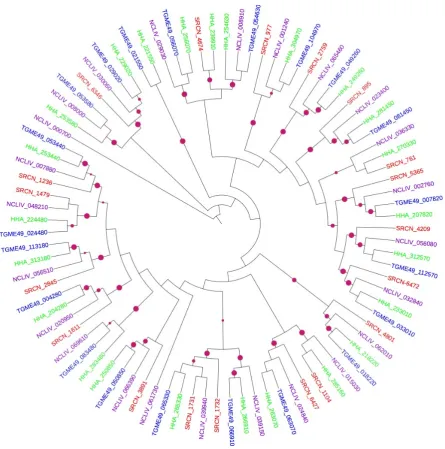
We phylogenetically deciphered evolutionary relationships among the various PK groups in the S. neurona kinomes with the PKs in other apicomplexans, which revealed that the S. neurona kinome has slightly fewer AGCs (n=9) compared to the kinomes of T. gondii (n=11), N. caninum (n=13) and H. hammondi (n=15). In general, the phylogenetic clustering of the S. neurona AGCs mirrored the homologies of these enzymes to those of the three apicomplexans used in this study (Figure 1; compare with Table 1).
14 of 34
Sequence analysis of S. neurona AGCs revealed significant divergence with only ~30% sequence similarity. Two S. neurona AGCs (SRCN_5610 and SRCN_3990) clearly cluster with T. gondii PKAs TGME49_028420 and TGME49_015670 [53] (Figure 1). Moreover, SRCN_5610 shares ~60% full length sequence identity with its ortholog. Further, two other S. neurona putative PKAs (SRCN_5165 and SRCN_3339), which also distinctly clustered with their T. gondii orthologs (TGME49_026030 and TGME49_086470, respectively) (Figure 1), contained the characteristic PKA’s ‘GxGxxG’ motif harboring a glycine triad that structurally forms a hairpin around the ATP binding pocket [53]. It is also notable that the single putative PKG (SRCN_4518) distinctly clustered with its T. gondii PKG ortholog (TGME49_111360) (Figure 1).
Figure 1. Mid-point rooted ML phylogenetic tree of apicomplexan AGCs. The terminal branches
are color-coded for AGCs in the kinomes of S. neurona (SRCN; red), T. gondii, ME-49 strain (TGME49; blue), H. hammondi (HHA; green) and N. caninum, Liverpool strain (NCLIV; purple). A solid purple circles on a branch indicates bootstrap support greater than 70. The phylogenetic tree was inferred from a multiple sequence alignment using PhyML with LG amino acid substitution model and gamma model of substitution rate heterogeneity. The tree image was rendered with iTOL.
The SRCN_3339, SRCN_5165, SRCN_5610 and SRCN_4518 possessed an additional functional domain; the AGC-kinase C-terminal domain that contained two of the three conserved
15 of 34
phosphorylation sites in AGCs (data not shown). These conserved sites serve as phosphorylation-regulated switches to control both intra- and inter-molecular interactions [54]. A striking feature in S. neurona is that like in T. gondii, it lacks PKB and PKC. The putative PDK1 (SRCN_1312) clustered with the T. gondii PDPK (TGME49_068210) [53].
Despite the absence of PKC in S. neurona, CAMK family members were identified, which perhaps underscores the importance of Ca2+ regulation in this apicomplexan. Majority of the identified S. neurona CAMKs segregated with their orthologs in T. gondii, N. caninum and H. hammondi in clades with robust bootstraps (Figure 2), thus validating the annotation of the CAMKs. Amongst the CAMKs, SRCN_2544 clustered with T. gondii PK1 (TGME049_043500) of the AMPK/SNF1 sub-family. There were also three additional SNF1 members in S. neurona (SRCN_5410, SRCN_4815 and SRCN_2257), which clustered with T. gondii TGME49_115190, TGME49_033900 and TGME49_091050, respectively.
Figure 2. Mid-point rooted ML phylogenetic tree of apicomplexan CAMKs. The terminal branches
are color-coded for AGCs in the kinomes of S. neurona (SRCN; red), T. gondii, ME-49 strain (TGME49; blue), H. hammondi (HHA; green) and N. caninum, Liverpool strain (NCLIV; purple). A solid purple circles on a branch indicates bootstrap support greater than 70. The phylogenetic tree was inferred from a multiple sequence alignment using PhyML with LG amino acid substitution model and gamma model of substitution rate heterogeneity. The tree image was rendered with iTOL.
16 of 34
Based on the clustering with T. gondii CDPK orthologs, 10 S. neurona CDPKs, including CDPK1 (SRCN_3314), CDPK2 (SRCN_4390), CDPK2B (SRCN_2165), CDPK3 (SRCN_3701), CDPK4 (SRCN_6606), CDPK5 (SRCN_3583), CDPK6 (SRCN_3011), CDPK7 (SRCN_6597), CDPK8 (SRCN_5948) and CDPK9 (SRCN_5812) were identified (Figure 2). This result implies that S. neurona has potentially lost at least two CDPKs (compared to the 12 CDPK that have been reported in T. gondii [21]). The possible loss notwithstanding, S. neurona contained the 6 CDPKs that are expressed and are well-conserved in most apicomplexans (i.e. CDPK1, CDPK3, CDPK4, CDPK5, CDPK6, and CDPK7) [55]. Sequence analysis revealed that, like in other apicomplexans, all identified S. neurona CDPKs except CDPK7 (SRCN_6597) contained both a kinase domain and a Ca2+-binding domain known as the EF-hand domain [21]. Like its T. gondii ortholog, TGME49_028750 (TgCDPK7), the S. neurona CDPK7 (SRCN_6597) contains a pleckstrin-homology (PH) domain just upstream of its PK domain [55]. The domain architecture in CDPKs is such that kinase activity is stimulated upon Ca2+-binding. Finally, the clustering of SRCN_5227, SRCN_1071, SRCN_3011 and SRCN_4093 in Figure 2 imply possible species-specific expansion of the CAMKs in S. neurona.
Majority of the putative CMGCs identified in S. neurona clustered with robust bootstraps with the conserved CMGCs in T. gondii, N. caninum and H. hammondi (Figure 3). Notable were the clustering of the CDKs, including CDK7 (SRCN_4674, SRCN_2759 and SRCN_761), CDK10 (SRCN_895) and CDK11 (SRCN_977). In addition, comparing the phylogenetic clustering of the CDKs in Figure 3 and the annotations presented in Table 1, the S. neurona kinome also appeared to have putative CDK5 (SRCN_4801, SRCN_1104 and SRCN_6346), all of which were supported by robust bootstraps. It should be noted that CDKs are amongst the principle molecular switches that regulate cell cycle progression in the apicomplexan parasites [56]. Also notable was the clustering in the same clade of three MAPKs (SRCN_6472, SRCN_4209 and SRCN_5365) and two GSKs (SRCN_1731 and SRCN_1732) (Figure 3).
17 of 34
Figure 3. Mid-point rooted ML phylogenetic tree of apicomplexan CMGCs. The terminal branches
are color-coded for AGCs in the kinomes of S. neurona (SRCN; red), T. gondii, ME-49 strain (TGME49; blue), H. hammondi (HHA; green) and N. caninum, Liverpool strain (NCLIV; purple). A solid purple circles on a branch indicates bootstrap support greater than 70. The phylogenetic tree was inferred from a multiple sequence alignment using PhyML with LG amino acid substitution model and gamma model of substitution rate heterogeneity. The tree image was rendered with iTOL.
In the OPK family, SRCN_4528 and SRCN_2630 are putative NEKs given their clustering with T. gondii NEK kinases TGME49_119700 and TGME49_094260, respectively (Figure 4). The S. neurona SRCN_108 and SRCN_3444 are putative ULKs due to their clustering with their T. gondii ULK orthologs (TGME49_035750 and TGME49_040630, respectively). SRCN_3669 clustered with TGME49_066950, a TBC-domain (Tre-2/Bub2/Cdc16) containing kinase. Further, there seemed to be a species-specific expansion of S. neurona Aurora kinases, SRCN_1606, SRCN_6157 and SRCN_2403 which cluster with T. gondii aurora kinase TGME49_003010; the clade containing these kinases however was not supported by robust bootstrap values (see Figure 4). SRCN_286 is a putative Wee kinase given that it clusters with Wee kinases from T. gondii (TGME49_073690) as well as from H. hammondi (HHA_273690; compare Figure 4 with Table 1).
18 of 34
Figure 4. Mid-point rooted ML phylogenetic tree of apicomplexan OPKs. The terminal branches
are color-coded for AGCs in the kinomes of S. neurona (SRCN; red), T. gondii, ME-49 strain (TGME49; blue), H. hammondi (HHA; green) and N. caninum, Liverpool strain (NCLIV; purple). A solid purple circles on a branch indicates bootstrap support greater than 70. The phylogenetic tree was inferred from a multiple sequence alignment using PhyML with LG amino acid substitution model and gamma model of substitution rate heterogeneity. The tree image was rendered with iTOL.
The clustering of putative S. neurona ROPKs (Figure 4) was also notable. For instance, SRCN_7082 clustered with T. gondii ROP33 (TGME49_001130), implying that it is a ROP33 (Table 1). SRCN_6184 clustered with T. gondii ROP19A (TGME49_042240) and ROP38 (TGME49_042110). It should however be noted that the sequence similarities with the ROP38 was low, implying that this could be a ROP19A (based on annotations presented in Table 1). Other putative S. neurona ROPKs included SRCN_3247, which clustered with ROP27 (TGME49_113330), SRCN_2076 with ROP25 (TGME49_002780). Here, it is notable that the clustering of SRCN_2076 the T. gondii ROP25 was supported with low bootstrap value, potentially meaning that based on the annotations (Table 1), SRCN_2076 is an ortholog to ROP30. SRCN_2123 and SRCN_2183 clustered with ROP35 (TGME49_104740), while SRCN_7084 clustered with ROP37 (TGME49_094560). Notably, from the phylogenetic tree in figure 4, SRCN_7083 and SRCN_4410 are probably duplicated forms of ROP34 given their clustering with T. gondii TGME49_040090. There was also indication of species-expansion of the S. neurona putative ROP35 (SRCN_2123 and SRCN_2183; see Figure 4).Although annotations
19 of 34
(Table 1) showed that SRCN_4310 was a putative ROP33 (39% sequence similarity to H. hammondi ROP33), this enzyme clustered with a hypothetical protein (TGME49_039380) of T. gondii (Figure 4). This discrepancy between annotation and phylogeny could be due to differences in the T. gondii. Another apparent discrepancy was the annotation of SRCN_4503 as a putative eIF2K-B (74% sequence similarity to the T. gondii TgCatPRC2 [IF2K-B]; see Table 1); but phylogeny showed clustering of this protein with a putative T. gondii ROP30 (TGME49_027010) (see Figure 4). Note that in this figure, the phylogeny bootstraps were low in the segregation of SRCN_4503 with ROP30. Moreover, SRCN_3151 and SRCN_3075 both segregated with TGME49_018550, a PIK3R4 kinase-related protein; from the phylogeny, SRCN_3151 and SRCN_3075 probably represent duplications. SRCN_6572 falls in the same clade with TGME49_004100, an eIF2 kinase IF2K-C, and TGME49_111510, an eIF2 kinase IF2K-B. NEKs are involved in cell cycle regulation, while Aurora kinases play pivotal roles in endodyogeny, duplication rate and parasite virulence [28]. Taken together, the presence of a variety of ROPKs in S. neurona is interesting given the fact that in T. gondii, ROPKs are key virulence factors [57].
2.3 Structural modeling
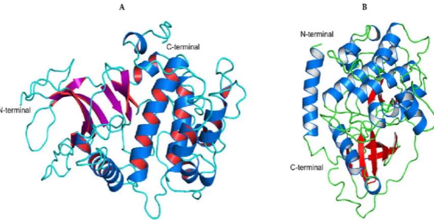
Using I-TASSER, we constructed the three-dimensional (3-D) homology models for SRCN_3247 using 4B6L, 4C0T, 1KOB, 4ZHX, 4IC7 and 4CZU as templates, and SRCN_5165 using 1CDK, 1SMK, 4WIH, 4Z84, 3PFQ, 4LQS and 1RDQ as templates. SRCN_3247 and SRCN_5165 are putative ROP27 and PKA respectively. From the predicted models for each of the sequences, the model with the highest C-score, a score that estimates the quality of the models produced, was used. The best SRCN_3247 model had a C-score of -0.46 and an estimated TM-score of 0.65 (Figure 5A) while the best SRCN_5165 model had a C-score of 1.6 and a TM-score of 0.94 (Figure 5B).
Figure 5. (A) Homology model of S. neurona SRCN_3247 (ROP27) generated based on template
structures 4B6L, 4C0T, 1KOB, 4ZHX, 4IC7 and 4CZU. (B) Homology model of SRCN_5165 generated based on template structures 1CDK, 1SMK, 4WIH, 4Z84, 3PFQ, 4LQS and 1RDQ.
Evaluation of the models’ quality and suitability for docking using PROCHECK revealed that SRCN_3247 and SRCN_5165 had 97.4% and 99.7% of the residues in the favored/allowed regions, respectively (Supplementary Figure 1A&B). The SRCN_3247 model contained 9 beta-sheets, 11 alpha and 5 3,10 helices while SRCN_5165 contains 9 beta-sheets, 11 alpha and 6 3,10 helices. Using the improved Protein Block Alignment (iPBA) webserver [58], structural superposition of the models to the top-ranking templates was performed for prediction of pair-wise structural alignment. The structural similarity score was measured using root mean square deviation (RMSD) between both Cα atom of the modeled protein and the corresponding template. The RMSD for the alignment of SRCN_3247 and 4B6L was 0.82 Å for the 276 aligned residues (Figure 6A) while that for
20 of 34
SRCN_5165 and 1CDK was 0.46 for 335 aligned residues (Figure 6B). The obtained RMSD values indicated that in both cases, the models and the templates had similar folds.
Figure 6. A. Superimposition of SRCN_3247 (marine) and 4B6L template (red). B. Superimposition of
SRCN_5165 (purple) and its template, 1CDK (red).
Analysis of putative binding pockets using DoGSiteScorer [59] revealed 14 potential pockets (P0-P13) on the surface of the SRCN_3247 model and 13 potential pockets (P0-P12) on the surface of the SRCN_5165 model. The largest pocket in SRCN_3247 (P0; Figure 7A) had a volume of 624.06 Å3 and a drug score of 0.79 while the largest pocket in SRCN_5165 (P0; Figure 8) had a volume of 760.51 Å3 and a drug score of 0.77.
Figure 7. A cartoon of the largest binding pockets of SRCN_3247 (Panel A) and SRCN_5165 (panel B).
Here, it should be noted that the presence of a binding pocket does not necessarily imply that a target protein is druggable. However, the binding volume is one of the important cavity properties
21 of 34
that influence the druggability of a particular target protein, of which most of druggable proteins have volumes of between 500-1000 Å3 [60].
2.4 Molecular docking of SRCN_3247 and SRCN_5165 with PK inhibitors
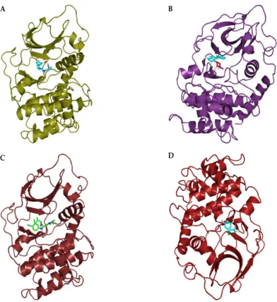
After the structural modeling, the binding modes of known kinase inhibitors to SRCN_3247 and SRCN_5165 was assessed by molecular docking of the chemical ligands into the predicted active site pockets of the modeled structures. For the docking analyses, the following four inhibitors were used: - BAY61-3606, an ATP-competitive inhibitor of mammalian spleen tyrosine kinase (Srk) [61]; Flavopiridol, a synthetic flavonoid that inhibits a wide range of cyclin-dependent kinases [62]; purvalanolB-B (PurB) is a potent CDK inhibitor [63]; and Piceatannol, a naturally occurring hydroxylated analogue of resveratrol that inhibits Syk kinase [64]. With a binding affinity of -8.4 kcal/mol, the overall binding pose of BAY61-3606 in SRCN_5165 is shown in Figure 8A. The binding affinity of flavopiridol to SRCN_5165 was -9.3 kcal/mol (Figure 8B). The binding energies for purB and piceatannol to SRCN_5165 were -6.1 kcal/mol (Figure 8C), and -6.0 kcal/mol (Figure 8D), respectively.
Figure 8. (A) Binding of BAY61-3606 to the active site pocket of SRCN_5165; (B) Binding of
flavopiridol to the active site pocket of SRCN_5165; (C) Binding of purvalanolB to the active site pocket of SRCN_5165; and (D) Binding of piceatannol to the active site pocket of SRCN_5165. The inhibitors and structural models are shown in stick and cartoon representations, respectively.
22 of 34
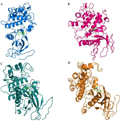
For SRCN_3247, BAY-61-3606 bound to the active site with an affinity of -7.2 kcal/mol (Figure 9A), while the affinities for flavopiridol, purB and piceatannol were -7.9 kcal/mol (Figure 9B), -6.9 kcal/mol (Figure 9C), and -7.3 kcal/mol (Figure 9D), respectively.
Figure 9. (A) Binding of BAY61-3606 to the active site pocket of SRCN_3247; (B) Binding of
flavopiridol to the active site pocket of SRCN_3247; (C) Binding of purvalanolB to the active site pocket of SRCN_3247; and (D) Binding of piceatannol to the active site pocket of SRCN_3247. The inhibitors and structural models are shown in stick and cartoon representations, respectively.
Taken together, by structural modeling and docking analysis, the above results indicate that the putative S. neurona PKA and ROP27 could be considered as drug targets. The data from the modeling and docking could pave way for further laboratory experiments to design potential drugs against EMP.
3. Discussion
We used different bioinformatics approaches to identify and classify the PKs of S. neurona, and docked four well-known inhibitors into modelled structures of two PK representatives. The critical roles performed by PKs make them attractive targets for novel therapeutics, and as such, the current study provides a rich source of potential drug targets, vaccine candidates or diagnostic proteins for developing new treatment and diagnostics strategies. PKs are considered viable drug targets because their catalysis mechanism and overall structure are conserved. Because small molecules can bind to the PKs’ catalytic clefts [65], many kinase inhibitors (e.g. imatinib, trastuzumab and lapatinib) have been developed to treat various human diseases [66].
23 of 34
The kinomes of apicomplexans range from 35 PKs (in Babesia bovis) to 135 PKs (in T. gondii) [35]. We identified a total of 92 putative PKs in the kinome of S. neurona, compared to the PKs reported in the kinomes of T. gondii (n=135), N. caninum (n=130) and H. hammondi (n=124) [15]. Although the total number of S. neurona PKs appeared markedly reduced compared to that of its close coccidian relatives (T. gondii and N. caninum [27]), taken as a percentage of total genome size, the proportion of S. neurona PKs is comparable to the 2% observed in humans [13] and other coccidians [27]. The contraction of the S. neurona kinome could be attributed to genome compaction, which occasionally offsets lineage-specific expansions of specific gene families. Notably, genome contraction is a common mode of genomic evolution in intracellular parasites, including apicomplexans [67, 68]. As such, the evolution of PKs may be in tandem to the overall genomic adaptive strategies of these parasites.
Using a hierarchical scheme based on the major PK groups, the S. neurona kinases could be classified and phylogenetically clustered into the various PK families. A complement of nine putative AGC kinases was identified in S. neurona, which is reduced compared with that of T. gondii, N. caninum and H. hammondi. Despite this potential gene loss, seven of the nine AGCs (SRCN_5165, SRCN_5610, SRCN_3339, SRCN_4249, SRCN_3990, SRCN_5430 and SRCN_1312) had orthologs in T. gondii, N. caninum and H. hammondi. In agreement with the observation that PKA is conserved in apicomplexans [27], two PKAs (SRCN_5610 and SRCN_3990) were identified in S. neurona. In eukaryotes, PKA signaling regulate various cellular responses such as DNA replication [69], cell growth and metabolism [70] and gene transcription by phosphorylating transcription factors such as the cAMP Response Element-Binding Protein (CREB) [71]. In T. gondii, increases in cytosolic cAMP levels activate PKA to trigger the developmental switch from the rapidly proliferating tachyzoites to the quiescent bradyzoites [72]. Additionally, two other S. neurona AGCs, (SRCN_5165 and SRCN_3339), were putative PKAs given that they contained the characteristic GxGxxG motif found in PKA [53]. The PKAs are attractive targets for the disruption of S. neurona growth. Notably, based on orthology, S. neurona contains a single putative PKG (SRCN_4518) that distinctly clustered with T. gondii PKG (TGME49_111360).
In apicomplexans, CAMKs modulate the intracellular Ca2+ concentration, which in turn regulates vital processes such as host-cell invasion, protein secretion and parasite differentiation. We identified four AMPK/SNF1 family members (SRCN_2544, SRCN_5410, SRCN_4815 and SRCN_2257). The AMP-activated PK cascade acts as a metabolic sensor that monitors cellular AMP and ATP levels and is activated by an elevation of the AMP:ATP ratio. In yeast, scarcity of nutrients and energy results in the activation of SNF1 [73]. When activated, AMPK restores energy balance by switching off ATP-consuming anabolic pathways and switching on ATP generating catabolic pathways such as fatty acid oxidation [74]. Further, we identified 10 putative CDPKs in the S. neurona, including CDPK1 (SRCN_3314), CDPK2 (SRCN_4390), CDPK2A (SRCN_2165), CDPK3 (SRCN_3701), CDPK4 (SRCN_6606), CDPK5 (SRCN_3583), CDPK6 (SRCN_3011), CDPK7 (SRCN_6597), CDPK8 (SRCN_5948) and CDPK9 (SRCN_5812). Compared to the 12 CDPKs reported in T. gondii [21], it appears that S. neurona had all the six well-conserved apicomplexan CDPKs (CDPK1, CDPK3, CDPK4, CDPK5, CDPK6 and CDPK7), which provide a link between Ca2+ signaling and parasite differentiation, motility, invasion and egress [55]. In T. gondii, down-regulation of CDPK1 interfered with parasite motility, host cell invasion and egress [44] while disruption of CDPK3 caused defective parasite egress [75]. Further, the essentiality of CDPK6 and CDPK7 in T. gondii have recently been demonstrated [76]. Indeed, TgCDPK1 has been targeted for the development of new drugs for toxoplasmosis [77]. Sequence analysis revealed that, similar to other apicomplexans, all identified S. neurona CDPKs except CDPK7 (SRCN_6597) contain both a PK domain and a EF-hand (Ca2+-binding) domain [21]. Similar to its T. gondii ortholog, TGME49_028750 (TgCDPK7), the S. neurona CDPK7 (SRCN_6597) contains a pleckstrin-homology (PH) domain just upstream of its PK domain [55]; the domain architecture is such that kinase activity is stimulated upon Ca2+ binding. Moreover, our phylogeny provided clues of possible gene duplications giving rise to SRCN_3990 and SRCN_3011 and well as SRCN_4093 and SRCN_1071). Interestingly, based on phylogenetic analysis, S. neurona probably contains four (SRCN_5227, SRCN_1071, SRCN_4093 and SRCN_3011) species- specific CAMKs.
24 of 34
The CMGCs, comprising of CDKs, MAPKs, GSKs, and CLKs coordinate a wide range of cellular functions in different species. For instance, members of the CDK subfamily are major coordinators of cell division in both mitosis and meiosis while the MAPKs subfamily is crucial in regulating cell proliferation, cell differentiation and cell death in eukaryotes [56, 78]. By both annotations and phylogenetic analyses, we identified four putative CDKs sub-family members; CDK5 (SRCN_4801 and SRCN_6346), CDK7 (SRCN_2759, SRCN_4674 and SRCN_761), CDK10 (SRCN_895) and CDK11 (SRCN_977). The finding of CDKs in S. neurona suggests that this parasite’s cell cycle regulation could be CDK-dependent and perhaps similar to that of higher eukaryotes [79]. The identification of three putative MAPKs (SRCN_4209, SRCN_6472 and SRCN_5365) in S. neurona points to the existence of MAPK regulated transduction pathway(s) in this pathogen. Similar to its T. gondii ortholog (TgMAPK1), which is a p38α MAPK homolog [80], SRCN_4209 may be potentially involved in parasite proliferation/stage differentiation, stress response and manipulation of the host immunity to enhance virulence. On the other hand, SRCN_6472 and SRCN_5365 may augment the roles of SRCN_4209 in the parasite. In T. gondii MAPK1/ERK7 is involved in intracellular proliferation [81]. We also identified two putative GSKs (SRCN_1731 and SRCN_1732). A genome-wide gene knockout approach in P. falciparum demonstrated PfGSK-3 to be critical for schizogony of the parasite [82]. Other S. neurona CMGC kinases identified include CLK (SRCN_1479), PRP4 (SRCN_2845), DYRK (SRCN_1611) CK2 (SRCN_6427) and SRPK (SRCN_1236).
ROPKs are secreted by T. gondii into the host cell and play roles in adhesion, motility and manipulation of immune responses [83]. We identified at 10 putative ROPK sub-family members in the S. neurona kinome; - ROP38 (SRCN_6184), ROP33 (SRCN_7082), ROP27 (SRCN_3247), ROP25 (SRCN_2076), ROP37 (SRCN_7084), ROP34 (SRCN_4410 and SRCN_7083), ROP35 (SRCN_2183), SRCN_2123 and ROP23 (SRCN_6812). It has been recently demonstrated that T. gondii ROP21 and ROP27 play a role in a constitutive pathway based on their localization in the PV and cyst matrix [84]. Moreover, T. gondii ROP35 has been shown to play a crucial role in chronic infection [85]. Although the S. neurona genome is more than twice the size of other coccidians whose genomes sequenced so far (e.g. Toxoplasma and Neospora), it has a considerably reduced number of ROPKs that nevertheless may have vital roles in the parasite’s virulence. Specifically, S. neurona is devoid of ROP5, ROP16, ROP18 and ROP38 that have been shown to confer virulence and alter the host’s cellular signaling pathways [81]. Putatively therefore, S. neurona ROPKs may have multiple roles in the survival of the parasite. In search of drug targets against S. neurona, the reduced ROPKs with possible multiple roles and absent in the vertebrate host are thus attractive candidates.
We explored the possibility of repositioning existing PK inhibitors as lead compounds in the development of new drugs for EPM, an approach that would substantially lower the cost of drug development. The lead compounds found via repositioning could be modified to increase efficacy. Molecular docking studies were therefore conducted to explore the potential binding of four inhibitors to the structural models of SRCN_3247 and SRCN_3314, representatives of putative S. neurona ROP27 and PKA, respectively. Probing the S. neurona kinome with small-molecule has many potential advantages. For instance, given that most kinase inhibitors in therapeutic use target multiple kinases with similar binding pockets [65], the promiscuity of such agents lead to polypharmacology [86] that would help minimize drug resistance. However, our modeling and docking analysis are only predictive, meaning that experimental validations are required.
4. Conclusion
The kinome of S. neurona contain members of the major classes of PKs, including AGC, CMGC, GSK, CAMK, CK, TKL, aPKs and several PKs in the OPK family. Similar to other apicomplexans, S. neurona kinome is devoid of PKC and the conventional TKs, but appears to have reduced MAPK family members. S. neurona kinome did not also have some of the ROPKs that have been implicated in the virulence of T. gondii. Known kinase inhibitors can be used as starting scaffolds in the search for drugs acting against EPM.