1
A coadunation of biological and mathematical
perspectives on the pandemic COVID-19: A Review
Sahar Qazi1, Kayenat Sheikh1, Mo Faheem2, Arshad Khan2, Khalid Raza1*1Bioinformatics & Computational Intelligence Lab., Department of Computer Science, Jamia Millia Islamia, New Delhi-110025
2Department of Mathematics, Jamia Millia Islamia, New Delhi-110025
kraza@jmi.ac.in*
March 30, 2020
Abstract:Coronaviruse disease (COVID-19) outbreak has created an emergency globally, and social distancing and isolation is the only solution to prevent its spread. Several countries have announced fully locked on to tackle this pandemic. The recent COVID-2019 has shaken the globe with incidence cases of more than half-million cases, and a mortality toll of more than twenty thousand to date. The coronavirus family is inclusive of pathogen of both – animal species and humans, encapsulating the isolated severe acute respiratory syndrome coronavirus (SARS-CoV). Researchers round the globe have been dexterously working to decode this lethal virus. Many mathematical frameworks have also been depicted which have helped to understand the dynamics of the COVID-19. Research on coronaviruses continues to explore various aspects of viral replication and pathogenesis to understanding the predilection of these viruses to switch between species, to develop an infection in a new host, and to identify significant reservoirs of coronaviruses will dramatically aid in our potential to prophesize when and where potential epidemics may occur. Many of the non-structural and accessory proteins encoded by the viruses remain unclear and unknown. This systematic review highlights the current situation of the pandemic, virus genomic composition, pathogenesis, symptomatology, diagnosis, and prognosis along with mathematical models of disease transmission and dynamics.
Keywords:nCov-19, COVID-19, coronavirus, SARS-CoV
1. Introduction
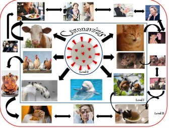
viral disease has latched itself to people and is growing rapidly in Europe, North America, Asia, and the Middle East, while the first confirmed cases identified in African and Latin American countries. The number of cases of COVID-19 has drastically grown up since 16th March, 2020 outside China. It is said that the carriers and reservoirs of this virus are bats and snakes (Leung, 2020; Zhou et al., 2020). Fig. 1 depicts the transmission of nCov-19 from Wuhan, China to the globe.
Fig. 1Diagrammatic representation nCoV-19 spread from Wuhan City, China to the globe
namely-Alphacoronavirus, Betacoronavirus, Gammacoronavirus, and Deltacoronavirus (Li, 2016). Alpha and Betacoronaviruses infect mammals while Gamma and Deltacoronaviruses infect birds (Tang et al., 2015). Few human infecting coronaviruses have been identified lately – for Alphacoronaviruses (HCoV-NL63 and HCoV229E) whereas, Betacoronaviruses are inclusive of HCoV-OC43, HCoVHKU1, severe acute respiratory syndrome coronavirus (SARS-CoV), and Middle East respiratory syndrome coronavirus (MERS-CoV) (Wu et al, 2020b).
Fig 2.Graphical representation of the COVID-19 statistics outside China during January 22 to March 25, 2020 (Data source: https://www.worldometers.info)
1.1 Outside China COVID-19 Scenario: A Worldometers Statistics
In order to write this review, we executed a meta-analysis to grasp the latest, relevant and significant information of COVID-19 cases outside China. Our main objective is to figure out the progression rate of COVID-19, recovery rate, and death rate, etc outside China. Fig 2
new cases versus recoveries, death rate versus recovery rate and distribution of cases in China and outside China during the period January 22 to March 25, 2020. There has been a huge exponential jump after 10thMarch, 2020 in COVID-19 cases in outside China (Fig. 2). These cases may grow even further shortly as per CDC estimation.
2. Genomic Composition
It has been observed that RNA viruses often carry out selective packaging of their genomes in a myriad of ways involving a genomic packaging signal. The first coronavirus packaging signal was studied thirty years back (Chen et al, 2020) howbeit; its preliminary functioning is still unknown. As previously mentioned, coronaviruses are composed of a non-segmented, positive-sense RNA genome of about ∼30 kb, which is inclusive of a 5’ cap entity in combination with a 3’ poly (A) tail which aids act as an mRNA for the translation of the replicase polyproteins (Fehr & Perlman, 2015). The replicase gene forming the non-structural proteins (NSP) occupies the majority (~20kb) of the genome when compared to the structural and accessory proteins which occupy the remaining 10kb. The 5’ end of the genome includes – a preliminary sequence and an untranslated region (UTR) containing multiple stem-loop structures necessary for RNA replication and transcription purpose. Moreover, many transcriptional regulatory sequences (TRSs) are attached at the initiation point of every structural gene important for their expression. The 3’ UTR is also composed of RNA structures mandatory for replication and production of the viral RNA. The symmetrical arrangement of the coronavirus genome is 5’-preliminary-UTR-replicase-S (Spike)–E (Envelope)-M (Membrane)-N (Nucleocapsid)-3’UTR-poly (A) tail in combination with accessory genes randomly dispersed within the structural genes at the 3’ end of the viral genome (Fig. 3). The accessory proteins may play an important role in viral pathogenesis even though not in replication (Fehr & Perlman, 2015).
other RNA species (Masters, 2019). Fig 3 represents the genomic composition of the novel coronavirus strain inclusive of initial primary ORFS and various accessory proteins.
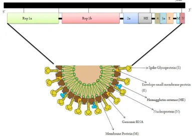
Fig 3.Genomic composition of the virus
3. Virus structure
Coronaviruses are large (diameter=~150-160 nm), round, bulbous, and pleiomorphic structures. Fig. 4 depicts the overall structure of SARS-CoV-2. The viral structure can be classified into two parts: theviral envelope, andnucleocapsid(Schoeman & Fielding, 2019).
3.1 The viral envelope
It is the outermost part of a virus, provides strength, rigidity and a definite shape. It comprises of Spike (S), Envelope (E) and Membrane (M). The subgroup of coronaviruses like Betacoronaviruses subgroup A has spike like protein structures embedded in the membrane called Hemagglutinin esterase (HE). SARS-CoV-2 differs from other viruses because it expresses HE (Schoeman & Fielding, 2019).
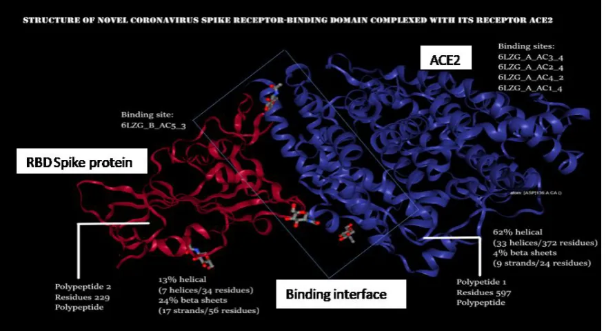
Glycosylated spike (S): For an easy entry inside the host cell, a densely glycosylated spike is used by the virus. S protein is a trimeric class I fusion protein that exists in a metastable prefusion conformation that undergoes a substantial structural rearrangement to fuse the viral membrane with the host cell membrane. The 3D structure of spike demonstrates that there exists a single Receptor Binding Domain (RBD) in the up conformation. As the RBD undergoes a hinge-like movement, S1 subunit becomes visible along with S2 unit (more stable). The S1 subunit determines the virus-host range and cellular tropism by RBD. The S2 mediates virus-cell membrane fusion by HR1 and HR2. This stochastic RBD movement is found in closely related beta-coronaviruses SARS-CoV and MERS-CoV and distantly related alpha-coronaviruses (PEDV). This mechanism in 2019-CoV indicates a similar trigger system in Coronaviridae, where receptor (ACE2) that binds to the RBDs forms 3 RBD up conformations (unstable) that results in the shredding of S1 and S2 refolding. Fig. 5
demonstrates the RBD of spike complexed with the ACE2 receptor (Protein Data Bank).
The overall spike structure of 2019-nCoV resembles to that of SARS-CoV spike. The minor difference between the two is the position of RBD in their respective down conformations. The spike of SARS-CoV-2 has a higher affinity for ACE2 receptor on the host, approximately 10 times more than the SARS-CoV affinity (Hoffmann et al, 2020). Polypeptide 1 (in blue color) is ACE2 (Angiotensin-converting Enzyme 2, Homo sapiens). It has four binding sites for spike protein namely, 6LZG_A_AC3_4, 6LZG_A_AC2_4, 6LZG_A_AC4_2, and 6LZG_A_AC1_4. Catalytic activity: (angiotensin II + H2O = angiotensin-1-7 + L-phenylalanine). Microbial infection: Interacts with Human coronavirus NL63/HCoV-NL63 spike glycoprotein. Polypeptide 2 (in red color) is the Spike protein of nCoV-19. It is the site of attachment to the ACE2 receptor of the host human cell for entry. Small molecules like NAG (N-acetyl-D-glucosamine) are found on both the chains whereas Zn ion is found only on the ACE2 receptor.
Envelope protein (E): Short integral membrane protein of 76-109 amino acids, from 8.4 to 12 kDa in size. Envelope has a short, hydrophilic amino terminus consisting of 7-12 amino acids, and then a large hydrophobic TMD of 25 amino acids and ends with a long, hydrophilic carboxyl terminus. The hydrophobic region of TMD has amphipathic alpha-helix that oligomerizes to form an ion-conductive pore in membranes. The TMD has 2 nonpolar, neutral amino acids valine and leucine, lending a strong hydrophobicity to the E protein, with an overall net charge zero. The middle region is uncharged and flanks on one side by N terminus and another side by C terminus (exhibits some hydrophobicity) of variable charge. The E protein has a bonding motif known as postsynaptic density protein 95 (PSD95) (PDZ)-binding-motif (PBM). The PDZ domain is a protein-protein interaction module that can bind to the C-terminus for target proteins such as cellular adapter proteins involved in host-cell processes important for viral infection (Schoeman & Fielding, 2019). The PBM region can either be deleted or mutated but it reverts to a pathogenic state. PBM mutations appear to be tolerated but that a reasonably intact PBM domain is still necessary to avoid revertant mutants. E protein interacts with M to form the viral envelope (Schoeman & Fielding, 2019).
incorporation is linked to particle size and that spikes cluster in regions where MLONG is generally present (Neuman & Kiss, 2011).
Hemagglutinin Esterase (HE): The hemagglutinin esterases (HEs) a homodimeric class I membrane protein are a family of viral envelope glycoproteins that causes reversible attachment to O-acetylated sialic acids by acting both as lectins and as receptor-destroying enzymes (RDEs). Enzymatic activity is associated with the hemagglutinin-esterase (HE), CoV HEs lack membrane fusion activity and, in the virion, they are accessory to the spike protein S (Zeng et al., 2008).
3.2 The nucleocapsid
It is protected by the viral envelope by strong surroundings. The positive-sense single-stranded RNA and nucleoprotein (N) are the genetic material ranging the size of 27- 34 kilobases. N protein is bound to the RNA genome to form nucleocapsid. The role of N protein is its potential ability to counter host immune responses as a viral suppressor protein of RNAi (VSR). The VSRs suppress the RNAi to overcome the host defense to establish infection (Wrapp et al., 2020).
4. The Life Cycle of Coronavirus
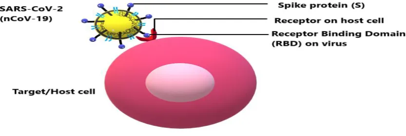
SARS-CoV-2 begins its life cycle when its glycosylated S protein binds to the cellular receptor ACE2 just like SARS-CoV.Fig. 6shows how the interaction for entry occurs (Jiang et al., 2012).
Fig. 6The interaction between the receptor-binding domain (RBD) present on the spike of the coronavirus and the corresponding host cell receptor
endosomal pathway (Hoffmann et al., 2020). However, camostat mesylate, an inhibitor of TMPRSS2 can block lung infection. The antibody response against SARS-CoV is efficient therefore it can be expected that similar kind of antibody neutralization might occur in the SARS-CoV-2 infection to some degree.SARS-CoV-2 then releases its genetic material i.e., +ssRNA into the host cell. Genome RNA composition is (5’UTR—Gene orf1ab--Gene S— Gene ORF3a—Gene E—Gene M—Gene ORF6—Gene ORF7a—Gene ORF8—Gene N— Gene ORF10--3’UTR).
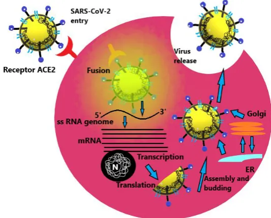
Genome RNA is translated into viral replicase polyproteins pp1a and 1ab, which are then cleaved into small products by viral proteinases. Fig. 7 depicts the life cycle of the virus inside a host.
Fig 7.The elaborative depiction of the life cycle pathway followed by the coronavirus inside a potent host cell
The polymerase produces a series of sub-genomic mRNAs by discontinuous transcription and finally translated into relevant viral proteins. Viral proteins and genome RNA are subsequently assembled into virions in the ER and Golgi and then transported via vesicles and released out of the cell. ACE2, angiotensin-converting enzyme 2; ER, endoplasmic reticulum; ERGIC, ER–Golgi intermediate compartment (Jiang et al., 2012).
5. Pathogenesis: Animal & Human
Animal Coronaviruses
persistent infection, mutation transforms the virus into a highly virulent strain of FCoV (Feline Infectious Peritonitis Virus, FIPV) progresses to the formation of a mortal disease namely – feline infectious peritonitis (FIP), which is of both the forms –wet and dry, quite similar to the sarcoidosis, which is a human disease. This virus is said to be lethal as it disturbs the cytokine and the chemokine expression leading to lymphocyte depletion (Perlman & Netland, 2009). Howbeit, this hypothesis is still not confirmed yet. On the other hand, bovine CoV, rat CoV, and infectious bronchitis virus (IBV) lead to benign to acute respiratory tract infections in cows, rats, and chickens respectively. Bovine CoV has been held responsible for an apex loss in the cattle industry as it infects cattle and spreads to other animals such – camel, deer, and elk, causing winter dysentery (diarrhoea), dehydration, weight loss, animal depression, lackadaisical attitude and also reduced milk production. IBV strains, namely – γ-coronavirus, are the janitors that affect the urogenital tract of chickens causing kidney disorders. Moreover, it reduces egg production in chicken and abnormal weight gain in chickens which is a significant loss in the poultry industry annually (Perlman & Nestland, 2009). Mihindukulasuriya et al (2008) identified a type of coronavirus from the liver of Beluga whales enduring respiratory infection namely – SW1, howbeit, electron microscopy failed to confirm whether it belonged to coronavirus family. There has always been much interest to study bat-based coronaviruses. The novel coronavirus (nCoV-19) is also said to be a BAT-SARS-Coronavirus, especially, Beta-BAT-SARS coronavirus. SARS and MERS coronaviruses and many other bat-based novel coronaviruses have been identified previously. Another coronavirus, Mesoniviridae, has been identified to infect insects (Nga et al, 2011 & Lauber et al, 2012). The most widely studied animal coronavirus is the murine hepatitis virus (MHV) responsible for neurological, respiratory, hepatic and enteric diseases in mice. For instance, MHV-1 leads to serious respiratory disease in A/J and C3H/HeJ mice, A59, while MHV-3 causes serious hepatitis. JHMV leads to encephalitis (Levy et al, 2000).
Human Coronaviruses
Perlamn, 2015). HCoV-NL63 has the onus of causing laryngotracheitis (van der Hoek et al, 2005). One interesting aspect of these viruses is variance in their genetic variability (Fehr & Perlman, 2015).
A group 2b β-coronavirus namely – SARS-CoV was first determined which was responsible forSevere Acute Respiratory Syndrome (SARS) spread in China during the year 2002–2003. During this outburst, around 8098 cases were observed with 774 lethalities leading to the death rate of 9%. Elderly people were morbid and mortality was also high in this section of individuals. This virus often attacks the epithelial cells within the lung and has the potential to penetrate the macrophages and dendritic cells (Peiris et al, 2003 & Spiegel et al, 2006). The novel coronavirus which has now become a pandemic around the globe is also said to belong from the beta-BAT-SARS-coronavirus family (Xu et al, 2020).
Another novel human CoV emerged in the Middle East back in 2012 whose nomenclature is –Middle East Respiratory Syndrome-CoV(MERS-CoV). It was observed to lead sequential pathogenic respiratory infections in Saudi Arabia and other countries in the Middle East (Zaki et al, 2012). With its rapid spread, it was assumed that it would lead to an apex and a very serious outburst, however, in 2013 newer cases never showed up. In 2014, an unpredictable outshoot of almost 200 cases and 40 mortalities from human-to-human transmission.Fig. 8 below represents the various levels of transmission and pathogenesis of the coronavirus. The level 0 represents the inactive coronavirus itself, which then enters an animal host, which is level I. Level II is a broader domain which is the transmission of the virus from animal-to-human and human-to-human respectively.
6. Signs and symptoms of COVID-19
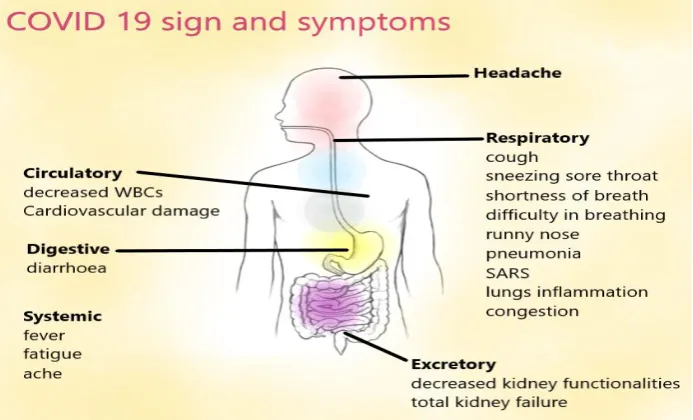
The most convincing mode of this virus transmission is said to be inhalation of contaminated/infectious aerosols whose incubation period is 3-14 days (CDC, 2020a). The disease caused by COVID-19 may range from asymptomatic to fatal disease. Underlying health conditions that enhance the susceptibility are hypertension, chronic obstructive pulmonary disease, diabetes, cardiovascular disease (Guo et al., 2020). Severe complications that might arise due to getting infected are respiratory distress syndrome, septic shock, metabolic acidosis which is hard to correct, coagulation dysfunction and multiple organ failure. Cytopathic effect (CPE) and cytokine storm or sustained inflammatory responses, hypoxia, septic shock etc. may be related to the critical conditions of SARS-CoV-2 infected patients (https://www.mayoclinic.org/).Fig. 9describes the symptoms of COVID-19.
Fig 9.Schematic diagram of signs, symptoms and affected organs in a human body infected with SARS-CoV-2 (Photo: Mikael Häggström, M.D./WikiCommons)
disorders of the brain, spinal cord, peripheral nerve, and muscle such as cerebral palsy, epilepsy (seizure disorders), stroke, intellectual disability, moderate to severe developmental delay, muscular dystrophy, or spinal cord injury are more prone to getting serious infection and reestablishment of health might be difficult. People belonging to older age groups, lacking efficient immune responses are also in close proximity to hazardous effects (Guo et al., 2020).
Manifestations are progressive in some cases to severe symptoms that can eventually lead to death are:
Fever associated with flu,
severe headache, dry cough
Upper/lower respiratory tract illness symptoms with shortness of breath and difficulty in breathing
Diarrhoea when the virus colonizes in the epithelial layer of the gastrointestinal tract,
general body fatigue/tiredness,
Severe pneumonia symptoms with abnormal chest CT, haemoptysis, and lymphopenia associated with SARS, acute cardiac injuries, kidney failure leads eventually to death.
7. Detection, Diagnosis & Prognosis
In order to diagnose COVID-19, like other viral diseases it composed of techniques that help to determine the presence of the virus and antibodies which develop in response to the infection. Because of limited screening, no countries had trustworthy data on the spread of the virus in their populace (Ioannidis, 2020).
7.1 Detection & Diagnosis Approaches
b) rRT-Polymerase Chain Reaction for virus detection: Deploying real-time reverse transcription-polymerase chain reaction (rRT-PCR) is the easiest way to figure out the infection in patients which can be done by taking the patients respiratory samples such as – nasopharyngeal swab or sputum sample (CDC, 2020). These results are available within a frame of either a few hours from the test to 2 days. This molecular biochemistry examination helps to identify the genomic composition. The initial PCR tests were produced at Charité in Berlin at the beginning of 2020 using real-time reverse transcription-polymerase chain reaction (rRT-PCR) which has now the main examination for CVD-19. Around 250,000 kits have been distributed by the WHO (Sheriden, 2020). These PCR-based kits are being deployed by Indian Council of Medical Research (ICMR) as well to diagnose infected patients. The most reliable diagnostic kits which have proved to have a high sensitivity and specificity, namely – RealStar SARS-CoV-2 PCR kit 1.0, Real Time Fluorescent RT-PCR Kit for detecting 2019- nCoV and TaqMan 2019-nCoV Control Kit v1 (ICMR, https://www.icmr.nic.in/content/covid-19#strategy).
c) Antibody Identification: Infections can also be diagnosed by determining the antibody production in an infected individual which mainly includes – antibody IgM and IgG. These are useful in identifying the immunity of a populace in the examination. Antibody identification tests can be performed by a point of care testing (PoCT). For identification by PoCT, a single blood sample is taken using puncturing the skin. It has been prophesized that the United States will launch an all-time PoCT by the 30thMarch, 2020 (FDA, 2020). In case of the Indian Council of Medical Research is concerned, they have employed MY LAB company’s PathoDetect assay which aids in identifying the coronavirus pathogens in the infected patients (ICMR, https://www.icmr.nic.in/content/covid-19#strategy).
7.2. Prognosis
of potential anti-viral drug leads namely – viral proteases, polymerases, and entry proteins. Unfortunately, much work yet remains in this domain of research. Approved vaccines are present for IBV, TGEV, and Canine CoV, however, they are not much effective in preventing the disease. As far as SARS-CoV is concerned, several potential vaccines such as – recombinant attenuated viruses, live virus vectors, or mono viral proteins expressed from DNA plasmids, are available but none of them are approved yet. Conventionally, it has been considered that live attenuated vaccines would be efficacious in targeting the coronaviruses. Vaccine generation for coronaviruses is an apex challenge and has seen many ups-downs in the past (Saif et al, 2004).
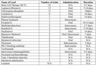
Since March, 2020, many drug candidates have been subjected tor clinical trials for their development of vaccines and treatments. These drugs mainly include compounds that are used to treat flu, malaria, Ebola disease (Khan et al., 2017). Table 1 summarises the conventional drug leads which have been subjected to clinical trials for COVID-19.
Table 1.Current drug leads subjected to clinical trials against COVID-19 (Li & Clercq, 2020)
Name of Lead Number of trials Administration Duration
Stem Cell Therapy (SCT) 23 N/A 5-7 days
Lopinavir/Ritonavir 15 Oral 7-14 days
Chloroquine phosphate 11 Oral 10days
Umifenovir 9 Oral 14 days
Hydroxychloroquine 7 Oral 14 days
Plasma treatment 7 Intravenous
-Favipiravir 7 Oral 10-14 days
Methylprednisolone 5 Intravenous 3-7 days
Remdesivir 5 Intravenous 5-10 days
Oseltamivir 2 Oral 14 days
Baloxavir Marboxil 2 Oral/ Intravenous 7 days
Thalidomide 2 Oral 14 days
Darunavir/cobicistat 2 Oral N/A
Thymosin 2 Subcutaneous 5 days
PD-1 blocking antibody 2 Intravenous N/A
Tocilizumab 2 N/A N/A
Intravenous Immunoglobulin 2 Intravenous 3-5 days
Ozonated autohemotherapy 2 N/A N/A
Type 1 Interferon injection 1 N/A N/A
Interferon nebulization 1 N/A N/A
Losartan N/A N/A N/A
the mathematical modeling for this virus (Li et al, 2020; Huang et al, 2020; Chan et al, 2020; Zhu et al, 2020; Bogosh et al, 2020; Wu et al, 2020c). The motive of these researches was to calculate the basic reproduction number ( ) by using Ordinary differential equations (ODEs), Markov Chain Monto Carlo methods (MCMC) (Wu et al, 2020c) or using intrinsic growth rate and serial intervals (Li et al 2020; Zhao et al, 2020a; Zhao et al, 2020b). In this section, we discuss five major models that exists in literatures that can help us understand the transmissibility of the SARS-CoV-2 from its natural reservoirs to humans.
8.1 The Bats-Hosts-Reservoir-People (BHRP) transmission network model
Chen et al. (2020) developed a mathematical model for simulating the phase-based transmissibility of the SARS-CoV-2. They used the data from the published literatures (Longini et al., 2005; Zhou et al, 2020; WHO, 2020(a); Japan MoH, 2020; WHO, 2020(b); WHO, 2020(c)). The assumption was that the virus first spreads among the bats and then is transmitted to some wild animals which were hunted and lastly delivered to the seafood market. These were now the ‘reservoir of the viruses’. People visited the market and therein got infected. The model is based on the following parameters:
1) The bats (B) and hosts (H) had been divided into quadrants namely – susceptible bats , exposed bats ( ), infectious bats ( ), removed bats and susceptible hosts , exposed hosts ( ), infectious hosts ( ), removed hosts respectively; while the people into five compartments – suspicious people , exposed people ( ) , symptomatic infected people ( ), symptomatic infected people ( ), removed people ( ).
2) The SARS-CoV-2 in a reservoir (the seafood market) was taken as . The retail purchase rate of the hosts in the market was assumed as . The transmission rate of infection through symptomatic infected people and asymptomatic infected people into was denoted by
and
3) , and denoted the birth rate of bats, hosts, and people, respectively, whereas and were taken as the death rate of the latter. The birth and the death rate of people during the outbreak were quite less. However, people who traveled, either migration or immigration across the Wuhan city reason being a bar, thus, and was taken as the rate of people coming into the city of Wuhan and going out of the city of Wuhan accordingly. and was taken as the total number of bats, hosts, and people, respectively.
4) , and denoted the new born bat, new born host and new born people, respectively and defined by
, and (1)
infection and human infection was denoted by t , t and t , respectivity. The transmission rate of infection through sufficient contact, from to , to and to were defined by and , respectively. The latent period of human infection was denoted by t and the transmission rate of infection through sufficient contact from to
was denoted by .
The dynamic and transmissibility of this disease modeled by the following system of first-order differential equations (Chen et al., 2020):
t
t
t t
t t
t t
t t
8.2 Susceptible-Exposed-Infectious-Recovered (SEIR) Model
The SEIR model was proposed by Tang et al. (2020a), the group studied the practical intercession of people’s health and measures were also included. The data used in this model has been taken from WHO situation report, the National Health Commission of the People’s Republic of China and the Health Commission of Wuhan City and Hubei Province (WHO, 2020d; WHO, 2020e). This research-based on the determination of the dynamic and epidemiology status of the disease by calculating the basic reproduction number. Let the susceptible population is , exposed population is , the pre-symptomatic population is , the infected population is , the hospitalised population is and the recovered population is The quarantined susceptible, isolated exposed and isolated infected population is denoted by , and respectively. The transmission and dynamic of the virus are modelled by the following system of differential equations:
t o
t 9
t t
t t
t
t
t t
t t
8.3 Time-Dependant Dynamic Model (TDDM)
Tang and collaborators updated their SEIR model using the latest available data on January 23, 2020 (Tang et al., 2020b) and called this model as TDDM. In TDDM model, it has been assumed that the contact rate is time dependant variable and given by,
t t t
Where initial contact is the rate on January 23, 2020, is the minimum contact rate by applying restrictions and control strategies, and denotes the exponential decreasing rate of contact rate. The transition rate of symptomatic infected individuals to the quarantined infected class t has been taken as an increasing function with respect to time,
t t t t t
The governing differential equation of this model is the same as the governing differential equation of the previous model, the only difference is the term is replaced by t andcis replaced byc(t). The effective daily reproduction number is defined by
t t t
t t
o
The same techniques and methods as in the SEIR model (Tang et al., 2020a) had been applied for simulating TDDM model. The estimated reproduction number by this study is 6.47.
8.4 Conceptual model
Lin et al. (2020) proposed the conceptual model where individual behavioral reaction and governmental actions were also included. Susceptible-Exposed-Infectious-Removed (SEIR) model has been carried out with two extra compartments, namely ‘D’ mimicking the public perception of risk regarding the 54 number of severe and critical cases and deaths, and ‘C’ represented the number of cumulative cases.
The following system of differential equations are used for modeling (Lin et al., 2020),
t
t
9
t
t
t
t
t t
t
t t
Where N is the total population, S, E, I, R denotes the susceptible, exposed, infected and removed population respectively, and t is the transmission rate that can be computed using the following relation,
t t
The basic reproduction number is calculated by employing next generation matrix approach and given by,
(27)
In this model, the estimated value of is 2.8.
8.5 Migration & Emigration Model
Wu et al. (2020c) proposed the migration and emigration model based on the data from December 31, 2019, to January 28, 2020. In this study, they assumed that the virus transmission occurred due to the migration and emigration of people from Wuhan city, China. Hence, the virus can be exported internationally. Following SEIR model is used for Wuhan epidemic,
t t t t t t t t t t o
t t t t t t t t t 9
t t t t t t
t t t
The expected number of exportation case on day ‘d’ is calculated by,
t t
The maximum likelihood estimation for reproduction number is given by,
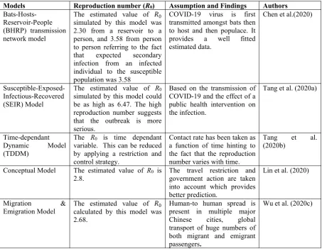
Markov Chain Monte Carlo method was applied for calculating the basic reproductive number. They simulated the SEIR model by putting the value of (Wu et al., 2020c). Table 2 summarises and compares the five discussed COVID-19 transmission models and Fig. 10 presents their simulated results. The SEIR model by Tang et al. (2020a) and TDDM model by Tang et al. (2020b) give better prediction. The reproduction number (R0) estimated by these two model is highest among all the models which suggest that this outbreak could be more serious than predicted in other models.
Table 2.Summarized comparative descriptions of the five COVID-19 transmission models
Models Reproduction number (R0) Assumption and Findings Authors
Bats-Hosts-Reservoir-People (BHRP) transmission network model
The estimated value of simulated by this model was 2.30 from a reservoir to a person, and 3.58 from person to person referring to the fact that expected secondary infection from an infected individual to the susceptible population was 3.58
COVID-19 virus is first transmitted amongst bats then to host and then populace. It provides a well fitted estimated data.
Chen et al.(2020)
Susceptible-Exposed-Infectious-Recovered (SEIR) Model
The estimated value of R0 simulated by this model could be as high as 6.47. The high reproduction number suggests that the outbreak is more serious.
Based on the transmission of COVID-19 and the effect of a public health intervention on the infection.
Tang et al. (2020a)
Time-dependant Dynamic Model (TDDM)
The R0 is time dependant variable. This can be reduced by applying a restriction and control strategy.
Contact rate has been taken as a function of time hinting to the fact that the reproduction number varies with time.
Tang et al. (2020b)
Conceptual Model The estimated value of R0 is
2.8. The travel restriction andgovernment action are taken into account which provides better prediction.
Lin et al. (2020)
Migration &
Emigration Model The estimated value ofcalculated by this model was 2.68.
Human-to human spread is present in multiple major Chinese cities, global transport of huge numbers of both migrant and emigrant passengers.
Discussion and Conclusion
The deadliest pandemic of severe acute respiratory syndrome coronavirus 2 (SARS-CoV-2), also called coronavirus disease (COVID-19), began in early December 2019 from the Wuhan city of China and spread to the world during January-March, 2020. During February-March 2020, the apex spread of this viral disease has latched itself to people and is growing rapidly in Europe, North America, Asia, and the Middle East. Although it is originated from China, the statistical analysis of the period January 22 to March 25, 2020, states an exponential growth of COVID-19 cases in outside China as well, which is most likely to further grow shortly. Coronaviruses lead to acute respiratory tract infections in 15-30% of the infected human populace and have been observed to be very serious in the elderly population and in individuals who endure cancer or any other immunological disorder. Severe complications may arise due to its infection includingrespiratory distress syndrome,septic shock,metabolic acidosis, coagulation dysfunction, and multiple organ failure. Further, cytopathic effect, cytokine storm, sustained inflammatory response, hypoxia, septic shock and so on may be related to the critical conditions of SARS-CoV-2 infected patients. Furthermore, severe pneumonia symptoms with abnormal chest CT, haemoptysis, and lymphopenia associated with SARS, acute cardiac injuries, kidney failure may eventually lead to death.
Coronaviruses are large (diameter=~150-160 nm), round, bulbous, and pleiomorphic structures whose outer surface provides strength, rigidity and a definite shape. These belong to Nidovirales order and are enveloped single-stranded positive-strand RNA virus with a genome of ~30kb. Human coronaviruses were first described in the 1960s and are said to have the onus for severe respiratory and gastrointestinal tract infections. The RNA viruses often carry selective packaging of their genomes and the first coronavirus packaging signal was studied thirty years back however, its preliminary functioning is still unclear. The packaging of coronavirus is quite a challenge to obtain a specific recognition and selection of gRNAs and other RNA species.
To understand the COVID-19 epidemic dynamics, several mathematical models were applied where basic reproduction number (R0) is computed by using Ordinary differential equations (ODEs), Markov Chain Monto Carlo methods (MCMC), intrinsic growth rate and serial intervals. Some of the models covered in the review are the Bats-Hosts-Reservoir-People (BHRP) transmission network model (Chen et al. 2020), the Susceptible-Exposed-Infectious-Recovered (SEIR) model (Tang et al. 2020a), Time-Dependant Dynamic Model (TDDM) (Tang et al., 2020b), conceptual model (Lin et al. 2020) and Migration & Emigration Model (Wu et al. 2020c). Out of these models, the SEIR model and TDDM model give a better prediction. Further, the reproduction number estimated by these two models is highest among all the models which suggest that this outbreak could be more serious than predicted in other models.
recombinant attenuated viruses, live virus vectors, or mono viral proteins expressed from DNA plasmids, are available but none of them are yet approved. Vaccine generation for coronaviruses is an apex challenge to date. Since March 2020, many drug candidates have been subjected to clinical trials.
Acknowledgements
SQ is supported by DST-Inspire Fellowship by the Department of Science & Technology (DST), MF is supported by CSIR-Junior Research Fellowship by the Council of Scientific and Industrial Research (CSIR), Government of India. Kayenat and KR acknowledge the Bioinformatics Infrastructure Facilities (BIF) at the Department of Computer Science, Jamia Millia Islamia funded by Department of Biotechnology (DBT), Government of India.
Conflict of Inerest
Authors declare that there is no any conflict of interst in the publication of this manuscript.
References
Ai, T. & Yang, Z. (2020).Correlation of Chest CT and RT-PCR Testing in Coronavirus Disease 2019 (COVID-19) in China: A Report of 1014 Cases. Radiology. Radiological Society of North America: 200642.https://doi.org/10.1148%2Fradiol.2020200642
Bai, H. X., Hsieh, B., et al. (2020). Performance of radiologists in differentiating COVID-19 from viral pneumonia on chest CT.Radiology: 200823.https://doi.org/10.1148%2Fradiol.2020200823
Bogoch, I. I., Watts, A., Thomas-Bachli, A., Huber, C., Kraemer, M. U., & Khan, K. (2020). Pneumonia of Unknown Etiology in Wuhan, China: Potential for International Spread Via Commercial Air Travel.Journal of Travel Medicine.https://doi.org/10.1093/jtm/taaa008
Bradburne, A. F., Bynoe, M. L., & Tyrrell, D. A. (1967). Effects of a" new" human respiratory virus
in volunteers. British medical journal, 3(5568), 767.
https://dx.doi.org/10.1136%2Fbmj.3.5568.767
CDC (2020a). Community mitigation strategies, Centers for Disease Control and Prevention, USA.
https://www.cdc.gov/coronavirus/2019-ncov/downloads/community-mitigation-strategy.pdf
(accessed on March 25, 2020).
CDC.(2020). Real-Time RT-PCR Panel for Detection 2019-nCoV Centers for Disease Control and Prevention.https://www.cdc.gov/coronavirus/2019-ncov/summary.html (accessed on March 25, 2020)
Chan, J. F. W., Yuan, S., Kok, K. H., To, K. K. W., Chu, H., Yang, J., ... & Tsoi, H. W. (2020). A familial cluster of pneumonia associated with the 2019 novel coronavirus indicating person-to-person transmission: a study of a family cluster. The Lancet, 395(10223), 514-523.
https://doi.org/10.1016/S0140-6736(20)30154-9
suggests a mechanism for helical packaging of viral RNA.Journal of molecular biology,368(4), 1075-1086.https://doi.org/10.1016/j.jmb.2007.02.069
Chen, T. M., Rui, J., Wang, Q. P., Zhao, Z. Y., Cui, J. A., & Yin, L. (2020). A mathematical model for simulating the phase-based transmissibility of a novel coronavirus. Infectious Diseases of Poverty,9(1), 1-8.https://doi.org/10.1186/s40249-020-00640-3
China-CDC (2020). Tracking the Epidemic. China CDC Weekly.
http://weekly.chinacdc.cn/news/TrackingtheEpidemic.htm (accessed on March 25, 2020)
Cinatl, J., Morgenstern, B., Bauer, G., Chandra, P., Rabenau, H., & Doerr, H. W. (2003). Treatment of SARS with human interferons. The Lancet, 362(9380), 293-294. https://doi.org/10.1016/S0140-6736(03)13973-6
Dong, L., Hu, S., & Gao, J. (2020). Discovering drugs to treat coronavirus disease 2019 (COVID-19).Drug Discoveries & Therapeutics,14(1), 58-60.http://doi.org/10.5582/ddt.2020.01012
Escors, D., Ortego, J., Laude, H., & Enjuanes, L. (2001). The membrane M protein carboxy terminus binds to transmissible gastroenteritis coronavirus core and contributes to core stability.Journal of virology,75(3), 1312-1324.http://doi.org/10.1128/JVI.75.3.1312-1324.2001
Fehr, A. R., & Perlman, S. (2015). Coronaviruses: an overview of their replication and pathogenesis. In Coronaviruses (pp. 1-23). Humana Press, New York, NY. https://doi.org/10.1007/978-1-4939-2438-7_1
Gui, M., Liu, X., Guo, D., Zhang, Z., Yin, C. C., Chen, Y., & Xiang, Y. (2017). Electron microscopy studies of the coronavirus ribonucleoprotein complex. Protein & cell, 8(3), 219-224.
https://doi.org/10.1007/s13238-016-0352-8
Guo, Y. R., Cao, Q. D., Hong, Z. S., Tan, Y. Y., Chen, S. D., Jin, H. J., ... & Yan, Y. (2020). The origin, transmission and clinical therapies on coronavirus disease 2019 (COVID-19) outbreak–an update on the status.Military Medical Research,7(1), 1-10. https://doi.org/10.1186/s40779-020-00240-0
Hamre, D., & Procknow, J. J. (1966). A new virus isolated from the human respiratory tract. Proceedings of the Society for Experimental Biology and Medicine, 121(1), 190-193.
https://doi.org/10.3181%2F00379727-121-30734
Hoffmann, M., Kleine-Weber, H., Schroeder, S., Krüger, N., Herrler, T., Erichsen, S., ... & Müller, M. A. (2020). SARS-CoV-2 cell entry depends on ACE2 and TMPRSS2 and is blocked by a clinically proven protease inhibitor.Cell.https://doi.org/10.1016/j.cell.2020.02.052
Huang, C., Wang, Y., Li, X., Ren, L., Zhao, J., Hu, Y., ... & Cheng, Z. (2020). Clinical features of patients infected with 2019 novel coronavirus in Wuhan, China. The Lancet, 395(10223), 497-506.https://doi.org/10.1016/s0140-6736(20)30183-5
Ioannidis, John P.A. (2020). A fiasco in the making? As the coronavirus pandemic takes hold, we are making decisions without reliable data. STAT News, March 17, 2020.
Jiang, S., Lu, L., Liu, Q., Xu, W., & Du, L. (2012). Receptor-binding domains of spike proteins of emerging or re-emerging viruses as targets for development of antiviral vaccines. Emerging microbes & infections,1(1), 1-8.https://doi.org/10.1038/emi.2012.1
Khan, F.N., Qazi, S., Tanveer, K. & Raza, K.. (2017). A Review on the Antagonist Ebola: A Prophylactic Approach. Biomedicine & Pharmacotherapy, Elsevier, 96: 1513-1526.
https://doi.org/10.1016/j.biopha.2017.11.103
Lauber, C., Ziebuhr, J., Junglen, S., Drosten, C., Zirkel, F., Nga, P. T., ... & Gorbalenya, A. E. (2012). Mesoniviridae: a proposed new family in the order Nidovirales formed by a single species of mosquito-borne viruses. Archives of virology, 157(8), 1623-1628. https://doi.org/10.1007/s00705-012-1295-x
Leung, C. (2020). The difference in the incubation period of 2019 novel coronavirus (SARS-CoV-2) infection between travelers to Hubei and non-travelers: The need of a longer quarantine period.Infection Control & Hospital Epidemiology, 1-8.https://doi.org/10.1017/ice.2020.81
Levy, G. A., Liu, M., Ding, J., Yuwaraj, S., Leibowitz, J., Marsden, P. A., ... & Phillips, M. J. (2000). Molecular and functional analysis of the human prothrombinase gene (HFGL2) and its role in viral hepatitis. The American journal of pathology, 156(4), 1217-1225.
https://doi.org/10.1016/S0002-9440(10)64992-9
Li, F. (2016). Structure, function, and evolution of coronavirus spike proteins. Annual review of virology,3, 237-261.https://doi.org/10.1146/annurev-virology-110615-042301
Li, G., & De Clercq, E. (2020). Therapeutic options for the 2019 novel coronavirus (2019-nCoV). Nature Reviews Drug Discovery, 19: 149-150.http://doi.org/10.1038/d41573-020-00016-0
Li, Q., Guan, X., Wu, P., Wang, X., Zhou, L., Tong, Y., ... & Xing, X. (2020). Early transmission dynamics in Wuhan, China, of novel coronavirus–infected pneumonia.New England Journal of Medicine, 382:1199-1207. https://doi.org/10.1056/NEJMoa2001316
Lin, Q., Zhao, S., Gao, D., Lou, Y., Yang, S., Musa, S. S., ... & He, D. (2020). A conceptual model for the outbreak of Coronavirus disease 2019 (COVID-19) in Wuhan, China with individual reaction and governmental action. International Journal of Infectious Diseases, 93: 211-216.
https://doi.org/10.1016/j.ijid.2020.02.058
Longini, I. M., Nizam, A., Xu, S., Ungchusak, K., Hanshaoworakul, W., Cummings, D. A., & Halloran, M. E. (2005). Containing pandemic influenza at the source.Science,309(5737), 1083-1087.http://doi.org/10.1126/science.1115717
Masters, P. S. (2006). The molecular biology of coronaviruses.Advances in virus research,66, 193-292.https://doi.org/10.1016/S0065-3527(06)66005-3
Masters, P.S. (2019). Coronavirus genomic RNA packaging. Virology, 537: 198-207.
https://doi.org/10.1016/j.virol.2019.08.031
McIntosh, K., Becker, W. B., & Chanock, R. M. (1967). Growth in suckling-mouse brain of" IBV-like" viruses from patients with upper respiratory tract disease. Proceedings of the National
Academy of Sciences of the United States of America, 58(6), 2268.
Mihindukulasuriya, K. A., Wu, G., Leger, J. S., Nordhausen, R. W., & Wang, D. (2008). Identification of a novel coronavirus from a beluga whale by using a panviral microarray.Journal of virology,82(10), 5084-5088.http://doi.org/10.1128/JVI.02722-07
Neuman, B. W., Adair, B. D., Yoshioka, C., Quispe, J. D., Orca, G., Kuhn, P., ... & Buchmeier, M. J. (2006). Supramolecular architecture of severe acute respiratory syndrome coronavirus revealed by electron cryomicroscopy. Journal of virology, 80(16), 7918-7928.
http://doi.org/10.1128/JVI.00645-06
Neuman, B. W., Kiss, G., Kunding, A. H., Bhella, D., Baksh, M. F., Connelly, S., ... & Siddell, S. G. (2011). A structural analysis of M protein in coronavirus assembly and morphology. Journal of structural biology,174(1), 11-22.https://doi.org/10.1016/j.jsb.2010.11.021
Nga, P. T., del Carmen Parquet, M., Lauber, C., Parida, M., Nabeshima, T., Yu, F., ... & Ichinose, A. (2011). Discovery of the first insect nidovirus, a missing evolutionary link in the emergence of the
largest RNA virus genomes. PLoS pathogens, 7(9): e1002215.
http://doi.org/10.1371/journal.ppat.1002215
Peiris, J. S. M., Chu, C. M., Cheng, V. C. C., Chan, K. S., Hung, I. F. N., Poon, L. L., ... & Chan, K. H. (2003). Clinical progression and viral load in a community outbreak of coronavirus-associated
SARS pneumonia: a prospective study. The Lancet, 361(9371), 1767-1772.
https://doi.org/10.1016/S0140-6736(03)13412-5
Perlman, S., & Netland, J. (2009). Coronaviruses post-SARS: update on replication and pathogenesis.Nature reviews microbiology,7(6), 439-450.https://doi.org/10.1038/nrmicro2147
Saif, L. J. (2004). Animal coronavirus vaccines: lessons for SARS.Developments in biologicals,119, 129-140.https://www.ncbi.nlm.nih.gov/pubmed/15742624
Salehi, S., Abedi, A., Balakrishnan, S., & Gholamrezanezhad, A. (2020). Coronavirus Disease 2019 (COVID-19): A Systematic Review of Imaging Findings in 919 Patients.American Journal of Roentgenology, 1-7.https://doi.org/10.2214%2FAJR.20.23034
Schoeman, D., & Fielding, B. C. (2019). Coronavirus envelope protein: current knowledge. Virology journal,16(1), 69.https://doi.org/10.1186/s12985-019-1182-0
Sheridan, C. (2020). Coronavirus and the race to distribute reliable diagnostics. Nat Biotechnol.
http://doi.org/10.1038/d41587-020-00002-2
Spiegel, M., Schneider, K., Weber, F., Weidmann, M., & Hufert, F. T. (2006). Interaction of severe acute respiratory syndrome-associated coronavirus with dendritic cells. Journal of general virology,87(7), 1953-1960.https://doi.org/10.1099/vir.0.81624-0
Tang, B., Bragazzi, N. L., Li, Q., Tang, S., Xiao, Y., & Wu, J. (2020b). An updated estimation of the risk of transmission of the novel coronavirus (2019-nCov). Infectious Disease Modelling,5, 248-255.https://doi.org/10.1016/j.idm.2020.02.001
Tang, B., Wang, X., Li, Q., Bragazzi, N. L., Tang, S., Xiao, Y., & Wu, J. (2020a). Estimation of the transmission risk of the 2019-nCoV and its implication for public health interventions. Journal of Clinical Medicine,9(2), 462.https://doi.org/0.3390/jcm9020462
van der Hoek, L., Pyrc, K., Jebbink, M. F., Vermeulen-Oost, W., Berkhout, R. J., Wolthers, K. C., ... & Berkhout, B. (2004). Identification of a new human coronavirus. Nature medicine, 10(4), 368-373.https://doi.org/10.1038/nm1024
van der Hoek, L., Sure, K., Ihorst, G., Stang, A., Pyrc, K., Jebbink, M. F., ... & Überla, K. (2005).
Croup is associated with the novel coronavirus NL63. PLoS medicine, 2(8): e240.
http://doi.org/10.1371/journal.pmed.0020240
Wang, M., Cao, R., Zhang, L., Yang, X., Liu, J., Xu, M., ... & Xiao, G. (2020). Remdesivir and chloroquine effectively inhibit the recently emerged novel coronavirus (2019-nCoV) in vitro.Cell research,30(3), 269-271.https://doi.org/10.1038/s41422-020-0282-0
WHO (2020a). World Health Organization. Novel Coronavirus – China. Available:
https://www.who.int/csr/don/12-january-2020-novel-coronavirus-china/en/(accessed on March 20, 2020).
WHO (2020b). World Health Organization. Novel Coronavirus – Thailand (ex-China). Available:
https://www.who.int/csr/don/14-january-2020-novel-coronavirus-thailand/en/ (accessed on March 20, 2020).
WHO (2020c). World Health Organization. Novel Coronavirus – Japan (ex-China). Available:
https://www.who.int/csr/don/17-january-2020-novel-coronavirus-japan-ex-china/en/ (accessed on March 20, 2020).
WHO (2020d). World Health Organization. Novel Coronavirus—China, Disease Outbreak News: Update. Available: https://www.who.int/csr/don/12-january-2020-novel-coronavirus-china/en/. (accessed on January 23, 2020).
WHO (2020e). World Health Organization. Situation Report. Available:
https://www.who.int/docs/default-source/coronaviruse/situation-reports/20200123-sitrep-3-2019-ncov.pdf(accessed on January 23, 2020).
Woo, P. C., Lau, S. K., Chu, C. M., Chan, K. H., Tsoi, H. W., Huang, Y., ... & Poon, L. L. (2005). Characterization and complete genome sequence of a novel coronavirus, coronavirus HKU1, from patients with pneumonia.Journal of virology,79(2), 884-895. http://doi.org/10.1128/JVI.79.2.884-895.2005
Wrapp, D., Wang, N., Corbett, K. S., Goldsmith, J. A., Hsieh, C. L., Abiona, O., ... & McLellan, J. S.
(2020). Cryo-EM structure of the 2019-nCoV spike in the prefusion
conformation.Science,367(6483), 1260-1263.http://doi.org/10.1126/science.abb2507
Wu, A., Peng, Y., Huang, B., Ding, X., Wang, X., Niu, P., ... & Sheng, J. (2020b). Genome composition and divergence of the novel coronavirus (2019-nCoV) originating in China.Cell host & microbe, 27(3): 325-328.https://doi.org/10.1016/j.chom.2020.02.001
Wu, J. T., Leung, K., & Leung, G. M. (2020c). Nowcasting and forecasting the potential domestic and international spread of the 2019-nCoV outbreak originating in Wuhan, China: a modelling study.The Lancet,395(10225), 689-697.https://doi.org/10.1016/S0140-6736(20)30260-9
Xu, Y., Li, X., Zhu, B., Liang, H., Fang, C., Gong, Y., ... & Zhang, H. (2020). Characteristics of pediatric SARS-CoV-2 infection and potential evidence for persistent fecal viral shedding.Nature Medicine, 1-4.https://doi.org/10.1038/s41591-020-0817-4
Zaki, A. M., Van Boheemen, S., Bestebroer, T. M., Osterhaus, A. D., & Fouchier, R. A. (2012). Isolation of a novel coronavirus from a man with pneumonia in Saudi Arabia. New England Journal of Medicine,367(19), 1814-1820.http://doi.org/10.1056/NEJMoa1211721
Zeng, Q., Langereis, M. A., van Vliet, A. L., Huizinga, E. G., & de Groot, R. J. (2008). Structure of coronavirus hemagglutinin-esterase offers insight into corona and influenza virus evolution. Proceedings of the National Academy of Sciences, 105(26), 9065-9069.
https://doi.org/10.1073/pnas.0800502105
Zhao, S., Lin, Q., Ran, J., Musa, S. S., Yang, G., Wang, W., ... & Wang, M. H. (2020a). Preliminary estimation of the basic reproduction number of novel coronavirus (2019-nCoV) in China, from 2019 to 2020: A data-driven analysis in the early phase of the outbreak. International Journal of Infectious Diseases,92, 214-217.https://doi.org/10.1016/j.ijid.2020.01.050
Zhao, S., Musa, S. S., Lin, Q., Ran, J., Yang, G., Wang, W., ... & Wang, M. H. (2020b). Estimating the unreported number of novel coronavirus (2019-nCoV) cases in China in the first half of January 2020: a data-driven Modelling analysis of the early outbreak. Journal of clinical medicine,9(2), 388.https://doi.org/10.3390/jcm9020388
Zhou, P., Yang, X. L., Wang, X. G., Hu, B., Zhang, L., Zhang, W., ... & Chen, H. D. (2020). A pneumonia outbreak associated with a new coronavirus of probable bat origin. Nature, 1-4.
https://doi.org/10.1038/s41586-020-2012-7