The human-COVID-19 Tango: Connecting the Dots
Hamdi K Hamdi
1*and Ensaf M AbdAldayem
21Molecular Sciences Department, Ensafco USA, the Biomedical Research Institute, Orange, California, USA 2Middle East University, Faculty of Educational Sciences, Department of Educational Technology, Amman, Jordan
Corresponding Author: Hamdi K Hamdi, [email protected]
As the world grapples with a pandemic with various and expanding epicenters, a flurry of medical and scientific activity has gained speed and momentum in a race to halt COVID-19. A controversial topic has been the connection between COVID-19 and the Renin-Angiotensin system (RAS). COVID-19, like Sars before it, enters by way of the Angiotensin Converting Enzyme 2 (ACE2). ACE2 is ubiquitously expressed in many tissues in the body serving as the doorway by which the virus can enter and spread causing inflammatory havoc. Demographic evidence coming out of China and other locations make it clear that the elderly and those suffering cardiovascular complications such as hypertension etc are most at risk. The connection to RAS and the demographic nature of the data coming out has led many to advance hypothesis, recommendations and even therapies based on existing RAS inhibitors and other components of the renin-Angiotensin system. It is pertinent to review the literature in the context of our understanding of the renin-angiotesnin system to allow better judgements to be made as well as lines of research initiated advancing a quick resolution to COVID-19. Covid-19 appears invincible as if dipped in the river Styx, but even Achilles had a vulnerable heel. Understanding the homeostatic balance that the coronavirus disrupts, we can discover the arrow in corona’s
heel.
Key words: COVID-19, ACE, ACE2, Angiotensin II, Bradykinin, RAS inhibitors, ACE inhibitors, AT1 receptor blockers, Losartan, Bradykinin Antagonists, Ang II loading,Giapreza, Icatibant.
Introduction
As the scientific community focuses on the genome of COVID-19 overlooking that it takes two to tango. COVID-19, like Sars before it, enters by way of ACE2 (Angiotensin Converting Enzyme 2). ACE2 is a member of the of the Renin-Angiotensin system (RAS). Due to the urgencyof the situation, it has becomes pertinent to connect the dots of the human-covid tango. This review will focus on RAS from the point of view of COVID-19. We apologize in advance if we did not include important papers on RAS in the context of the cardiovascular and other fields.
Survey Methodology
A literature search was conducted in the PubMed
database that included the terms: “Angiotensin and COVID-19”. Other search terms that included
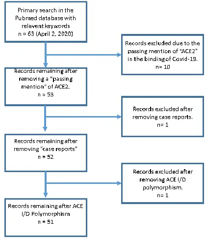
ACE2 in particular yielded the same set of articles. The search was conducted on April 2, 2020. These key words were used because they yielded the appropriate topic under study. The articles where chosen in sequential order and reviewed for major ideas. Exclusion criteria were articles that mentioned ACE2 in passing as the COVID-19 receptor. We also excluded case reports and the ACE I/D polymorphism. A MOOSE flow diagram is presented in Figure 1. Since most articles were published recently and in open access journals, we were able to obtain full texts of the articles. Upon reviewing the articles, it became apparent that there were different schools of thought on the use of RAS inhibitors in the context of the Coronavirus outbreak. These different perspectives are
discussed in this review along with an accompanying discussion of the pertinent aspects of the renin-Angiotensin system with respect to COVID-19 for clarification.
Background on the Renin-Angiotensin System as it pertains to COVID-19 for clarification purposes
The Renin-Angiotensin system involves several human organs working in concert to ensure the
function of this multi-faceted system. Components of RAS whether hormone peptides or receptors are wide spread in the body and are found in all organs and organ systems (Fagerberg et al 2014). RAS is a complex system of biochemical pathways involving various proteins, enzymes, peptides and receptors. RAS regulates homeostasis systemically (Azushima et al., 2020) as well as locally (De Mello & Frohlich, 2011). When discussing RAS we tend to think of vasorestriction (Benjamin, Collier, & Webb, 1988) and dilation of blood vessels (Vanhoutte e al.,1989) and assign negative and positive connotations to them respectively. In biological processes, however both play a role and it is the dysregulation of the balance between them that
leads to a disease process. RAS is also associated with inflammation (Duprez, 2006), cell growth (Dai et al., 2019 ) and cell death (Wang et al., 2013) again forming negative connotations about necessary biological processes that lead to negative consequences because of a homeostatic imbalance. RAS has been known to play a major role in cardiovascular physiology (Unger, Azizi, & Belz, 2000), however in the past decade it has become a major player in cellular physiology dictating cell growth or death (apoptosis) by its two main receptors (Jacques et al., 2019; Cai et al., 2019; Namsolleck et al., 2014): the Angiotensin II receptor 1 (AT-1) and Angiotensin II receptor 2 (AT-2).
RAS is composed of two main pathways (axis) that are mutually antagonistic in maintaining homeostasis, despite the constant flow of mediators and perturbations to the local and systemic environments. The first axis represented by ACE/AngII/AT1 promotes inflammation, cell proliferation and vasoconstriction (Iwai & Horiuchi, 2009). The second pathway represented by ACE2/Ang(1-7)/MAS is an anti-growth pathway inhibiting inflammation and inducing vasodilation (Santos et al.,2013). Another important component of RAS that is often overlooked and in fact did not appear to be discussed (in COVID-19 context) in any of the articles that we have reviewed is the Bradykinin pathway (Marceau et al.,2020). Bradykinin is a pro-inflammatory proliferative peptide hormone inducing vasodilation, vascular permeability and edema (Kempe et al.,2020; Lopatko et al.,2019; Péterfi et al.,2019). Bradykinin is upregulated by ACE inhibitors (Bakhle, 2020) and may be playing a significant role that has gone unnoticed in the COVID-19 crisis. We will review all three components with regards to COVID-19, discussing the main ideas being published and conclude with an elucidation of future directions in this field.
COVID-19 and the RAS connection
Unlike larger viruses (mimivirus-400nm) that require actin-dependent phagocytosis (Yaakov et al., 2019) COVID-19 (125 nm) enters the cell by clatherin mediated (CM) endocytosis (Wang et al, 2020). CM Endocytosis is a membrane process that needs a protein coat to induce curvature leading to the invagination of the memebrane (Lherbette et al.,2019; Narasimhan et al.,2020). It is a complex process requiring many cellular mediators. COVID-19 targets ACE2 positive cells (Wan et al.,2020; Tai et al.,2020). ACE 2 is a membrane bound enzyme peptidase on the surface of cells found in the lung, arteries, heart, kidney and intestines (Fagerberg et al.,2014; Xu et al.,2020; Qi et al., 2020; Zou et al., 2020). ACE2 converts the peptide Angiotensin II to Ang 1-7 (Burrell et al.,2004). The catalytic domain of the enzyme faces the extracellular surface where it binds Angiotensin II and releases Ang 1-7. In the literature ACE2 has been called the COVID-19 receptor, since COVID-19, like Sars before it has been shown to bind specifically to ACE2 initiating the curvature needed for the endocytosis of the virus along with the endocytosis of ACE2. This process allows COVID-19 to enter the cell, depleting ACE2 from the cellular membrane as in Sars (Kuba et al 2005). Papers published in this category stressed the structural relationship between ACE2 and its viral binding protein as well as the homology in the viral proteins from various species (Liu et al.,2020; Chen et al.,2020; Tai et al.,2020; Qiu et al.,2020; Li, Qiao, & Zhang, 2020). Chen et al. 2020, using sequence alignments, three-dimensional structural analyses, protein structure simulations and molecular docking were able to compare and contrast the interaction of human ACE2 with both Sars and COVID-19. Performing molecular simulations, they superimposed the predicted structure of the RBD protein of COVID-19 on the x-ray crystallographic structure of Sars with some structural variation. The inferred RBD (COVID-19) structure had a similar pattern of interaction with ACE2 as was seen with Sars and a more stable Gibbs free
energy. In their discussion, they noted that the viral binding domain of ACE2 is different from the catalytic domain. They also hypothesized on the inhibition of ACE2 by ACE1 inhibitors and or other novel inhibitors. They speculated that the binding of ACE2 by specific catalytic domain inhibitors might change its conformation preventing it from binding the virus. To our knowledge, ACE1 inhibitors have not been shown to inhibit ACE2. This does not preclude their use in the context of the COVID-19 pandemic but more on this in the conclusion section of this review.
Other papers focused on the homologous relationship, the similarities and differences between COVID-19 and Sars (Liu et al.,2020; Li, Qiao, & Zhang, 2020; Tai et al.,2020). For example,
Taiet al. showed the similarity between COVID-19 and Sars through the cross-reactivity of Sars-RBD specific antibodies with the RBD protein of COVID-19. The RBD protein of COVID-19 bound to human ACE2 expressing cells and did not bind to non-ACE2 expressing cells. The RBD protein from COVID-19 also bound tighter to ACE2 expressing cells than the RBD of Sars. Their results also showed that COVID-19 RBD bound to bat ACE2 expressing cells with the same intensity as it bound to the human ACE2 expressing cells alluding to the origin of COVID-19. The tighter binding they concluded explained the rapid transmission of COVID-19 in comparison to Sars. Using RBD protein from COVID-19 they were able to inhibit a pseudovirus construct from entering human expressing ACE2 cells alluding to a possible therapeutic mechanism.
ACE/AngII/AT1 Pathway (pro-inflammation) vs. ACE2/Ang(1-7)/MAS (anti-inflammation)
bloodstream, where Renin cleaves the decapeptide Angiotensin I from Angiotensinogen (Wu et al., 2011). Ang I travels by blood to the lungs (pertinent to COVID-19) where it is converted by another enzyme called the Angiotensin converting enzyme (ACE) to produce the octapeptide (active hormone) Angiotensin II (Corvol, Williams, & Soubrier, 1995). Depending on the availability of the Ang II receptors on human cells, Ang II induces inflammation through various biochemical and physiological mechanisms (Benigni, Cassis, & Remuzzi, 2010). This well studied ACE/Ang II axis is now complemented by the discovery of a second enzyme in RAS called ACE2 (Marian, 2013) which is also widely distributed in the body and is also found in the lungs (Fagerberg et al.,2014). ACE2 binds to Ang II converting it to the peptide Ang (1-7). This peptide in turn binds to its own receptor (MAS) inhibiting the inflammatory response (Varagic et al.,2014). In a twist of fate it is ACE2 that is the receptor for COVID-19 (Chen et al.,2020).
The ACE/AngII/AT1 (ACE axis) is a pro-inflammatory pathway characterized by cell proliferation and vasoconstriction. The driving force of this pathway is the production of Angioteinsin II by the catalytic activity of the Angiotensin Converting Enzyme (ACE) upon the decapeptide Angiotensin I. ACE is an enzyme peptidase located on the cell surface but is also found in a soluble form (Beldent et al.,1993). ACE is widely distributed in all human tissues including the lungs (Fagerberg et al.,2014) . ACE also degrades the peptide hormone Bradykinin. Bradykinin is a blood vessel dilator and a powerful pro-inflammatory agent. The pro-inflammatory actions of the ACE pathway is initiated by the binding of Angiotensin II to the AT1 receptor. Many studies have revealed that Ang II facilitates the inflammatory cascade by regulating cells and factors involved in inflammation. Bernstein et al (2013) presents an excellent and comprehensive review on ACE. ACE is a key regulator of the
Danser , Epstein, & Batlle, 2020; Kuster et al.,2020; Liu et al., 2020; Meng et al.,2020; Vaduganathan et al., 2020). There is evidence that Ang II levels are elevated in COVID-19 patients (Liu, et al., 2020) and this can be explained by the down regulation of ACE2 as a result of viral entry and the endocytosis of ACE2 in the process. ACE2 is the main backbone of the anti-inflammatory axis of RAS and is known to reduce lung inflammation (Calò, Rigato, & Bertoldi,2019; Meng et al.,2014), which is the main target organ of COVID-19. ACE2, is a peptidase that binds Ang II (pro-inflammatory) converting it into the Ang 1-7 peptide (anti-inflammatory). Ang 1-7 binds to the MAS receptor, which is found in the epithelium and bronchial smooth muscle and mediates the anti-inflammatory response (Magalhães et al., 2015). Although the exact mechanisms of this anti-inflammatory effect are not fully understood, it could involve numerous components of the inflammatory cascade. For example, reductions of
NFκB-related signaling (Yu et al.,2018), transforming growth factor (TGF)-β (Chappell & Al Zayadneh, 2017; Chou et al.,2013) and cytokine modulation (Villalobos et al., 2016) have all been seen. Infusions of Ang 1-7 in lung injury models protected the lungs from acute lung lesions and decreased lung edema (Klein et al., 2013), which can be crucial for COVID-19 patients. Ang 1-7 has also been shown to protect alveolar epithelial cells from apoptosis (Gopallawa & Uhal, 2016) a serious consequence that leads to lung fibrosis. Perhaps the insidious nature of COVID-19 lies in its targeting and reduction of ACE2 levels causing an imbalance in RAS and favoring the inflammatory pathway. Sun et al, stressed that the “exhaustion”
of ACE2, as a result of viral entry, inhibited the ACE2 pathway leading to acute severe pneumonia. They further argued that the use of ACE inhibitors and AT1 blockers would reverse this event. In this regard, we agree with half of Sun et al’s argument.
While AT1 blockers might be beneficial in reducing acute severe pneumonia, the use of ACE inhibitors
might be self-defeating due to their induction of Bradykinin a pro-inflammatory agent.
Although none of the papers, we reviewed, discussed Bradykinin in the context of COVID-19. We feel that it is pertinent to the COVID-19 story and its introduction is crucial at this time. Although the effect of ACE inhibition lowers Ang II, it raises Bradykinin levels (Taddei & Bortolotto, 2016) and desensitizes the Bradykinin receptor (Benzing et al.,1999 ;Minshall et al., 1998). Bradykinin is a peptide hormone composed of nine amino acids and cleaved by ACE between the eighth and the ninth amino acid rendering it inactive. In the absence of an inflammatory trigger, the rise of Bradykinin levels promotes vasodilation. Bradykinin is a potent vasodilator (Hornig, Kohler, & Drexler, 1997) and is down regulated by the action of ACE and upregulated by ACE inhibition. Bradykinin is also a powerful mediator of inflammation (Hofman et al., 2016), vascular permeability (Minnear, Kivlen, & Malik, 1983) and edema in lung tissue (Hao et al., 2001). Bradykinin has been shown to induce endothelial cell contraction promoting water and protein transport (Miyamoto, Ishiguro, & Nishio, 1999). These properties of Bradykinin probably play a major but yet undiscovered role leading to the medical complications seen in COVID-19 patients.
The fate of ACE2, Ang II and Bradykinin in the context of COVID-19
Renin-Angiotensin system. This however is changing due to the discovery of other active peptides and receptors adding to the complexity of RAS in systemic and local biological processes. Despite the added complexities, Ang II retains its prominent role in RAS as evidenced by the successful and widespread use of RAS inhibitors (ACEi and ARBs) targeting the production or function of Ang II. Besides having endocrine functions, regulating vasoconstriction, fluid homeostasis, cardiac output, Ang II affects local tissue by controlling many facets of cellular function such as cellular growth and death. In fact, the cellular role of Ang II is unconnected to its systemic role as a hormone. In this regard and relevant to our discussion in this review, Ang II was shown to mediate alveolar epithelial cell (AEC) death (apoptosis) via the AT1 receptor (Papp et al., 2002). Thus, Ang II is an endocrine as well as a paracrine and autocrine hormone effecting biological processes via the binding of receptors on the surface of cells. In summary, the concentration of Ang II systemically or locally depends on the concentration (gene expression, genetic control), and activity (enzyme kinetics) of various gene products. These include Renin, Angiotensinogen, ACE, and ACE2. Thus, the role of Ang II in the COVID-19 saga will depend on the concentration (Ang II concentration vs virus titer) of both actors, since both Ang II and COVID-19 bind to the same enzyme protein, ACE2. COVID-19 opportunistically binds to ACE2 gaining entry into the cell. In this case, both the virus and ACE2 are endocytosed. This depletes the levels of ACE2 on the cell surface, inhibiting the production of Ang 1-7, the anti-inflammatory peptide, and in the meantime raising the concentration of Ang II, the pro-inflammatory peptide. Indeed, it has been shown that COVID-19 patients have an elevated level of Ang II peptide (Liu et al., 2020). The rise in Ang II is understandable in the context of ACE2 depeletion after viral infection and does not give us a baseline for the patient before infection. It would be interesting to see whether increasing
3). This demographic fact necessitates that we study the effect of increasing Ang II on the entry of virus in ACE2+ cells. It also necessitates that we consider Bradykinin inhibitors in the collection of
drugs being investigated for COVID-19.
Conclusion
To appreciate the dynamics of COVID-19’s
connection to RAS and its resultant infection and ensuing inflammatory response, we need to envision the two axes of the renin-Angiotensin system: the pro-inflammatory axis choreographed by ACE/Ang II via the AT1/AT2 receptors and Bradykinin, vs the ACE2/Ang 1-7 promoting the anti-inflammatory axis via the Mas receptor. By attacking the anti-inflammatory axis, COVID-19 down regulates ACE2, since the cells engulf the virus particle along with the ACE2 enzyme, leaving the pro-inflammatory axis unopposed resulting in severe lung inflammation. This has led to the proposition of opposing views on RAS inhibitors. In one view, Lei Fang and colleagues advocated withholding Angiotensin converting enzyme inhibitors (ACEIs) or Angiotensin receptor blockers (ARBs) due to the possibility that they can
upregulate ACE2, COVID-19’s port of entry.
Demographically the group with the highest risk of Infection complications was hypertensive
patients, which tends to support Lei Fang et. al’s
hypothesis. Christopher J Tignanelli et al. 2020, published an opposing view, that withholding RAS inhibitors is premature, stressing the protective nature of ACE2 and losartan infusions in preventing damage by induced lung injury in various animal studies. We believe both views have merit and are not necessarily in opposition when we consider the dynamic relationship between COVID-19 and the pro-inflammatory and anti-inflammatory axes of RAS. Perhaps the
Figure 2. In children, the higher Ang II levels serve as a guard preventing wide-spread and rapid infection of the cells. This attenuates the infection and does not trigger an inflammatory response leading to a benign infection.
Cell
N
ACE2 Ang II Ang II
Ang II Ang II AT1
Ang II Ang II Ang II Ang II
solution lies in the timing. ACE and ACE2 are mutually antagonistic. RAS inhibitors are not identical in action nor targets. ACE
inhibitors target ACE and Bradykinin and are ineffective against ACE2. ACE inhibitors decrease the production of the hormone Ang II inhibiting both axes by decreasing the binding of Ang II to AT1 and ACE2. The decrease in Ang II and increase in Bradykinin in hypertensive elderly patients using ACEI meds is in contrast to the high Ang II
levels in children. This dichotomy in children (High Ang II, low Bradykinin, mild infection) and the elderly (low Ang II, high Bradykinin, lethal infection) can help us elucidate a working hypothesis when considering COVID-19 infection. ACE inhibitors reduce Ang II, while raising ACE2 and Bradykinin levels. Does COVID-19 bind tighter to ACE2 in the absence of Ang II? Does COVID-19 produce a malignant inflammatory response in the presence of Bradykinin? Could this be partly responsible for the virulence of the virus in the elderly? In this regard, we need to distinguish between patients on ACE inhibitors vs AT1 blockers. Hypertensive elderly patients on AT1 receptor blockers would have an inhibited pro-inflammatory pathway, while keeping Ang II levels high (as in children) occupying ACE2 and preventing COVID-19 binding. In the event of virus entry into the cells and the down regulation of ACE2, would leave Ang II unopposed to bind the AT1 receptor thereby driving the proinflammatory response in lung tissue. At this stage, the use of AT1 receptor blockers can indeed lesson the inflammatory response without inhibiting ACE and upregulating Bradykinin. This scenario would support the hypothesis of the use of AT1 blockers to minimize damage and inflammation to the lungs and other affected organs. We would venture to guess and propose the use of Bradykinin antagonists to prevent the pro-inflammatory response, vascular permeability and edema that could develop in the course of infection. Perhaps both hypotheses have merit when considering the timing of RAS inhibition prior to viral infection and during the course of viral infection. We propose the continuation of RAS inhibitor use due to its crucial medical benefit to the elderly until an infection has been confirmed. Once infection has occurred and progresses, we favor the use of AT1 receptor blockers, which would be beneficial in protecting lung tissue from inflammation and damage. We would also urge the medical and scientific community to test the possibility of using
Bradykinin inhibitors or in combination with AT1 receptor blockers to minimize the damaging inflammatory response and pulmonary edema associated with COVID-19. Finally, we propose a novel approach and that is the use of intravenous
“Ang II loading” in these patients in combination
with AT1 receptor blockers to prevent the further infection of tissues by the budding off viral particles (Figure 4). Ang II in this case would serve as a guardian of ACE2 in non-infected cells. A battle dictated by binding constants, conformational maneuvers and gibbs free energy.
Ang II might turn out to be the arrow in Corona’s
heel.
Future Directions
To elucidate future directions of research in this field, we propose a working model that can be tested and applied. COVID-19 produces a virulent infection in the elderly (hypertensive patients) and a benign infection in children. In children there appears to be high Ang II/low Bradykinin levels coupled with low ACE2. We propose that this combination of effectors leads to a benign COVID-19 infection. The exact mechanism remains obscure but we can predict that virus entry in the cells of children is attenuated due to the lower ACE2 and the higher basal Ang II levels prior to infection. Ang II, by occupying the catalytic site of ACE2, might change ACE2 conformation enough to interfere with viral binding. This can be tested in the laboratory by increasing the concentration of Ang II in the medium of ACE2+ cells and determine whether this inhibits viral entry. In the elderly population, ACE inhibitors lower Ang II, upregulate ACE2 and elevate Bradykinin. Under normal circumstances, this is an ideal health condition. If we consider COVID-19 infection, however, this
“state” of RAS might predispose the elderly to a
virulent infection. Since COVID-19 uses the ACE2 as a port of entry and ACE2 is elevated in this population accompanied by a depression in Ang II. The decrease in Ang II might give the virus an open target (ACE2) with the proper conformation for
binding. A further complication in the elderly would be the elevated Bradykinin levels. Under normal circumstances, Bradykinin would promote vasodilation and heart health. During viral infection, however, Bradykinin becomes a potent inflammatory mediator promoting edema and vascular permeability. Inhibiting Bradykinin might mitigate the virulent inflammatory response in these patients. Perhaps a perfect therapy for COVID-19 patients, besides the combination of AT1 receptor blockers and Bradykinin antagonists, would be the counterintuitive use of “Ang II loading” in these patients. This would mimic the
RAS “state” in children and perhaps abort the continuing infection of the budding viral particles. We urge the scientific and medical communities to test this working model in the search for a solution for COVID-19.
Acknowledgements
We thank our kids for being patient in these trying times. We also thank Professor Tahrir Hamdi for her critical reading of this manuscript.
References
Azushima K, Morisawa N, Tamura K, Nishiyama A. Recent Research Advances in Renin-Angiotensin-Aldosterone System Receptors. Curr Hypertens Rep. 2020;22(3):22. Published 2020 Feb 29. doi:10.1007/s11906-020-1028-6
Bakhle YS. How ACE inhibitors transformed the renin-Angiotensin system [published online ahead of print, 2020 Mar 6]. Br J Pharmacol. 2020;10.1111/bph.15045.
doi:10.1111/bph.15045
and aging. EMBO Mol Med. 2010;2(7):247–257. doi:10.1002/emmm.201000080
Benjamin N, Collier JG, Webb DJ. Angiotensin II augments sympathetically induced venoconstriction in man. Clin Sci (Lond). 1988;75(4):337–340. doi:10.1042/cs0750337 Benzing T, Fleming I, Blaukat A, Müller-Esterl W, Busse R. Angiotensin-converting enzyme inhibitor ramiprilat interferes with the sequestration of the B2 kinin receptor within the plasma membrane of native endothelial cells. Circulation. 1999;99(15):2034–2040.
doi:10.1161/01.cir.99.15.2034
Bernstein KE, Ong FS, Blackwell WL, et al. A modern understanding of the traditional and nontraditional biological functions of Angiotensin-converting enzyme [published correction appears in Pharmacol Rev. 2013;65(1):544]. Pharmacol Rev. 2012;65(1):1–46. Published 2012 Dec 20. doi:10.1124/pr.112.006809
Bock HA, Hermle M, Brunner FP, Thiel G. Pressure dependent modulation of renin release in isolated perfused glomeruli. Kidney Int. 1992;41(2):275–
280. doi:10.1038/ki.1992.39
Broughton Pipkin F, Smales OR, O'Callaghan M. Renin and Angiotensin levels in children. Arch Dis Child. 1981;56(4):298–302. doi:10.1136/adc.56.4.298
Burrell LM, Johnston CI, Tikellis C, Cooper ME. ACE2, a new regulator of the renin-Angiotensin system. Trends Endocrinol Metab. 2004;15(4):166–169.
doi:10.1016/j.tem.2004.03.001
Cai W, Zhong S, Zheng F, et al. Angiotensin II confers resistance to apoptosis in cardiac myofibroblasts through the AT1/ERK1/2/RSK1 pathway. IUBMB Life. 2019;71(2):261–276. doi:10.1002/iub.1967
Calò LA, Rigato M, Bertoldi G. ACE2/Angiotensin 1-7 protective antiinflammatory and antioxidant
role in hyperoxic lung injury: support from studies in Bartter's and Gitelman's syndromes [published online ahead of print, 2019 Dec 18]. QJM. 2019;hcz319. doi:10.1093/qjmed/hcz319
Chappell MC, Al Zayadneh EM. Angiotensin-(1-7) and the Regulation of Anti-Fibrotic Signaling Pathways. J Cell Signal. 2017;2(1):134. doi:10.4172/2576-1471.1000134
Chen Y, Guo Y, Pan Y, Zhao ZJ. Structure analysis of the receptor binding of 2019-nCoV [published online ahead of print, 2020 Feb 17]. Biochem Biophys Res Commun. 2020;. doi:10.1016/j.bbrc.2020.02.071
Chen Y, Guo Y, Pan Y, Zhao ZJ. Structure analysis of the receptor binding of 2019-nCoV [published online ahead of print, 2020 Feb 17]. Biochem Biophys Res Commun. 2020;. doi:10.1016/j.bbrc.2020.02.071
Chou CH, Chuang LY, Lu CY, Guh JY. Interaction between TGF-β and ACE2-Ang-(1-7)-Mas pathway in high glucose-cultured NRK-52E cells. Mol Cell Endocrinol. 2013;366(1):21–30. doi:10.1016/j.mce.2012.11.004
Corvol P, Williams TA, Soubrier F. Peptidyl dipeptidase A: Angiotensin I-converting enzyme. Methods Enzymol. 1995;248:283–305. doi:10.1016/0076-6879(95)48020-x
Crowley SD, Frey CW, Gould SK, et al. Stimulation of lymphocyte responses by Angiotensin II promotes kidney injury in hypertension [published correction appears in Am J Physiol Renal Physiol. 2010 May;298(5):F1286. Dosage error in article text]. Am J Physiol Renal Physiol. 2008;295(2):F515–F524.
doi:10.1152/ajprenal.00527.2007
remodeling. Atherosclerosis. 2019;288:124–136. doi:10.1016/j.atherosclerosis.2019.07.010
Danser AHJ, Epstein M, Batlle D. Renin-Angiotensin System Blockers and the COVID-19 Pandemic: At Present There Is No Evidence to Abandon Renin-Angiotensin System Blockers [published online ahead of print, 2020 Mar 25]. Hypertension.
2020;HYPERTENSIONAHA12015082.
doi:10.1161/HYPERTENSIONAHA.120.15082 De Mello WC, Frohlich ED. On the local cardiac renin Angiotensin system. Basic and clinical implications. Peptides. 2011;32(8):1774–1779. doi:10.1016/j.peptides.2011.06.018
Duprez DA. Role of the renin-Angiotensin-aldosterone system in vascular remodeling and inflammation: a clinical review. J Hypertens. 2006;24(6):983–991.
doi:10.1097/01.hjh.0000226182.60321.69
Ehlers MR, Kirsch RE. Catalysis of Angiotensin I hydrolysis by human Angiotensin-converting enzyme: effect of chloride and pH. Biochemistry. 1988;27(15):5538–5544.
doi:10.1021/bi00415a023
Esler M, Esler D. Can Angiotensin receptor-blocking drugs perhaps be harmful in the COVID-19 pandemic?. J Hypertens. 2020;38(5):781–782. doi:10.1097/HJH.0000000000002450
Fagerberg L, Hallström BM, Oksvold P, et al. Analysis of the human tissue-specific expression by genome-wide integration of transcriptomics and antibody-based proteomics. Mol Cell Proteomics. 2014;13(2):397–406. doi:10.1074/mcp.M113.035600
Fang L, Karakiulakis G, Roth M. Are patients with hypertension and diabetes mellitus at increased risk for COVID-19 infection? [published online ahead of print, 2020 Mar 11]. Lancet Respir Med. 2020;. doi:10.1016/S2213-2600(20)30116-8
Gilliam-Davis S, Gallagher PE, Payne VS, et al. Long-term systemic Angiotensin II type 1 receptor blockade regulates mRNA expression of dorsomedial medulla renin-Angiotensin system components. Physiol Genomics. 2011;43(13):829–
835. doi:10.1152/physiolgenomics.00167.2010 Gopallawa I, Uhal BD. Angiotensin-(1-7)/mas inhibits apoptosis in alveolar epithelial cells through upregulation of MAP kinase phosphatase-2. Am J Physiol Lung Cell Mol Physiol. 2016;310(3):L240–L248.
doi:10.1152/ajplung.00187.2015
Gurwitz D. Angiotensin receptor blockers as tentative SARS-CoV-2 therapeutics [published online ahead of print, 2020 Mar 4]. Drug Dev Res. 2020;10.1002/ddr.21656. doi:10.1002/ddr.21656 Hao Y, Okamura S, Wang LM, Mineshita S. The involvement of Bradykinin in adrenaline-induced pulmonary edema in rats. J Med Dent Sci. 2001;48(3):79–85.
Hofman Z, de Maat S, Hack CE, Maas C. Bradykinin: Inflammatory Product of the Coagulation System. Clin Rev Allergy Immunol. 2016;51(2):152–161. doi:10.1007/s12016-016-8540-0
Hornig B, Kohler C, Drexler H. Role of Bradykinin in mediating vascular effects of Angiotensin-converting enzyme inhibitors in humans. Circulation. 1997;95(5):1115–1118. doi:10.1161/01.cir.95.5.1115
Iwai M, Horiuchi M. Devil and angel in the renin-Angiotensin system: ACE-renin-Angiotensin II-AT1 receptor axis vs. ACE2-Angiotensin-(1-7)-Mas receptor axis. Hypertens Res. 2009;32(7):533–
536. doi:10.1038/hr.2009.74
AT2 receptor 1. Can J Physiol Pharmacol. 2019;97(6):581–588. doi:10.1139/cjpp-2018-0592 Kang YS, Park YG, Kim BK, et al. Angiotensin II stimulates the synthesis of vascular endothelial growth factor through the p38 mitogen activated protein kinase pathway in cultured mouse podocytes. J Mol Endocrinol. 2006;36(2):377–388. doi:10.1677/jme.1.02033
Kawano H, Cody RJ, Graf K, et al. Angiotensin II enhances integrin and alpha-actinin expression in adult rat cardiac fibroblasts. Hypertension. 2000;35(1 Pt 2):273–279. doi:10.1161/01.hyp.35.1.273
Kempe S, Fois G, Brunner C, Hoffmann TK, Hahn J, Greve J. Bradykinin signaling regulates solute permeability and cellular junction organization in lymphatic endothelial cells. Microcirculation. 2020;27(2):e12592. doi:10.1111/micc.12592 Klein N, Gembardt F, Supé S, et al. Angiotensin-(1-7) protects from experimental acute lung injury. Crit Care Med. 2013;41(11):e334–e343. doi:10.1097/CCM.0b013e31828a6688
Kono T, Taniguchi A, Imura H, Oseko F, Khosla MC. Biological activities of Angiotensin II-(1-6)-hexapeptide and Angiotensin II-(1-7)-heptapeptide in man. Life Sci. 1986;38(16):1515–
1519. doi:10.1016/0024-3205(86)90565-5
Kuba K, Imai Y, Rao S, et al. A crucial role of Angiotensin converting enzyme 2 (ACE2) in SARS coronavirus-induced lung injury. Nat Med. 2005;11(8):875–879. doi:10.1038/nm1267 Kuster GM, Pfister O, Burkard T, et al. SARS-CoV2: should inhibitors of the renin-Angiotensin system be withdrawn in patients with COVID-19? [published online ahead of print, 2020 Mar 20]. Eur Heart J. 2020;ehaa235. doi:10.1093/eurheartj/ehaa235
Larsson PT, Schwieler JH, Wallén NH. Platelet activation during Angiotensin II infusion in healthy
volunteers. Blood Coagul Fibrinolysis. 2000;11(1):61–69.
Lherbette M, Redlingshöfer L, Brodsky FM, Schaap IAT, Dannhauser PN. The AP2 adaptor enhances clathrin coat stiffness. FEBS J. 2019;286(20):4074–
4085. doi:10.1111/febs.14961
Li R, Qiao S, Zhang G. Analysis of Angiotensin-converting enzyme 2 (ACE2) from different species sheds some light on cross-species receptor usage of a novel coronavirus 2019-nCoV. J Infect. 2020;80(4):469–496.
doi:10.1016/j.jinf.2020.02.013
Lin SY, Goodfriend TL. Angiotensin receptors. Am J Physiol. 1970;218(5):1319–1328. doi:10.1152/ajplegacy.1970.218.5.1319
Liu Y, Yang Y, Zhang C, et al. Clinical and biochemical indexes from 2019-nCoV infected patients linked to viral loads and lung injury. Sci China Life Sci. 2020;63(3):364–374. doi:10.1007/s11427-020-1643-8
Liu Y, Yang Y, Zhang C, et al. Clinical and biochemical indexes from 2019-nCoV infected patients linked to viral loads and lung injury. Sci China Life Sci. 2020;63(3):364–374. doi:10.1007/s11427-020-1643-8
Liu Z, Xiao X, Wei X, et al. Composition and divergence of coronavirus spike proteins and host ACE2 receptors predict potential intermediate hosts of SARS-CoV-2 [published online ahead of print, 2020 Feb 26]. J Med Virol. 2020;10.1002/jmv.25726. doi:10.1002/jmv.25726 Lopatko Fagerström I, Ståhl AL, Mossberg M, et al. Blockade of the kallikrein-kinin system reduces endothelial complement activation in vascular inflammation. EBioMedicine. 2019;47:319–328. doi:10.1016/j.ebiom.2019.08.020
Pharmacol. 2015;172(9):2330–2342. doi:10.1111/bph.13057
Marceau F, Bachelard H, Bouthillier J, et al. Bradykinin receptors: Agonists, antagonists, expression, signaling, and adaptation to sustained stimulation [published online ahead of print, 2020 Feb 24]. Int Immunopharmacol. 2020;82:106305. doi:10.1016/j.intimp.2020.106305
Marian AJ. The discovery of the ACE2 gene. Circ Res. 2013;112(10):1307–1309. doi:10.1161/CIRCRESAHA.113.301271
Meng J, Xiao G, Zhang J, et al. Renin-Angiotensin system inhibitors improve the clinical outcomes of COVID-19 patients with hypertension. Emerg Microbes Infect. 2020;9(1):757–760. doi:10.1080/22221751.2020.1746200
Meng Y, Yu CH, Li W, et al. Angiotensin-converting enzyme 2/Angiotensin-(1-7)/Mas axis protects against lung fibrosis by inhibiting the MAPK/NF-κB
pathway. Am J Respir Cell Mol Biol. 2014;50(4):723–736. doi:10.1165/rcmb.2012-0451OC
Minnear FL, Kivlen CM, Malik AB. Effect of Bradykinin on lung vascular permeability in sheep. J Appl Physiol Respir Environ Exerc Physiol. 1983;55(4):1079–1084.
doi:10.1152/jappl.1983.55.4.1079
Minshall RD, Tan F, Nakamura F, et al. Potentiation of the actions of Bradykinin by Angiotensin I-converting enzyme inhibitors. The role of expressed human Bradykinin B2 receptors and Angiotensin I-converting enzyme in CHO cells [published correction appears in Circ Res 1998 Jan 9-23;82(1):137]. Circ Res. 1997;81(5):848–856. doi:10.1161/01.res.81.5.848
Miyamoto A, Ishiguro S, Nishio A. Stimulation of Bradykinin B2-receptors on endothelial cells induces relaxation and contraction in porcine basilar artery in vitro. Br J Pharmacol.
1999;128(1):241–247. doi:10.1038/sj.bjp.0702783
Nabah YN, Mateo T, Estellés R, et al. Angiotensin II induces neutrophil accumulation in vivo through generation and release of CXC chemokines. Circulation. 2004;110(23):3581–
3586. doi:10.1161/01.CIR.0000148824.93600.F3 Namsolleck P, Recarti C, Foulquier S, Steckelings UM, Unger T. AT(2) receptor and tissue injury: therapeutic implications. Curr Hypertens Rep. 2014;16(2):416. doi:10.1007/s11906-013-0416-6 Narasimhan M, Johnson A, Prizak R, et al. Evolutionarily unique mechanistic framework of clathrin-mediated endocytosis in plants. Elife. 2020;9:e52067. Published 2020 Jan 23. doi:10.7554/eLife.52067
Ohkubo H, Kageyama R, Ujihara M, Hirose T, Inayama S, Nakanishi S. Cloning and sequence analysis of cDNA for rat Angiotensinogen. Proc Natl Acad Sci U S A. 1983;80(8):2196–2200. doi:10.1073/pnas.80.8.2196
Papp M, Li X, Zhuang J, Wang R, Uhal BD. Angiotensin receptor subtype AT(1) mediates alveolar epithelial cell apoptosis in response to ANG II. Am J Physiol Lung Cell Mol Physiol. 2002;282(4):L713–L718.
doi:10.1152/ajplung.00103.2001
Patel AB, Verma A. COVID-19 and Angiotensin-Converting Enzyme Inhibitors and Angiotensin Receptor Blockers: What Is the Evidence? [published online ahead of print, 2020 Mar 24]. JAMA. 2020;10.1001/jama.2020.4812. doi:10.1001/jama.2020.4812
P-selectin upregulation. Circulation. 2000;102(17):2118–2123.
doi:10.1161/01.cir.102.17.2118
Pueyo ME, Gonzalez W, Nicoletti A, Savoie F, Arnal JF, Michel JB. Angiotensin II stimulates endothelial vascular cell adhesion molecule-1 via nuclear factor-kappaB activation induced by intracellular oxidative stress. Arterioscler Thromb Vasc Biol. 2000;20(3):645–651. doi:10.1161/01.atv.20.3.645 Qi F, Qian S, Zhang S, Zhang Z. Single cell RNA sequencing of 13 human tissues identify cell types and receptors of human coronaviruses [published online ahead of print, 2020 Mar 18]. Biochem Biophys Res Commun. 2020;S0006-291X(20)30523-4.
doi:10.1016/j.bbrc.2020.03.044
Qiu Y, Zhao YB, Wang Q, et al. Predicting the Angiotensin converting enzyme 2 (ACE2) utilizing capability as the receptor of SARS-CoV-2 [published online ahead of print, 2020 Mar 19]. Microbes Infect. 2020;S1286-4579(20)30049-6. doi:10.1016/j.micinf.2020.03.003
Report of the WHO-China Joint Mission on Coronavirus Disease 2019 (COVID-19) [Pdf] - World Health Organization, Feb. 28, 2020
Sadoshima J. Cytokine actions of Angiotensin II. Circ Res. 2000;86(12):1187–1189. doi:10.1161/01.res.86.12.1187
Santos RA, Ferreira AJ, Verano-Braga T, Bader M. Angiotensin-converting enzyme 2, Angiotensin-(1-7) and Mas: new players of the renin-Angiotensin system. J Endocrinol. 2013;216(2):R1–R17. Published 2013 Jan 18. doi:10.1530/JOE-12-0341 Santos RA, Simoes e Silva AC, Maric C, et al. Angiotensin-(1-7) is an endogenous ligand for the G protein-coupled receptor Mas. Proc Natl Acad Sci U S A. 2003;100(14):8258–8263. doi:10.1073/pnas.1432869100
Simões e Silva AC, Silveira KD, Ferreira AJ, Teixeira MM. ACE2, Angiotensin-(1-7) and Mas receptor
axis in inflammation and fibrosis. Br J Pharmacol. 2013;169(3):477–492. doi:10.1111/bph.12159 Sun ML, Yang JM, Sun YP, Su GH. Inhibitors of RAS Might Be a Good Choice for the Therapy of COVID-19 Pneumonia. 2020;43(3):219–222. doi:10.3760/cma.j.issn.1001-0939.2020.03.016 Taddei S, Bortolotto L. Unraveling the Pivotal Role of Bradykinin in ACE Inhibitor Activity. Am J Cardiovasc Drugs. 2016;16(5):309–321. doi:10.1007/s40256-016-0173-4
Tai W, He L, Zhang X, et al. Characterization of the receptor-binding domain (RBD) of 2019 novel coronavirus: implication for development of RBD protein as a viral attachment inhibitor and vaccine [published online ahead of print, 2020 Mar 19]. Cell Mol Immunol. 2020;10.1038/s41423-020-0400-4. doi:10.1038/s41423-020-0400-4 Tai W, He L, Zhang X, et al. Characterization of the receptor-binding domain (RBD) of 2019 novel coronavirus: implication for development of RBD protein as a viral attachment inhibitor and vaccine [published online ahead of print, 2020 Mar 19]. Cell Mol Immunol. 2020;10.1038/s41423-020-0400-4. doi:10.1038/s41423-020-0400-4 Tayeh MA, Scicli AG. Angiotensin II and Bradykinin regulate the expression of P-selectin on the surface of endothelial cells in culture. Proc Assoc Am Physicians. 1998;110(5):412–421.
The Epidemiological Characteristics of an Outbreak of 2019 Novel Coronavirus Diseases (COVID-19) - China CCDC, February 17 2020 Tian X, Li C, Huang A, et al. Potent binding of 2019 novel coronavirus spike protein by a SARS coronavirus-specific human monoclonal antibody. Emerg Microbes Infect. 2020;9(1):382–
385. Published 2020 Feb 17. doi:10.1080/22221751.2020.1729069
26]. Lancet Respir Med. 2020;S2213-2600(20)30153-3. doi:10.1016/S2213-2600(20)30153-3
Timmermans PB, Wong PC, Chiu AT, et al. Angiotensin II receptors and Angiotensin II receptor antagonists. Pharmacol Rev. 1993;45(2):205–251.
Toffelmire EB, Slater K, Corvol P, Menard J, Schambelan M. Response of plasma prorenin and active renin to chronic and acute alterations of renin secretion in normal humans. Studies using a direct immunoradiometric assay. J Clin Invest. 1989;83(2):679–687. doi:10.1172/JCI113932 Turner AJ, Tipnis SR, Guy JL, Rice G, Hooper NM. ACEH/ACE2 is a novel mammalian metallocarboxypeptidase and a homologue of Angiotensin-converting enzyme insensitive to ACE inhibitors. Can J Physiol Pharmacol. 2002;80(4):346–353. doi:10.1139/y02-021 Unger T, Azizi M, Belz GG. Blocking the tissue renin-Angiotensin system: the future cornerstone of therapy. J Hum Hypertens. 2000;14 Suppl 2:S23–S31. doi:10.1038/sj.jhh.1001070
Vaduganathan M, Vardeny O, Michel T, McMurray JJV, Pfeffer MA, Solomon SD. Renin-Angiotensin-Aldosterone System Inhibitors in Patients with COVID-19 [published online ahead of print, 2020 Mar 30]. N Engl J Med. 2020;10.1056/NEJMsr2005760.
doi:10.1056/NEJMsr2005760
Vanhoutte PM, Auch-Schwelk W, Biondi ML, Lorenz RR, Schini VB, Vidal MJ. Why are converting enzyme inhibitors vasodilators?. Br J Clin Pharmacol. 1989;28 Suppl 2(Suppl 2):95S–104S. doi:10.1111/j.1365-2125.1989.tb03585.x
Varagic J, Ahmad S, Nagata S, Ferrario CM. ACE2: Angiotensin II/Angiotensin-(1-7) balance in cardiac and renal injury. Curr Hypertens Rep. 2014;16(3):420. doi:10.1007/s11906-014-0420-5
Villalobos LA, San Hipólito-Luengo Á, Ramos-González M, et al. The Angiotensin-(1-7)/Mas Axis Counteracts Angiotensin IIDependent and -Independent Pro-inflammatory Signaling in Human Vascular Smooth Muscle Cells. Front Pharmacol. 2016;7:482. Published 2016 Dec 15. doi:10.3389/fphar.2016.00482
Wan Y, Shang J, Graham R, Baric RS, Li F. Receptor Recognition by the Novel Coronavirus from Wuhan: an Analysis Based on Decade-Long Structural Studies of SARS Coronavirus. J Virol. 2020;94(7):e00127-20. Published 2020 Mar 17. doi:10.1128/JVI.00127-20
Wang G, Jiang L, Wang J, et al. The G Protein-Coupled Receptor FFAR2 Promotes Internalization during Influenza A Virus Entry. J Virol. 2020;94(2):e01707-19. Published 2020 Jan 6. doi:10.1128/JVI.01707-19
Wang X, Dai Y, Ding Z, Khaidakov M, Mercanti F, Mehta JL. Regulation of autophagy and apoptosis in response to Angiotensin II in HL-1 cardiomyocytes. Biochem Biophys Res Commun. 2013;440(4):696–700.
doi:10.1016/j.bbrc.2013.09.131
Wolf G, Ziyadeh FN, Thaiss F, et al. Angiotensin II stimulates expression of the chemokine RANTES in rat glomerular endothelial cells. Role of the Angiotensin type 2 receptor. J Clin Invest. 1997;100(5):1047–1058. doi:10.1172/JCI119615 Wrapp D, Wang N, Corbett KS, et al. Cryo-EM structure of the 2019-nCoV spike in the prefusion conformation. Science. 2020;367(6483):1260–
1263. doi:10.1126/science.abb2507
Published 2020 Feb 24. doi:10.1038/s41368-020-0074-x
Yaakov LB, Mutsafi Y, Porat Z, Dadosh T, Minsky A. Kinetics of Mimivirus Infection Stages Quantified Using Image Flow Cytometry. Cytometry A. 2019;95(5):534–548. doi:10.1002/cyto.a.23770 Yu X, Cui L, Hou F, et al. Angiotensin-converting enzyme 2-Angiotensin (1-7)-Mas axis prevents pancreatic acinar cell inflammatory response via inhibition of the p38 mitogen-activated protein
kinase/nuclear factor-κB pathway. Int J Mol Med. 2018;41(1):409–420.
doi:10.3892/ijmm.2017.3252