This author’s accepted manuscript may be used for non-commercial purposes in accordance with Wiley Terms and Conditions for Self-Archiving.
The full details of the published version of the article are as follows:
TITLE: Can malignant and inflammatory pleural effusions in dogs be distinguished using computed tomography
AUTHORS: Thom C. Watton, Ana Lara-Garcia, Christopher R. Lamb
JOURNAL TITLE: Veterinary Radiology & Ultrasound
PUBLISHER: Wiley
PUBLICATION DATE: 16 July 2017 (online)
Can malignant and inflammatory pleural effusions in dogs be distinguished using 1
computed tomography? 2
Thom C. Watton, Ana Lara-Garcia, Christopher R. Lamb 3
Department of Clinical Sciences and Services, The Royal Veterinary College, University of 4
London 5
6
Address correspondence to: C. R. Lamb, Department of Clinical Sciences and Services, The 7
Royal Veterinary College, Hawkshead Lane, North Mymms, Hertfordshire AL9 7TA, UK. 8
Tel: 01707-666234 9
Email: [email protected] 10
Key words: computed tomography, chylothorax, dog, mesothelioma, pleural disease, 11
pyothorax 12
Running head: CT of pleural effusions 13
Funding Sources: The authors received no financial support for the research, authorship or 14
Abstract 16
Computed tomography (CT) is the primary imaging modality used to investigate human 17
patients with suspected malignant or inflammatory pleural effusion, but there is a lack of 18
information about the clinical use of this test in dogs. In order to identify CT signs that could 19
be used to distinguish pleural malignant neoplasia from pleuritis, a retrospective case-20
control study was done based on dogs that had pleural effusion, pre- and post-contrast 21
thoracic CT images, and cytological or histopathological diagnosis of malignant or 22
inflammatory pleural effusion. There were 20 dogs with malignant pleural effusion (13 23
mesothelioma, 6 carcinoma; 1 lymphoma), and 32 dogs with pleuritis (18 pyothorax; 14 24
chylothorax). Compared to dogs with pleuritis, dogs with malignant pleural effusions were 25
significantly older (median 8.5 years versus 4.9 years, p=0.001), more frequently had CT 26
signs of pleural thickening (65% versus 34%, p= 0.05), tended to have thickening of the 27
parietal pleura only (45% versus 3%, p=0.002) and had more marked pleural thickening 28
(median 3mm versus 0mm, p=0.03). CT signs of thoracic wall invasion were observed only in 29
dogs with malignant pleural effusions (p= 0.05). There were no significant differences in 30
pleural fluid volume, distribution or attenuation, degree of pleural contrast accumulation, 31
amount of pannus, or prevalence of mediastinal adenopathy. Although there was 32
considerable overlap in findings in dogs with malignant pleural effusion and pleuritis, 33
marked thickening affecting the parietal pleural alone and signs of thoracic wall invasion on 34
CT support diagnosis of pleural malignant neoplasia, and may help prioritize further 35
Introduction 37
Pleural fluid accumulation may occur as a result of several different pathologic mechanisms 38
that determine the nature of the fluid.1 Inspection of pleural fluid is the basis for tentative
39
diagnosis in many instances2; however, pleural fluid containing blood, moderate protein and
40
non-specific cellular content can occur with neoplasia, inflammatory or idiopathic 41
conditions3,4, and fluid analysis alone may be insufficient for diagnosis. Although low in
42
prevalence, the neoplasms that most frequently cause pleural effusion in dogs by direct 43
seeding and invasion of the pleura are mesothelioma and carcinoma. Mesothelioma, a 44
primary malignant neoplasm of the pleura5-7, pericardium8-10 or peritoneum10-12, can be
45
particularly difficult to diagnose on routine cytologic preparations because malignant 46
mesothelial cells may appear similar to the reactive mesothelial cells seen with 47
inflammatory pleural conditions.1,6,13,14 Carcinoma metastasis to the pleura can also be
48
difficult to diagnose cytologically and may require immunocytochemical analysis of the 49
effusion or histopathology.15 Metastasis to the pleura could occur with any type of
50
carcinoma, but in the dog this condition is mainly associated with primary epithelial lung 51
tumors, mammary carcinoma, prostatic carcinoma and transitional cell carcinoma of the 52
urinary bladder.16 Dogs with pleural carcinoma (or sarcoma) usually have a primary tumor
53
elsewhere, the detection of which aids diagnosis of the pleural effusion. 54
Diagnostic imaging is indicated for patients with pleural effusion of unknown cause. 55
Depending on clinical signs, ultrasonography and/or radiography may be performed first 56
and may enable detection of various well-recognized predisposing causes of pleural 57
effusion, such as thoracic masses, lung lobe torsion, pericardial disease, and cardiac failure. 58
computed tomography (CT) is indicated to examine the thorax in more detail.17 CT is the
60
primary imaging modality used for humans with suspected pleural neoplasia.18-21 There have
61
been numerous studies of the CT features of pleural mesothelioma and other pleural 62
malignancies in humans.18-24 CT features that support a diagnosis of pleural malignant
63
neoplasia rather than pleuritis include pleural thickening >1cm, nodular thickening, 64
interlobar distribution of thickening, and thoracic volume contraction.18-26
65
There have been fewer reports of imaging findings in dogs with malignant neoplasia 66
affecting primarily the pleura. The radiographic signs primarily represent pleural or 67
pericardial fluid accumulation, although pleural masses due to mesothelioma may be visible 68
in radiographs made after fluid drainage.6 Few reports describe use of CT to examine the
69
pleura in dogs. Multifocal, irregular thickening of the parietal pleura on CT was illustrated in 70
a report of a dog with pleural mesothelioma.27 Pleural thickening in CT images was also
71
reported in 8/12 (67%) dogs28 and 3/10 (30%) dogs29 with pyothorax, but was not described
72
in detail. A more recent study30 described the CT findings in 7 dogs with various pleural
73
conditions including primary and metastatic neoplasia and pleuritis. Masses and nodular 74
lesions affecting the pleura were observed in 5/7 (71%) dogs, but there was no apparent 75
association between the morphologic features of pleural lesions and the specific diagnosis.30
76
Even on gross inspection, pleural masses due to mesothelioma may resemble granulation 77
tissue.10 For patients in which pleural masses are suspected but not visualized clearly or
78
imaging findings are non-specific, thoracoscopy or thoracotomy for pleural biopsy is 79
The aim of the present study was to compare the results of CT in a larger series of dogs with 81
pleural effusion secondary to pleural malignant neoplasia or pleuritis in order to identify 82
signs that could be used to distinguish these conditions. 83
84
Materials and Methods 85
Ethical approval was granted by the Clinical Research Ethical Review Board at the Royal 86
Veterinary College. For this retrospective case-control study, medical records from the 87
Queen Mother Hospital for Animals (QMHA) in the period 2010-2016 were searched by one 88
observer (TCW) for dogs that had pleural effusion, pre- and post-contrast thoracic CT 89
images, and cytologic or histopathologic diagnosis of pleural effusion secondary to pleural 90
malignant neoplasia or pleuritis. For the purposes of this study, pleuritis included pyothorax 91
and chylothorax. Although chylothorax has various primary causes, the effect of chyle on 92
the pleura is inflammatory.31 Dogs with primary neoplasia affecting non-pleural thoracic
93
structures were not included. Dogs in which a migrating thoracic foreign body was visible in 94
CT images were also not included. 95
Diagnosis of malignant pleural neoplasia was based on compatible cytologic, histologic 96
and/or immunohistochemical findings. Criteria for pleural malignant neoplasia were 97
characteristic cellular morphology on cytologic16 or histologic preparations and, when
98
diagnosis was uncertain, immunocytochemistry or immunohistochemistry for vimentin and 99
cytokeratin.15 Diagnosis of pyothorax was based on cytologic evidence of suppurative
100
bacterial infection of pleural fluid with or without positive culture. Diagnosis of chylothorax 101
pleural fluid triglyceride concentration (>2.84mmol/L). In all instances, diagnosis by a board-103
certified veterinary clinical pathologist was required for inclusion in the study. 104
As part of the inclusion criteria, all CT images were acquired using the same multi-slice 105
scanner (MX8000 IDT, Phillips Best, the Netherlands), and transverse images were reviewed 106
using a DICOM viewer (OsiriX version 7.01). Studies lacking pre- and post-contrast series 107
obtained with optimal settings for soft tissue examination were excluded. For the purposes 108
of this study, optimal settings were helical acquisition, slice thickness up to 3mm, medium 109
frequency (‘soft tissue’) reconstruction algorithm, and with post-contrast CT images 110
acquired 60 seconds after the start of intravenous injection of 2ml/kg of iohexol 300mg/ml 111
(Omnipaque 300, GE Healthcare, Oslo, Norway). Studies with evidence of excessive motion 112
blur were also excluded. 113
All CT studies were reviewed by a single board-certified radiologist (CRL) without 114
knowledge of signalment, clinical history or diagnosis. In cases where multiple CT studies 115
had been done, only the first CT study with evidence of pleural effusion was selected for 116
review. CT images were reviewed using soft tissue (width 320 HU; level 80 HU) and lung 117
(width 1500 HU; level -500 HU) windows with reference to several subjective and objective 118
criteria. The presence of pericardial, pleural or mediastinal fluid, the distribution of pleural 119
fluid (symmetrical or asymmetrical), presence of pleural thickening, presence of pannus, 120
mediastinal lymphadenopathy, and evidence of thoracic wall invasion were recorded. 121
Pleural fluid average attenuation measurements (Hounsfield units, HU) were made using a 122
single circular region of interest placed on the largest visible collection of pleural fluid in pre-123
contrast images. Pleural thickening was defined as a hyperdense line at the border of 124
parietal or both) and morphology (diffuse, lobar, nodular, mass-like and/or calcified). 126
Attenuation measurements using a point region of interest and a thickness measurement 127
(mm) were recorded at the site of maximal pleural thickening where applicable. The term 128
pannus refers to fibrovascular tissue within the pleural cavity that tends to form sheets and 129
exhibits enhancement following intravenous contrast administration. Subjective assessment 130
of pleural fluid volume, amount of pannus, and degree of mediastinal lymphadenopathy 131
were recorded using an ordinal scale (0, none; 1, slight; 2, marked). Diagnosis of thoracic 132
wall invasion was based on observing thickening of intercostal muscles, loss of 133
intermuscular fat planes, streaking of intercostal or sub-cutaneous fat, periosteal reaction 134
on ribs or sternebrae and/or lysis of ribs or sternebrae. 135
Data were analyzed using a commercial statistical software package (SPSS 22, IBM). Fisher’s 136
exact test was used to test differences in categorical data, and Mann-Whitney tests were 137
utilized to test differences in continuous data between dogs that had pleural malignant 138
neoplasia or pleuritis. Differences with p<0.05 were considered significant. Binomial 95% 139
confidence intervals (CI) for estimates of likelihood ratios were determined using the 140
statistical calculator provided by the Centre for Evidence Based Medicine 141
(http://ktclearinghouse.ca/cebm/practise/ca/calculators/statscalc). 142
143
Results 144
Fifty-two dogs satisfied all criteria for inclusion in the study. There were 24 females (14 145
neutered) and 28 males (18 neutered) representing 22 different pedigree dog breeds plus 6 146
crossbred dogs. Twenty dogs had pleural malignant neoplasia (13 mesothelioma, 6 147
Diagnosis of mesothelioma was based on histology in 8 dogs, immunohistochemistry in one, 149
immunocytochemistry in one and cytology in 3 dogs. Diagnosis of carcinoma or lymphoma 150
was based on cytology in each instance. Median age of dogs that had pleural malignant 151
neoplasia was 8.5 years (range 4.0-12.8 years) compared to 4.9 years (range 1.3-13.0 years) 152
for dogs with pleuritis (p=0.001). 153
Results of CT are summarized in Table 1. Pleural thickening was the sign most frequently 154
observed in dogs with malignant pleural effusion (figure 1) whereas enlarged mediastinal 155
lymph nodes was the sign most frequently observed in dogs with pleuritis. Dogs with 156
malignant pleural effusion more frequently had CT signs of pleural thickening (65% versus 157
34%, p= 0.03), tended to have thickening of the parietal pleura only (65% versus 13%, 158
p=0.01) and had more marked pleural thickening (median 3mm versus 0mm, p=0.01). CT 159
signs of thoracic wall invasion were observed only in dogs with malignant pleural effusions 160
(p=0.05) (figure 2). Likelihood ratios for pleural malignant neoplasia for categorical CT signs 161
of significance or borderline significance were: pleural thickening 1.9 (95% CI 1.1-3.0); 162
parietal pleural thickening only 5.2 (95% CI 2.0-13.7); and thoracic wall invasion 11.0 (95% CI 163
0.6-202.4). The likelihood ratio for visceral pleural thickening as a signs of pleuritis was 3.1 164
(95% CI 0.8-12.8). The criterion pleural thickening >1cm was not significantly associated with 165
pleural malignant neoplasia. There were also no significant differences in pleural fluid 166
volume, distribution or attenuation, degree of pleural contrast accumulation, amount of 167
pannus (figure 3) or prevalence of mediastinal adenopathy or pulmonary nodules. Cause of 168
pulmonary nodules in dogs with pleuritis was not determined: none had signs of malignant 169
neoplasia affecting non-thoracic structures, but none were examined pathologically. 170
Discussion 172
In this study there was marked overlap in the CT signs observed in dogs with malignant 173
pleural effusion and dogs with pleuritis. As reported in humans18-26, pleural thickening may
174
be observed in patients with either malignant effusion or pleuritis, hence although CT is 175
indicated as an aid to differential diagnosis of pleural effusion, it appears to be inaccurate. 176
In the dogs in the present series, the most discriminating CT sign (i.e. that with the highest 177
likelihood ratio for pleural malignancy) was thoracic wall invasion; however, this was 178
observed in only 15% dogs with mesothelioma, which suggests it is not a sensitive sign, and 179
was of borderline statistical significance because of the wide confidence interval associated 180
with small number of affected dogs. Parietal pleural thickening in the absence of visceral 181
pleural thickening also appears to be a useful discriminating sign. This was observed in 45% 182
dogs with malignant pleural effusion and only 3% dogs with pleuritis. Conversely, visceral 183
pleural thickening was observed in 10% dogs with malignant pleural effusion and 31% dogs 184
with pleuritis, although this difference was not significant. Malignant pleural effusion was 185
associated with a greater median pleural thickening that pleuritis, but there were no 186
significant differences in the prevalence of nodular thickening or calcified pleural lesions. 187
Foci of calcification or ossification in mesotheliomas has been reported infrequently in 188
dogs8, hence the potential diagnostic value of this sign appears to be limited.
189
The normal pleura of humans is too thin to be visible in CT images.20 On the basis of
190
unpublished observations in a limited number of dogs with pleural transudates, we believe 191
the same is true in dogs; therefore, observing a hyperdense line in post-contrast CT images 192
at the border of a pleural fluid collection was considered to be evidence of pleural 193
attenuation value. Visceral pleural thickening is most clearly visible in animals that also have 195
pneumothorax (usually because of pleural drain placement). 196
The mechanisms that promote pleural effusion are similar in animals with neoplastic and 197
inflammatory pleural conditions, including increased permeability of the pleural 198
microvasculature and impaired lymphatic drainage from the pleural cavity because of tumor 199
or fibrosis obstructing lymphatic vessels.1,20 The presence of a thoracostomy tube can also
200
induce pleural effusion, with potential for secondary infection when tubes have been in 201
placed more than a few days.32
202
Markedly asymmetrical (including unilateral) distribution of pleural fluid was observed in 203
the present study only in dogs with pleuritis, and has been noted in an earlier study of dogs 204
with pyothorax.28 This finding could reflect restricted flow of pleural fluid by increased
205
viscosity and/or obstruction of normal routes by fibrin ‘peel’ that coats the pleura. 206
Organization of fibrin peel with ingrowth of capillaries and fibroblasts occurs within 7 days 207
of the onset of pleuritis.20 Pannus is a term used for fibrovascular tissue that occurs in
208
inflammatory conditions that tends to form sheets over structures, such as the cornea.33
209
This term is also applicable to the sheet- or mass-like tissue that replaces fibrin peel in dogs 210
with inflammatory or reactive pleural effusion.34 In CT images, pannus may be distinguished
211
from fibrin peel because it enhances after intravenous contrast administration, and 212
distinguished from true pleural thickening when it occupies the pleural cavity with minimal 213
contact with the pleural surfaces; however, without detailed imaging-pathologic correlation, 214
it is possible that some masses due to pleural neoplasia in this series could have been 215
misinterpreted as pannus, and vice versa, particularly when pannus is thick and/or in broad 216
distinguishing these entities because real-time imaging displaying motion of sheet-like tissue 218
would support diagnosis of pannus; however, sessile, immobile pleural masses may remain 219
difficult to diagnose on the basis of their imaging features alone. 220
Diagnosis of malignant pleural effusion is most challenging in patients in which no primary 221
neoplasm can be found elsewhere in the body. Whereas most dogs with pleural carcinoma 222
will have a primary neoplasm in the lung or abdomen, no other primary neoplasm will be 223
found in dogs with mesothelioma, hence mesothelioma is the more challenging diagnosis. In 224
the present study, only 3 dogs with mesothelioma were diagnosed on the basis of cytology 225
alone; most required immunological testing or histology. When mesothelioma is suspected 226
there is a need for detailed examination of the pleura. The importance of the present study 227
is that it provides new information about use of CT to examine the pleura, which should 228
help address this clinical problem. 229
To minimize bias, and to replicate the indication for detailed imaging examination of the 230
pleura, we did not include dogs in this study whose CT images contained signs that strongly 231
suggested either neoplasia or inflammatory conditions, such as intrathoracic masses or 232
foreign material. Presence of a pulmonary mass, for example, could bias an observer 233
towards an assumption of malignant pleural effusion, and to overemphasize related findings 234
such as adenopathy or pleural thickening. The occurrence of pulmonary nodules in a similar 235
proportion of dogs with malignant effusion and dogs with pleuritis emphasizes the non-236
specific nature of that finding. 237
The main limitation of the present study is the small number of dogs included. This mainly 238
reflects the difficulty collecting larger numbers of dogs with primary pleural neoplasia, but is 239
will be imprecise, which limits the statistical power of the tests done to compare the 241
neoplastic and pleuritis groups. 242
Neoplastic processes resulting in pleural effusion can present a significant diagnostic 243
challenge and have been associated historically with a poor prognosis.6,7,13 Recent advances
244
in malignant effusion management with pleural ports and intracavitary chemotherapy 245
appear to provide an improved prognosis.35,36 Definite diagnosis of mesothelioma
246
sometimes requires pleural biopsy via thoracoscopy or thoracotomy. Invasive procedures 247
such as these may not be favored by veterinarians or owners in the absence of supportive 248
imaging findings. On the basis of the present study, it may be concluded that CT signs of 249
marked thickening affecting the parietal pleural alone and signs of thoracic wall invasion 250
support diagnosis of pleural malignant neoplasia whereas visceral pleural thickening 251
supports a diagnosis of pleuritis. These results may help prioritize further diagnostic testing 252
of dogs with pleural effusion. 253
254
List of Author Contributions 255
Category 1 256
(a) Conception and Design 257
Author name(s) Thom C. Watton, Ana Lara-Garcia, Christopher R. Lamb 258
(b) Acquisition of Data 259
Author name(s) Thom C. Watton, Ana Lara-Garcia, Christopher R. Lamb 260
(c) Analysis and Interpretation of Data 261
Category 2 263
(a) Drafting the Article 264
Author name(s) Thom C. Watton, Ana Lara-Garcia, Christopher R. Lamb 265
(b) Revising Article for Intellectual Content Author name(s) Thom C. Watton, Ana Lara-266
Garcia, Christopher R. Lamb 267
Category 3 268
(a) Final Approval of the Completed Article Author name(s) Thom C. Watton, Ana Lara-269
References 271
1. Dempsey SM, Ewing PJ. A review of the pathophysiology, classification, and analysis of 272
canine and feline cavitary effusions. J Am Anim Hosp Assoc 2011;47: 1-11. 273
2. Tyler RD, Cowell RL. Evaluation of pleural and peritoneal effusions. Vet Clin North Am-274
Small Anim Pract 1989;19: 743-768. 275
3. Nakamura RK, Rozanski EA, Rush JE. Non-coagulopathic spontaneous hemothorax in 276
dogs. J Vet Emerg Crit Care 2008;18: 292-297. 277
4. Vogtli T, Gaschen F, VogtliBurger R, Lombard C. Hemorrhagic pericardial effusion in the 278
dog: A retrospective study of 10 cases (1989-1994) with a review of the literature. 279
Schweizer Archiv fur Tierheilkunde 1997;139: 217-224. 280
5. Dubielzig RR. Sclerosing mesothelioma in 5 dogs. J Am Anim Hosp Assoc 1979;15: 745-281
748. 282
6. Thrall DE, Goldschmidt MH. Mesothelioma in dog - 6 case-reports. J Am Vet Radiol Soc 283
1978;19: 107-115. 284
7. Morrison WB, Trigo FJ. Clinical characterization of pleural mesothelioma in 7 dogs. 285
Compend Contin Educ Pract Vet 1984;6: 342-348. 286
8. Yamamoto S, Fukushima R, Kobayashi M, Machida N. Mixed form of pericardial 287
mesothelioma with osseous differentiation in a dog. J Comp Pathol 2013;149: 229-232. 288
9. Ceribasi S, Ozkaraca M, Ceribasi AO, Ozer H. Pericardial mesothelioma in a German 289
Shepherd dog: a case report. Vet Med 2013;58: 594-598. 290
10. Smith DA, Hill FWG. Metastatic malignant mesothelioma in a dog. J Comp Pathol 291
11. D'Angelo AR, Di Francesco G, Quaglione GR, Marruchella G. Sclerosing peritoneal 293
mesothelioma in a dog: histopathological, histochemical and immunohistochemical 294
investigations. Vet Ital 2014;50: 301-305. 295
12. Gumber S, Fowlkes N, Cho DY. Disseminated sclerosing peritoneal mesothelioma in a 296
dog. J Vet Diagn Invest 2011;23: 1046-1050. 297
13. Mott FE. Mesothelioma: a review. Ochsner J 2012;12:70-79. 298
14. Hirsch VM, Farrow CS. A scirrhous anaplastic carcinoma in two dogs resembling 299
mesothelioma. Can Vet J 1981;22:271-276. 300
15. Przeździecki R, Sapierzyński R. Using of immunocytochemistry in differential diagnosis 301
of neoplasms of serosal cavities in dogs. Pol J Vet Sci.2014;17(1):149-59. 302
16. Rebar AH, Thopson CA. Body cavity effusions. In: RE Raskin, DJ Meyer (Eds.) Atlas of 303
canine and feline cytology. WB Saunders, Philadelphia; 2010: 185-187. 304
17. Prather AB, Berry CR, Thrall DE. Use of radiography in combination with computed 305
tomography for the assessment of noncardiac thoracic disease in the dog and cat. Vet 306
Radiol Ultrasound 2005;46:114-121. 307
18. Benamore RE, O’Doherty MJ, Entwistle JJ. Use of imaging in the management of 308
malignant pleural mesothelioma. Clin Radiol 2005;60:1237-1247. 309
19. Traill ZC, Davies RJO, Gleeson FV. Thoracic computed tomography in patients with 310
suspected malignant pleural effusions. Clin Radiol 2001;56:193-196. 311
21. Metintas M, Ucgan I, Elbek O, Erginel S, Metintas S, Kolsuz M, et al. Computed 313
tomography features in malignant pleural mesothelioma and other commonly seen 314
pleural diseases. Eur J Radiol 2001;41:1-9. 315
22. Sahin AA, Çöplü L, Selçuk ZT, Eryilmaz M, Emri S, Akhan O, et al. Malignant pleural 316
mesothelioma caused by environmental exposure to asbestos or erionite in rural 317
Turkey: CT findings in 84 patients. Am J Roentgenol 1993;161:533-537. 318
23. Kawashima A, Libshitz HI. Malignant pleural mesothelioma: CT manifestations in 50 319
cases. Am J Roentgenol 1990;155:965-969. 320
24. Wang ZJ, Reddy GP, Gotway MB, Higgins CB, Fablons DM, Ramaswamy M, et al. 321
Malignant mesothelioma: evaluation with CT, MR imaging, and PET. RadioGraphics 322
2004;24:105-119. 323
25. Aquino SL, Webb WR, Gushiken BJ. Pleural exudates and transudates: diagnosis with 324
contrast-enhanced CT. Radiology 1994;192:803-808. 325
26. Waite RJ, Carbonneau RJ, Balikian JP, Umali CB, Pezzela AT, Nash G. Parietal pleural 326
change in empyema: appearances at CT. Radiology 1990;175:145-150. 327
27. Echandi RL, Morandi F, Newman SJ, Holford A. Imaging diagnosis – Canine thoracic 328
mesothelioma. Vet Radiol Ultrasound 2007;48:243-245. 329
28. Swinbourne F, Baines EA, Baines, SJ, Halfacree ZJ. Computed tomographic findings in 330
canine pyothorax and correlation with findings at exploratory laparotomy. J Small Anim 331
29. Caivano D, Birettoni F, Rishniw M, Bufalari A, De Monte V, Proni A, et al. 333
Ultrasonographic findings and outcomes of dogs with suspected migrating intrathoracic 334
grass awns: 43 cases (2010-2013). J Am Vet Med Assoc 2016;248: 413-421. 335
30. Reetz JA, Buza EL, Krick EL. CT features of pleural masses and nodules. Vet Radiol 336
Ultrasound 2012;53:121-127. 337
31. Fossum TW, Evering WN, Miller MW, Forrester SD, Palmer DR, Hodges CC. Severe 338
bilateral fibrosing pleuritis associated with chronic chylothorax in 5 cats and 2 dogs. J 339
Am Vet Med Assoc 1992; 201:317-324. 340
32. Hung GC, Gaunt MC, Rubin JE, Starrak GS, Sakals SA. Quantification and characterization 341
of pleural fluid in healthy dogs with thoracostomy tubes. Am J Vet Res 2016;77: 1387-342
1391. 343
33. Bedford PGC, Longstaffe JA. Corneal pannus (chronic superficial keratitis) in the German 344
Shepherd dog. J Small Anim Pract 1979; 20:41-56. 345
34. Vansteenkiste DP, Lee KCL, Lamb CR. Computed tomographic findings in 44 dogs and 10 346
cats with grass seed foreign bodies. J Small Anim Pract 2014;55:579-584. 347
35. Charney SC, Bergman PJ, McKnight JA, Farrelly J, Novosad CA, Leibman NF, et al. 348
Evaluation of intracavitary mitoxantrone and carboplatin for treatment of 349
carcinomatosis, sarcomatosis and mesothelioma, with or without malignant effusions: 350
retrospective analysis of 12 cases (1997–2002). Vet Comp Oncol 2005;3:171-181. 351
36. Lara-Garcia A, Kosarek C, Hosoya K, Alvarez F, Smeak D, Pozzi A, et al. Palliative 352
management of malignant effusion with implantable vascular access ports in dogs. 353
Table 1. Computed tomographic features of malignant and inflammatory pleural effusions in 355
52 dogs 356
357
Malignant Inflammatory p-value
358
Computed tomographic feature (n=20) (n=32)
359
360
Pleural fluid median (range) attenuation (HU) 19 (12-32) 20 (7-45) NS
361
Asymmetrical distribution of fluid 0 5 (16%) NS
362
Pleural thickening 17 (85%) 14 (44%) 0.03
363
Parietal/visceral/both 13/1/1 4/6/4 0.01
364
Diffuse 6 (30%) 7 (22%) NS
365
Nodular 6 (30%) 7 (22%) NS
366
Median maximal thickness (mm) 3 (0-40) 0 (0-38) 0.01
367
Pleura >1cm thick 6 (30%) 4 (13%) NS
368
Median pre-/post-C (HU) 38/98 38/77 NS
369
Median difference 61 (12-98) 35 (10-66) NS
370
Calcification of pleura 2 (10%) 0 NS
371
Pannus 5 (25%) 10 (31%) NS
372
Thoracic wall invasion 3 (15%) 0 0.05
373
Pericardial fluid 1 (5%) 1 (3%) NS
374
Mediastinal fluid 2 (10%) 3 (6%) NS
375
Mediastinal adenopathy 11 (55%) 21 (66%) NS
376
Pulmonary nodules 5 (25%) 4 (13%) NS
377
____________________ 378
Legends 380
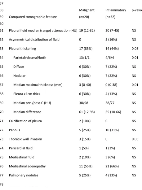
Figure 1. Examples of diffuse pleural thickening. A) Slight diffuse thickening of the parietal 381
pleura (arrowheads) in a dog with mesothelioma; B) Nodular thickening of the parietal 382
pleura (arrowheads) in a dog with mesothelioma; C) Marked irregular thickening of the 383
parietal and mediastinal pleura (arrowheads) in a dog with pyothorax; D) Slight diffuse 384
thickening of the visceral pleura (arrowheads) in a dog with pyothorax. A-C soft tissue 385
window (width 320 HU; level 80 HU); D lung window (width 1500 HU; level -500 HU). 386
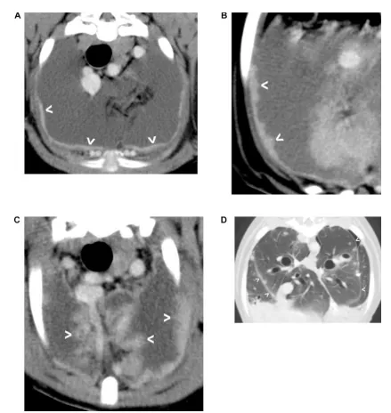
Figure 2. Examples of thoracic wall invasion by mesothelioma. A) Broad mass (*) involving 388
parietal pleura and adjacent intercostal muscles; B) Locally invasive mass (large arrowheads) 389
thickening the inner layer of thoracic wall (small arrowheads) and obliterating the fat plane 390
between muscles of the thoracic wall. Both images displayed using a soft tissue window 391
(width 320 HU; level 80 HU). 392
393
394
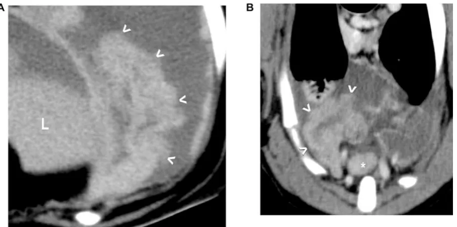
Figure 3. Examples of pannus (A and B) in dogs with pyothorax. In each instance, the 396
morphology of pannus is a thick, folded sheet of tissue (arrowheads) that appears separate 397
from the pleura. An enlarged sternal lymph node (*) is visible in B. Both images displayed 398
using a soft tissue window (width 320 HU; level 80 HU). 399