1535-9778/05/$08.00⫹0 doi:10.1128/EC.4.3.526–535.2005
Copyright © 2005, American Society for Microbiology. All Rights Reserved.
Sex-Specific Homeodomain Proteins Sxi1
␣
and Sxi2
a
Coordinately
Regulate Sexual Development in
Cryptococcus neoformans
Christina M. Hull,
1† Marie-Josee Boily,
1and Joseph Heitman
1,2*
Department of Molecular Genetics and Microbiology1and the Howard Hughes Medical Institute,2
Duke University Medical Center, Durham, North Carolina
Received 14 January 2005/Accepted 14 January 2005
Homeodomain proteins are central regulators of development in eukaryotes. In fungi, homeodomain pro-teins have been shown to control cell identity and sexual development.Cryptococcus neoformansis a human fungal pathogen with a defined sexual cycle that produces spores, the suspected infectious particles. Previously, only a single homeodomain regulatory protein involved in sexual development, Sxi1␣, had been identified. Here we present the discovery of Sxi2a, a predicted but heretofore elusive cell-type-specific homeodomain protein essential for the regulation of sexual development. Our studies reveal that Sxi2a is necessary for proper sexual development and sufficient to drive this development in otherwise haploid␣cells. We further show that Sxi1␣ and Sxi2a interact with one another and impart similar expression patterns for two key mating genes. The discovery of Sxi2a and its relationship with Sxi1␣leads to a new model for how the sexual cycle is controlled inC. neoformans, with implications for virulence.
Patterning during development in animals is controlled by a special set of coordinately regulated genes, the homeobox genes. These genes encode homeodomain proteins that act in concert with other cellular factors to establish patterns in di-verse organisms. For example, in the fruit flyDrosophila
mela-nogaster, where homeobox genes were first discovered,
expres-sion of homeodomain proteins specifies identity along the anteroposterior axis (17). In plants, homeodomain proteins regulate, among other things, the patterning of petals in flow-ers (25). These proteins are also important in controlling sex-ual differentiation, as has been documented in the mouse (45). The roles of homeodomain proteins have also been studied in fungi. In the yeast Saccharomyces cerevisiae, homeodomain proteins establish cell identity (27), and in the mushroom
Cop-rinus cinereus, myriad homeodomain proteins are involved in
mating and sexual development (11).
Sexual reproduction is a central part of eukaryotic life cycles and appears to be important for long-term success. Organisms with strictly clonal life cycles go extinct more rapidly than those with sexual cycles, suggesting that sex confers an advantage, although the reasons for the dominance of sexual reproduction are controversial (7). There are two prevailing hypotheses about how advantage is conferred: sexual recombination either allows the propagation of beneficial mutations in a population or facilitates the purging of deleterious mutations (37, 50). In either case, the majority of eukaryotes utilize a sexual phase in their life cycles. Even in organisms previously thought to not engage in this form of reproduction, cryptic or rarely utilized
sexual cycles have been identified, emphasizing the importance of maintaining the machinery to carry out sexual reproduction. The ascomycetous pathogenic yeast Candida albicans was thought to reproduce only clonally, until a cryptic mating pro-cess was identified (24, 36) and the genome sequence revealed conservation of nearly all of the genes required for complete sexual development (47). Another fungus pathogenic for hu-mans,Cryptococcus neoformans, is a divergent basidiomycete that has also maintained the ability to undergo sexual devel-opment (31). In this case, although sexual reproduction has not been observed directly in the environment, strains of opposite mating types undergo sexual development in the laboratory (1, 23). This complex process inC. neoformansis initiated by the fusion of two haploid cells of different mating types (aand␣). Fusion results in both an altered cell type and a change in growth pattern from yeast cells to dikaryotic filaments. The filamentous dikaryon has the capacity to form specialized mei-otic structures and produce haploid spore products (30), sus-pected to represent the infectious particles inhaled into the lungs of human hosts.
Spores can be analyzed in the laboratory, providing a pow-erful genetic tool that has been invaluable in the identification of virulence traits that allowC. neoformansto promote disease.
C. neoformansis known for causing fungal meningitis in
im-munocompromised patients, and an important virulence at-tribute is mating type (9). Almost all isolates ofC. neoformans
are of a single mating type (␣). Animal experiments have shown that in some genetic backgrounds, ␣ cells are more virulent thanacells (32, 33). This difference betweenaand␣ cells has prompted numerous studies to determine the molec-ular differences between the two mating types in an effort to understand the virulence process.
We previously discovered a key ␣-specific protein that es-tablishes cell identity and controls sexual development inC.
neoformans. Sex inducer 1␣(Sxi1␣) was shown to be necessary
for proper sexual differentiation and sufficient to drive sexual development when expressed in a cells (22). Although this * Corresponding author. Mailing address: 322 CARL Building, Box
3546, Department of Molecular Genetics and Microbiology, 100 Re-search Dr., Duke University Medical Center, Durham, NC 27710. Phone: (919) 684-2824. Fax: (919) 684-5458. E-mail: heitm001@duke .edu.
† Present address: Departments of Biomolecular Chemistry and Medical Microbiology & Immunology, University of Wisconsin—Mad-ison, MadWisconsin—Mad-ison, WI 53706.
526
on September 8, 2020 by guest
http://ec.asm.org/
␣-specific factor can promote sexual development, it was clear from genetic studies that a factor fromacells was also required for appropriate completion of the sexual cycle. Using a com-bination of molecular genetics and bioinformatics, we identify here a gene in the mating-type locus ofa cells, SXI2a, with similarity to homeodomain DNA binding proteins. We show that thisa-specific factor is essential for proper sexual devel-opment and, likeSXI1␣inacells, can change the identity of␣ cells, causing them to adopt an a/␣ cell fate. Furthermore, two-hybrid and gene expression studies suggest that Sxi2a functions in concert with Sxi1␣through a direct interaction to regulate transcription of key mating genes and induce sexual development. These studies reveal that an interaction between homeodomain proteins of two dissimilar classes governs sexual development in C. neoformans, reminiscent of previous and classic studies in both ascomycetes and basidiomycetes (S.
cer-evisiae,Ustilago maydis,C. cinereus, andC. albicans) involving
homeodomain protein complexes, includinga1/␣2 and bE/bW. These discoveries establish a foundation upon which further insights into the control of cell identity and cell differentiation in this ubiquitous human fungal pathogen can be derived.
MATERIALS AND METHODS
Constructingsxi2adeletion strains.To create thesxi2a⌬deletion strain, a
PCR overlap approach was used as described previously (13). The 5⬘flanking
region was amplified with primers JOHE9021 (CAGGCAGCCAGATACAG AGG) and JOHE9023 (GGTCGAGCAACTTCGCTCGGTGATGGTAGAACT
GGAGA), the 3⬘flanking region was amplified with primers JOHE9024 (CCACC
TCCTGGAGGCAAGTAGGAGATTTGTATGCAATAC) and JOHE9026 (GAT
TGTGTGTAACATTGGAG), and theURA5selectable marker was amplified with
primers JOHE9022 (TCTCCAGTTCTACCATCACCGAGCGAAGTTGCTCG ACC) and JOHE9025 (GTATTGCATACAAATCTCCTACTTGCCTCCAGGA
GGTGG). The PCR product was introduced into theMATaserotype Dura5strain
JEC34 by biolistic transformation, and transformants were selected on medium
lacking uracil (⫺ura) and containing 1 M sorbitol (14, 46). Transformants were
screened by PCR for the proper integration of the deletion construct, positive clones
were confirmed by Southern blot analysis, and the resulting independentsxi2a⌬
strains were designated CHY766 to CHY770.
PCR amplification.All PCR amplifications were performed with an MJ Re-search DNA engine DYAD thermal cycler and the ExTaq PCR system
(Inter-gen). Primers were used at a concentration of 0.4M, and templates for PCRs
were titrated and evaluated empirically for each product. For diagnostic PCR to
establish linkage ofSXI2awithMATaprimers to theSXI2aandSXI1␣genes and
a control sequence were used on genomic DNA templates. The following
prim-ers were used to show␣-specific segregation:SXI1␣, JOHE6710 (CGAAGGG
CAAAGTCGAAAACG) and JOHE6711 (CCGAAATAATGGGAACTCC);
SXI2a, JOHE9028 (ATGGGCAGCAACCTTGACATC) and JOHE9872 (GGT
GAATGCAGCATGTTGGTTG); and for the cell type-independent control fragment, JOHE6712 (CTTACCAGTTTGGCTCCTTA) and JOHE6713 (CCT TCTTGGCTAAACCTTTC). The following primers were used to generate
Northern probes:SXI1␣, JOHE6710 and JOHE8207 (CGAGGATCCTTAAC
ACGCTAGGCGCGG);SXI2a, JOHE9028 and JOHE9870 (GGATAGATCT
TACCCCCTGAGGACTGT); MFa, JOHE6683 (TTCTTCGGCAGCCTCAC
TAT) and JOHE6684 (GAAAAGAGGTACGAGTAGAT);MF␣; JOHE1204
(TTTTACGCTTTTTGCAGATTCCGCCAAA) and JOHE3242 (GACCACTG
TTTCTTTCGTTCT); andGPD1(glyceraldehyde-3-phosphate dehydrogenase),
JOHE6524 (CGTCGTTGAATCTACCGGTG) and JOHE6525 (CACCAGCA ATGTAAGAGATG). The amplification conditions for PCR were 30 cycles of 95°C for 30 s, 55°C for 30 s, and 72°C for 30 s. All PCR samples were visualized by standard DNA electrophoretic techniques (38).
Strain manipulations and media.All strains used were of the serotype D background and are described in the relevant procedures. All were handled by standard techniques and with standard media as described previously (2, 40). Mating and self-filamentation assays were conducted on V8 medium at room temperature in the dark for 2 to 4 days. Filamentation was evaluated by observ-ing the periphery of test spots on V8 medium. The matobserv-ing tester strains used
were JEC20 (a) and JEC21 (␣) (33). For confrontation assays, strains were
streaked after 2 days on yeast extract-peptone-dextrose (YPD) agar near one another on solid filament agar plates and incubated at room temperature in the dark for 6 days before they were photographed. Fusion assays were carried out with strains after growth for 2 days on YPD agar. Cells were resuspended in 1 ml of phosphate-buffered saline, quantitated in a spectrophotometer, and diluted to
108cells/ml. Equal numbers of mating partners were mixed, a V8 agar plate was
spotted with 10l, and plates were incubated at room temperature in the dark.
After 24 h, the cells were scraped off the V8 plates, resuspended in 100l of
water, and spread on selective plates. Plates were incubated at 30°C for 5 days,
and the resulting colonies were counted. Test crosses were as follows: 1,aUra⫹
nat⫺(JEC20)⫻ ␣Ura⫺nat⫹(CHY621); 2, Ura⫹nat⫺(JEC20)⫻sxi1␣⌬Ura⫺
nat⫹(CHY618); 3,sxi2a⌬Ura⫹nat⫺(CHY768)⫻ ␣Ura⫺nat⫹(CHY621); and
4,sxi2a⌬Ura⫹nat⫺(CHY768)⫻sxi1␣⌬Ura⫺nat⫹(CHY618). Fusants were
selected on proline medium (1.7 g of yeast nitrogen base [without amino acids
and without ammonium sulfate], 1 g ofL-proline, 20 g of dextrose, 20 g of agar
per liter) containing 100g of ml nourseothricin per ml. A cross between strains
known to have fusion defects,acpk1Ura⫹Ade⫺(JEC171)⫻cpk1Ade⫹Ura⫺
(RDC3), resulted in no fusants after growth under selection on SD (synthetic
medium plus dextrose [40])⫺adenine⫺uracil plates. To test the effects of ectopic
expression ofSXI2a, theSXI2aopen reading frame (ORF) and approximately
300 bp of 3⬘untranslated region were liberated from plasmid pCH269 with BglII.
The resulting fragment was cloned into the BamHI site of the telomeric,
GPD1-containing plasmid pRCD85 to create pCH285. This plasmid was digested with
meganuclease I-SceI and transformed via electroporation into JEC43 (␣ura5),
JEC34 (a ura5), and CHY925 (sxi2a⌬ura5) to create the strains CHY1014,
CHY1022, and CHY927, respectively. To test ectopic expression ofSXI1␣ina/a
cells, plasmid pCH258 was transformed into strain CHY640 (a/aura5/ura5) via
electroporation and selected on SD plates without uracil to generate strain CHY815. Transformants were tested for self-filamentous behavior on V8 plates as described above. Spore analysis was carried out on strains CHY1014 and CHY815. Spores from V8 were microdissected and germinated on YPD. Each segregant was then tested for ploidy via fluorescence-activated cell sorter (FACS) analysis as described previously (41) and for auxotrophic markers. Twenty-two spores were evaluated from strain CHY1014, and all were found to
be Ura⫺␣haploids, indicating loss of theURA5-markedSXI2aplasmid.
Thirty-nine spores were evaluated from CHY815, and all wereahaploids whose marker
phenotypes are indicated in Fig. 4.
Two hybrid assays.Constructs for SXI1␣andSXI2awere generated with
coding regions excised from cDNA-containing plasmids. SXI1␣ cDNA was
cloned as a BamHI fragment from pCH271 into the BamHI site of pGAD-C1 (amino-terminal Gal4 activation domain [AD]), pGBD-C1 (amino-terminal Gal4 DNA binding domain [BD]), and pCDBD2 (carboxy-terminal Gal4 DNA
BD).SXI2acDNA was cloned as a BglII fragment from pCH274 into the BglII
sites of pGAD-C1and pGBD-C1 and into the BamHI site of pCDBD2. The
truncated versions ofSXI2awere generated by PCR with primer JOHE9028.1
(GGATAGATCTATGGGCAGCAACCTTGACATC) in combination with JOHE11774 (GGATAGATCTGCAGAAGACACCAGTTTATC), JOHE11775 (GGATAGATCTAGGGCGAGAGTGCGGGACTTG), or JOHE11776 (GGA TAGATCTAAGACCAAGTTCCATCTTTAG) to generate 1,725-, 671-, and 406-bp products, respectively. Products were digested with BglII and cloned into the BglII site of pGAD-C1. All constructs were sequenced across the ligation junctions or PCR-generated regions, and no deviations from the expected
se-quences were identified. Each pairwise combination ofSXI1␣andSXI2afusions
in the yeast two-hybrid vectors was transformed into cells of yeast strain PJ69-4a (26). Transformants were selected on medium lacking tryptophan and leucine
(SD⫺leu⫺trp) and then tested for growth on medium lacking adenine (SD
⫺leu⫺trp⫺ade) and medium lacking histidine (SD⫺leu⫺trp⫺his⫹3AT).
-Galactosidase assays were performed as described previously (8).
Northern blot analysis.RNA was prepared from C. neoformanscells with TRIZOL from Invitrogen. Strains were grown on solid V8 medium for 24 h (haploids) or 48 h (cocultures and diploids) at room temperature before they were harvested by scraping off the agar surface. Northern blots were carried out
according to standard protocols (4) with 10g of total RNA used for each
sample. TheMF␣,MFa,SXI2a,SXI1␣, andGPD1probes were generated by
PCR as described above, and radiolabeled probes (Rediprime II kit from Am-ersham Pharmacia Biotech), were used in hybridization reactions as described previously (12) at 65°C.
Sequence manipulations.Splice predictions of candidate gene sequences for
SXI2a were facilitated with a Softberry algorithm (www.softberry.com).
Se-quence comparisons were conducted with the BLAST algorithm (3) against the
StanfordC. neoformansgenome sequence (C. neoformans Genome Project,
Stanford Genome Technology Center, and The Institute for Genomic
on September 8, 2020 by guest
http://ec.asm.org/
search). Sequence analyses were conducted and alignments were generated with SeqWeb version 2 (Accelrys).
Nucleotide sequence accession number.TheSXI2asequence can be accessed in GenBank under accession no. AY911308.
RESULTS
Identification of an a-specific factor in C. neoformans. Ex-periments characterizing the role ofSXI1␣in sexual develop-ment revealed that although we had identified a key␣-specific factor, an unidentified factor(s) fromacells was also required for sexual development. Ectopic expression ofSXI1␣inacells resulted in the initiation and completion of the sexual devel-opment process; however, expression of this factor in haploid
␣or␣/␣ cells did not lead to sexual development, indicating that a signal fromacells was required for the process. To test the hypothesis that predicteda-specific factors controlling sex-ual development are encoded by the mating-type (MAT) locus in a cells, we constructed a complete deletion of the entire 120,246-bp MATa allele in diploid a/␣ cells. Cells in which
MATa was deleted no longer exhibited the self-filamentous phenotype characteristic ofa/␣cells, and in mating assays they behaved like haploid␣cells (data not shown). These findings confirm that at least one of the factors required to direct formation of mating structures resides inMATaand prompted an intensive bioinformatic analysis of the MATa region to identify genes with similarity to transcription factors found in other fungalMATloci.
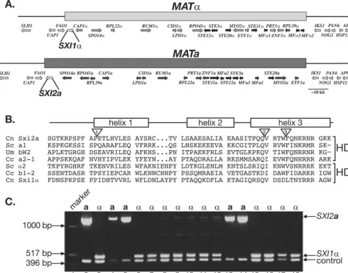
We surmised that a missing a-specific factor might be en-coded by a gene with synteny to theSXII␣gene in the␣allele of theMATlocus. We utilized the BESTORF gene prediction algorithm from Softberry, Inc., to electronically produce pre-dicted spliced cDNA products encoded by a 10-kb region. A candidate cDNA spanning a novel ORF was identified. This ORF, designatedSXI2a, for Sex Inducer 2a, is located in the
MATaallele and is the first gene in the locus, residing in the most-telomere-proximal position. This placement is analogous to the position of theSXI1␣gene in theMAT␣allele (Fig. 1A). We also evaluated the MATa allele from anotherC. neofor-mansvariety (var.grubii) and a sibling species (C. gattii) (16, 35) and found that this ORF was conserved in all three. The ORF is syntenic in only two, and in the third (C. neoformans
var. grubii) it now resides at an internal position, likely as a
result of gene inversions, which have been shown to be prev-alent in theMATlocus (16).
The predicted protein sequence encoded by theSXI2aORF contains a domain with similarity to known homeodomain pro-teins. Identification ofSXI2awas challenging because the most conserved region of the homeodomain (helix 3) sequence is interrupted by two introns, resulting in the production of a very small, 4-amino-acid exon (Fig. 1B). The presence of these clustered introns thwarted earlier attempts to identify ana -specific homeodomain factor by using conserved homeodo-main sequences from the third helix in BLAST searches againstC. neoformansgenomic DNA sequence.
Homeodomain proteins from eukaryotes generally fall into two categories: those that conform to a more traditional ho-meodomain structure designated HD2 (e.g., S. cerevisiae a1
and C. cinereus a2-1) and those that are characterized by a
3-amino-acid insertion between helices 1 and 2 of the
home-odomain, designated HD1 (e.g.,S. cerevisiae␣2 andC. cinereus
b1-2) (6, 10). In previous studies, heterodimeric regulatory complexes formed by homeodomain proteins usually include a member from each homeodomain category (11). The well-characterized homeodomain proteinsa1 and␣2 fromS.
cer-evisiaefall into the HD2 and HD1 classes and interact with one
another to form a transcriptional repressor (20). The predicted homeodomain region for Sxi2ais similar to the HD2 class, and the analogous region in Sxi1␣ is similar to the HD1 class of homeodomains (Fig. 1B). In addition, many HD2 homeobox genes contain a conserved intron in the coding region for the third helix that lies between conserved tryptophan and phenyl-alanine residues (18). This intron is also conserved inSXI2a. Finally, an analysis of meiotic progeny from a cross betweenC.
neoformans aand ␣ strains revealed that the SXI2a gene is
linked to the a mating type, supporting its location within
MATa and confirming thatSXI2ais ana-specific gene (Fig. 1C).
SXI2a controls sexual development. Under mating condi-tions, haploida and ␣ cells ofC. neoformans fuse with one another and undergo sexual development (the formation of filaments, basidia, and spores), as diagrammed in Fig. 2A. We deleted the predicted ORF for the SXI2a gene and tested
sxi2a⌬ strains in a series of assays to determine the role of Sxi2ain sexual development. In a coculture assay in which the deletion strain was mixed with a wild-type␣ mating partner under filamentation conditions, there was a dramatic reduction in filament formation (Fig. 2B, top row). This striking result mirrored that of asxi1␣ deletion strain cocultured with ana mating partner. In both cases, thesxi⌬strains failed to undergo proper sexual development and produced only rudimentary filaments lacking basidia and spores, which were largely devoid of nuclei and clamp connections. In these respects, the fila-ments produced most closely resemble conjugation tubes nor-mally produced during the initial stages of mating that facili-tate partner recognition and cell-cell fusion.
To further understand the nature of this mating defect, confrontation assays were conducted. In this assay, wild-type cells grown on low-nutrient solid medium respond to one an-other at a distance:␣cells form filamentous projections toward acells andacells become enlarged (Fig. 2, bottom row). We testedsxi1␣⌬ andsxi2a⌬strains for the ability to respond to mating partners. In both cases, the cells responded like wild-type cells, indicating that their defects in filamentation do not reflect deficiencies in sensing or communicating with mating partners via mating pheromones (a-factor and␣-factor). This was also the case in assays in which both thesxi1␣⌬andsxi2a⌬ strains were tested with each other (Fig. 2, bottom row). These observations suggest that the rudimentary filaments produced during coculture of thesxi1␣andsxi2amutants with strains of the opposite mating type are indeed conjugation tubes (rather than true dikaryotic mating filaments or the monokaryotic fil-aments produced by diploids during self-filamentous growth). Finally, the sxi⌬ strains were tested for the ability to fuse with a mating partner. In this assay, differentially marked strains were cocultured under mating conditions for 24 h and then placed under selection for genetic markers from each parent. The frequency of cell fusion was measured as the number of colonies formed on selective medium. The effi-ciency of fusion between wild-typeaand␣cells was designated
on September 8, 2020 by guest
http://ec.asm.org/
100%, and the efficiency of fusion by the deletion strains was evaluated as the percentage of fusion events relative to the expected wild-type frequency (Fig. 2, middle row). By this assay, thesxi1␣⌬andsxi2a⌬strains exhibited no defects in cell fusion, while a mating-impaired, fusion-defective mutant strain (cpk1⌬) showed a ⬎1,000-fold decrease in cell fusion, as re-ported previously (15). Thus, the dramatic defects we observe in sexual development of thesxi⌬ strains are not a result of inefficient cell fusion but rather reflect a detrimental effect on subsequent steps in sexual development.
Expression of SXI2ain haploid ␣cells induces sexual de-velopment. We showed previously that Sxi1␣ is sufficient to drive filamentation and sporulation in haploidacells (22). We have carried out a similar analysis with Sxi2a. In this experi-ment,SXI2awas placed under the control of a constitutiveC.
neoformanspromoter on an autonomously replicating plasmid.
The plasmid was transformed intoa,␣, anda/␣cells, and the resulting transformants were tested for the ability to undergo sexual development under inducing conditions. Strikingly, the
␣cells containing theGPD1-SXI2aplasmid formed filaments, FIG. 1.MATacontains a gene encoding the predicted homeodomain protein Sxi2a. (A) TheSXI2agene is located in theMATlocus. A schematic representation of theC. neoformans MATlocus showsMAT␣on the top andMATaon the bottom. The shaded bars represent the region of nonidentical DNA present inMAT␣andMATa. Genes within the locus are represented by shaded arrows. Each gene within the locus, with the exception ofSXI1␣andSXI2a, has a counterpart allele that encodes a similar, but not identical, protein in the opposite mating type.SXI1␣and
SXI2aare unique to their respective mating types. (B) Sxi2ais a homeodomain protein. The predicted homeodomain region ofC. neoformans(Cn) Sxi2ais aligned with known homeodomains of other proteins:S. cerevisiae(Sc)a1,U. maydis(Um) bW2,C. cinereus(Cc) a2-1,S. cerevisiae␣2,
C. cinereusb1-2, andC. neoformansSxi1␣. A schematic representation of the homeodomain region shows the helices of a classic three-helix bundle found in homeodomains above the sequences.C. neoformansSxi2a,S. cerevisiaea1,U. maydisbW2, andC. cinereusa2-1 fall into the HD2 class of homeodomains, whereasS. cerevisiae␣2,C. cinereusb1-2, andC. neoformansSxi1␣are members of the HD1 family containing a 3-amino-acid insertion between helices 1 and 2. The inverted triangles denote the positions of introns in Sxi2a. Introns 1, 2, and 3, are 65, 66, and 294 bases, respectively. The third intron is conserved in many HD2 homeodomain proteins. (C)SXI2ais linked toMATa. Genomic DNA from segregants of a genetic cross was subjected to PCR analysis with primers to the ORF ofSXI2a.Lane 1, marker; lanes 2 and 3, DNA from controlacells and ␣cells, respectively; lanes 4, 5, 13, and 14, DNA from segregants that mate asacells; lanes 6 to 12 and 15 to 18, DNA from segregants that mate as␣cells.
on September 8, 2020 by guest
http://ec.asm.org/
basidia, and spores. A control strain containing an empty vec-tor did not and behaved like a wild-type␣strain, showing no filamentation under these conditions (Fig. 3A). This result shows that, likeSXI1␣,SXI2ais sufficient to induce filamen-tous behavior that is temporally delayed (data not shown) but morphologically indistinguishable from diploid sexual develop-ment. We note that the filaments produced were monokaryotic with unfused clamp connections like those produced by sexual reproduction ofa/␣diploid cells, rather than those produced by aand ␣ cell mating that are dikaryotic with fused clamp connections (data not shown). In contrast, when theSXI1␣and
SXI2agenes were introduced into thesxi2a⌬orsxi1␣⌬strains, respectively, neither was sufficient to induce self-filamentous growth (data not shown), indicating that the effects of Sxi1␣ and Sxi2aare dependent on the presence of both factors.
One caveat to these heterologous expression studies is that because the test strains were haploid, we could not exclude the possibility that the filamentation and sporulation we observed in the presence ofSXI1␣orSXI2awere due to the induction of
a haploid filamentation and sporulation pathway that has been observed primarily in␣cells. Known as monokaryotic fruiting this pathway is generally thought to be strictly mitotic (49). We evaluated this possibility by expressing the SXI1␣ or SXI2a gene in homozygous diploid cells. In the laboratory, stable, diploidC. neoformansstrains have been created that grow as mononucleate, budding yeast (41). We previously created ho-mozygousa/a and ␣/␣ diploid strains for ploidy studies and found that they behaved like their haploid counterparts:a/a cells mate with␣cells, and␣/␣cells mate withacells (22). We used these strains here to evaluate the effects of ectopic ex-pression ofSXI1␣orSXI2a. We expressedSXI1␣ina/acells heterozygously marked at three genomic loci and observed the induction of filamentation, basidia formation, and sporulation. We analyzed the spore products that were generated and dis-covered that they were all haploid based on FACS analysis (data not shown), indicating that a chromosomal reduction event had occurred. The spores all mated asacells and were recombinant for the markers tested. That is, the spores dis-FIG. 2.SXI2ais required for sexual development. (A) Sexual development inC. neoformans. When haploidacells encounter haploid␣cells at 25°C in the presence of either an unidentified plant factor in V8 medium or nitrogen limitation, the cells initiate a mating response and fuse with one another. The fused cells adopt a filamentous growth pattern in which the haploid nuclei do not fuse with one another. This dikaryotic filament grows in a polar manner, and adjacent filament cells are linked by fused clamp connections. Ultimately, in response to unknown signals, the terminal filament cell ceases extension and forms a rounded compartment at the distal end of the cell. It is in this basidium that nuclear fusion, meiosis, and sporulation occur. Meiotic products are packaged, and spores are extruded in long chains on the surface of the basidium. (B)sxi2a⌬ strains can respond to and fuse with a mating partner but fail to form normal filaments. Wild-type,sxi1␣⌬, andsxi2a⌬strains were tested in coculture (top row) (48 h), fusion (middle row) (24 h), and confrontation (bottom row) (48 h) assays to assess the nature of their defects in sexual development. Strain combinations are shown above the top row and indicate the strains used in each cross as well as the strains used in the fusion and confrontation assays. There was no significant difference in the extent of the limited rudimentary filaments produced in crosses by the mutant strains.
on September 8, 2020 by guest
http://ec.asm.org/
played phenotypes consistent with reassortment of the markers from the diploid parent: all of the predicted auxotrophic phe-notypes were represented in roughly the expected frequencies for a standard cross between two haploid strains (Fig. 3B). If the expression of SXI1␣ had activated a mitotic sporulation pathway, we would have expected the production of diploid, prototrophic spores. We observed the same pattern in recip-rocal experiments in whichSXI2awas expressed in␣/␣ cells (data not shown). Our findings support the hypothesis that the expression of Sxi1␣ and Sxi2ain the same cell induces com-plete sexual development, including meiosis and sporulation.
SXI1␣ and SXI2a impart similar patterns of pheromone gene regulation.To understand how Sxi1␣and Sxi2acontrol sexual development, we evaluated gene expression in the
de-letion strains. We analyzed mating conditions in three different settings: haploid cells grown alone, haploid cells grown in co-culture with mating partners, and diploid cells grown alone. In each case, RNA was extracted and subjected to Northern blot analysis with probes for five different genes:SXI1␣,SXI2a, the pheromone genesMF␣andMFa, and the control geneGPD1. We evaluated the expression patterns ofSXI1␣andSXI2ain wild-type and deletion strains. Our analysis confirms that, as expected, strains with gene deletions lack detectable levels of transcript from the targeted gene (Fig. 4, lanes 3, 4, 6 to 8, and 10 to 12). The data also show that in wild-type haploid cells, the transcript levels ofSXI1␣andSXI2aare barely detectable by Northern blotting (Fig. 4, lanes 1 and 2), although these tran-scripts were detectable by reverse transcription followed by FIG. 3.SXI2ainduces sexual development in␣cells. (A) Expression ofSXI2ain haploid␣cells results in filamentation and spore formation. Panels show the amount of filamentation achieved under mating conditions for each of the strain combinations indicated (from left to right): a wild-type cross between haploidaand␣strains (a⫻ ␣), a haploid␣strain (␣), and a haploid␣ strain expressing thea-specific geneSXI2a
(␣⫹SXI2a) (B) Schematic representation of filament formation ina/adiploid cells expressingSXI1␣and a table showing analysis of spore products. The “parent genotype” is that of the homozygousa/adiploid used in the experiment. The “spore phenotypes” are those combinations expected to result from sporulation of the parent diploid. The frequencies presented result from an analysis of 39 random spores that were micromanipu-lated, germinated, and analyzed from thea/a⫹SXI1␣strain under filamentation conditions. n/a, not applicable. The structure on the side of the filament cells represents an unfused clamp connection.
on September 8, 2020 by guest
http://ec.asm.org/
PCR (data not shown). In contrast, both transcripts are up-regulated during coculture and ina/␣heterozygous diploids, and they are readily detected by Northern blotting, indicating that gene products supplied by bothaand␣lead to the induc-tion ofSXI1␣andSXI2atranscription. This up-regulation was not induced by mating pheromones as has been observed in other basidiomycetes (48) and instead appears to require cell fusion (C. M. Hull and J. Heitman, unpublished data). This regulation is also not dependent upon the presence ofSXI1␣
andSXI2a. That is, strains in which SXI1␣has been deleted still exhibit induction ofSXI2aand vice versa (Fig. 4, lanes 6, 7, 10, and 11). These data indicate thatSXI1␣andSXI2ado not work in concert to activate their own transcription, and additional factors specific to a and ␣ cells or regulated by factors specific toaand␣cells must be responsible for induc-ingSXI1␣andSXI2a.
We also investigated transcription of the MF␣ and MFa pheromone genes, which are induced by exposure to cells of the opposite mating type. In haploid cells, the levels of both the aand␣pheromone transcripts are consistent with their mating type and do not appear to change in the sxi1␣⌬ or sxi2a⌬ strains (Fig. 4, lanes 1 to 4). Under coculture conditions and also in stable diploid strains, the levels of pheromone transcript change dramatically in response to the deletion of either
SXI1␣orSXI2a. As was observed previously, transcript levels of both the MFa and MF␣ pheromone genes increase in dikaryotic and diploid strains in whichSXI1␣has been deleted, indicating thatSXI1␣is a formal repressor of pheromone gene transcription (Fig. 4, lanes 6 and 10). This pattern is also observed in thesxi2a⌬strains; the levels of bothMFaandMF␣
pheromone transcript increase in dikaryotic and diploid strains in whichSXI2ahas been deleted (Fig. 4, lanes 7 and 11). The
level of derepression is not substantially different in crosses or strains in which bothSXI1␣andSXI2ahave been deleted (Fig. 4, lanes 8 and 12). These expression data suggest strongly that
SXI1␣andSXI2aregulate the same target genes and support the hypothesis that Sxi1␣ and Sxi2aact in concert to repress the transcript levels of mating genes and thereby govern sexual development.
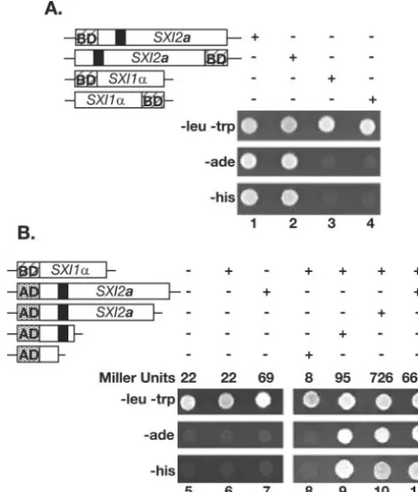
SXI1␣andSXI2ainteract in a two-hybrid assay.To test the model that Sxi1␣and Sxi2acoordinately regulate gene expres-sion by interacting with one another, we evaluated Sxi1␣and Sxi2afor the ability to interact, using a yeast two-hybrid assay. TheSXI1␣andSXI2aORFs were fused to theGAL4AD or DNA BD and tested inS. cerevisiaefor the ability to transac-tivate several different reporter genes. All combinations of fusion proteins were constructed: amino-terminal and carboxy-terminal fusions to either the AD or BD for each protein.
SXI2afused to BD results in transactivation in the absence of an AD construct (Fig. 5A, columns 1 and 2). The BD-SXI1␣
fusion construct did not activate the reporter genes on its own (Fig. 5A, columns 3 and 4) and was used to test for interaction FIG. 4. Sxi1␣and Sxi2a regulate pheromone gene expression. A
Northern blot showing the levels of selected transcripts in wild-type,
sxi1␣⌬, andsxi2a⌬strains after growth on V8 medium in three differ-ent contexts, indicated below the lane numbers: haploid strains alone, mixed mating cocultures, and diploid strains alone. The top two panels show RNA probed with theMFa andMF␣ORFs. The third panel shows the same blot probed with portions of theSXI1␣ andSXI2a
ORFs. The bottom panel shows the same blot probed with a portion of theGPD1ORF. Lane 1, wild-type haploida; lane 2, wild-type haploid ␣; lane 3, haploidsxi1␣⌬; lane 4, haploidsxi2a⌬; lane 5, wild-typea
cocultured with wild-type␣; lane 6, wild-typeacocultured withsxi1␣⌬; lane 7,sxi2a⌬cocultured with a wild-type␣; lane 8,sxi2a⌬cocultured with asxi1␣⌬; lane 9,a/␣diploid; lane 10,a/sxi1␣⌬diploid; lane 11,
sxi2a⌬/␣diploid; lane 12,sxi2a⌬/sxi1␣⌬diploid.
FIG. 5. Sxi1␣and Sxi2ainteract with one other. The top panel in each set (⫺leu⫺trp) confirms the presence of each two-hybrid plasmid under selection. The middle and bottom panels indicate the presence or absence of activation of a biosynthetic reporter gene (eitherADE2
orHIS3). The presence or absence of indicated constructs is repre-sented with⫹or⫺over each panel. The fusion proteins tested are indicated in schematic form: the hatched box BD represents the Gal4 DNA BD, the speckled box AD represents the Gal4 AD, and the black box represents the Sxi2ahomeodomain region. Growth in a test spot indicates activation of the reporter. (A) Lanes 1 to 4 reveal a transac-tivating property of Sxi2ain the absence of Sxi1␣when fused to a DNA BD. (B) Lanes 5 to 11 confirm an interaction between Sxi1␣and Sxi2a
and define the region of Sxi2anecessary for interaction. The results of liquid-galactosidase assays in each column are shown in Miller units.
on September 8, 2020 by guest
http://ec.asm.org/
between Sxi1␣and Sxi2a. Figure 5B shows that when a DNA BD was fused to the amino terminus of Sxi1␣and an AD was fused to the amino terminus of Sxi2a, there was activation of two independent reporter genes required for growth under selection (Fig. 5B, column 11), indicating an interaction be-tween Sxi1␣ and Sxi2a. Controls (Fig. 5B, columns 5 to 7) confirmed that activation of the reporter was dependent on the presence of bothGAL4fusion proteins, and liquid -galacto-sidase assays (reported in Miller units) confirmed the activa-tion of theGAL4-lacZgene as a third, independent reporter (Fig. 5B). Columns 9 and 10 show that this interaction is dependent on only a small portion of Sxi2a. C-terminal trun-cations of Sxi2a were tested for the ability to interact with Sxi1␣, and the smallest interacting fragment contained the first one-third of the protein, including the homeodomain region (column 9). A smaller fragment in which the homeodomain had been deleted did not interact with Sxi1␣(column 8). Al-though we cannot exclude models in which other proteins might also be present in Sxi1␣-Sxi2acomplexes, we conclude that the most likely model is that Sxi1␣ and Sxi2a interact directly, as is the case with other known HD1-HD2 het-erodimeric complexes. We propose that this interaction leads to the coordinate regulation by Sxi1␣-Sxi2aof sexual develop-ment inC. neoformans.
DISCUSSION
We describe here the discovery of a cell-type-specific home-odomain protein required for sexual development inC.
neo-formans. Previous work had predicted the presence of ana
-specific factor required for sexual development, but sequence analyses had failed to reveal its identity (22). Functional anal-yses narrowed the region of the genome in which this factor could reside to theaallele of theMATlocus, and bioinformatic approaches identified ana-specific gene located inMATawith similarity to homeobox-containing genes. Deletion of this gene,SXI2a, resulted in a profound defect in sexual develop-ment in C. neoformans, and ectopic expression of SXI2a in haploid␣cells resulted in the induction of sexual development in the absence of a mating partner. Sxi1␣ and Sxi2ainduce similar patterns of mating gene expression and interact with one another in a two-hybrid assay, supporting a model in which these proteins interact with one another to form a het-erodimeric homeodomain transcriptional regulatory complex that specifies the dikaryotic state and thus promotes sexual development. We hypothesize that the absence of either Sxi1␣ or Sxi2arenders newly fused cells incapable of initiating a new developmental pathway. The factors are both required for specifying the a ⫹ ␣ state of the newly formed dikaryon. Without these key proteins, fused cells are arrested and inca-pable of forming dikaryotic filaments, basidia, or spores. Thus, Sxi1␣and Sxi2aare factors that cooperate to change the iden-tities of fused mating partners and direct a new developmental program inC. neoformans.
Model for the regulation of sexual development. We pro-pose that Sxi1␣and Sxi2acarry out the coordinated regulation of sexual development through a direct protein-protein inter-action with one another. It has been shown in related fungi that the homeodomain proteins that control sexual development interact with one another to regulate gene expression. A
well-studied example of this type of gene regulation is represented by thea1 and␣2 proteins fromS. cerevisiae(27). In this bud-ding yeast,a1 is produced inacells and ␣2 is produced in␣ cells. When a and ␣ cells mate, the proteins form a het-erodimeric complex that alters gene regulation: haploid-spe-cific genes are repressed, and this change allows diploid cells to undergo meiosis and sporulation. This regulatory scheme is also found inU. maydis, where the homeodomain proteins bE and bW form a regulatory heterodimer and promote sexual development (19, 29, 39). In these systems as well as others, the proteins in the heterodimeric complex each fall into a different class of homeodomains and the complexes consist of one HD1 protein and one HD2 protein. In C. neoformans, Sxi1␣ and Sxi2aare the only predicted cell-type-specific homeodomain DNA binding proteins in the MAT locus required for the control of sexual development, and they fall into the two classes of homeodomains: Sxi1␣is an HD1 protein, and Sxi2a is an HD2 protein. These features, along with our data that the proteins interact in a two-hybrid assay, make a compelling argument for a regulatory complex inC. neoformans develop-ment. Finally, both factors control expression of the phero-mone genes in a similar way, further suggesting that they re-press pheromone gene exre-pression in a coordinated manner either directly or indirectly.
Of note regarding Sxi2awas its ability in a two-hybrid assay to activate transcription without a partner protein when fused to the Gal4 DNA BD. Homeodomain partners have been observed to carry out different functions in the heterodimeric complex, suggesting that the strategies for achieving regulation differ between complexes. For example, inC. cinereus, it ap-pears that only one of the heterodimeric partners binds DNA and the other is essential for nuclear localization of the com-plex, and its DNA BD is not required (42). InS. cerevisiae
binding to DNA is primarily mediated by contacts of thea1 protein, and the role of␣2 appears to be to enhance DNA binding bya1 (43) and to recruit the Tup1-Ssn6 corepressor complex (28). Perhaps inC. neoformans, the most important role for Sxi1␣ is to interact with Sxi2a and shuttle it to the DNA of regulated genes to activate transcription. Although the formal roles for Sxi1␣and Sxi2ain pheromone gene reg-ulation are as transcriptional repressors, this effect could be indirect, and the complex could be acting upstream to activate repressors of pheromone genes. Understanding the nature of the interaction between Sxi1␣, Sxi2a, and their targets will elucidate the mechanisms by which these key regulators regu-late transcription and control development.
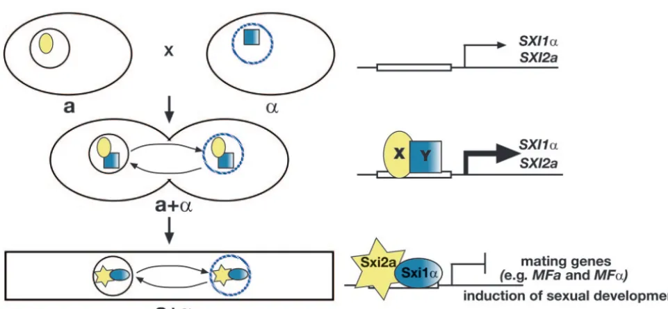
SXI1␣ andSXI2aare up-regulated during sexual develop-ment.It is clear from our studies that Sxi1␣and Sxi2afunction after cell fusion. Deletion studies in diploids show that sexual development in these strains cannot take place in the absence of either Sxi1␣ or Sxi2a, even though both cell and nuclear fusion have taken place. Both proteins are clearly required subsequent to cell fusion for sexual development to continue. This is consistent with the finding that transcription of both
SXI1␣ and SXI2a is up-regulated during mating and in dip-loids. These findings lead to a model of regulation in which Sxi1␣and Sxi2aplay very little, if any, role in haploid cells (Fig. 6). However, in response to the fusion of a and ␣ mating partners,SXI1␣andSXI2aare up-regulated. Interestingly, this regulation is independent of the presence of both factors,
on September 8, 2020 by guest
http://ec.asm.org/
dicating that an Sxi1␣-Sxi2aregulatory heterodimer is not re-sponsible for inducing expression of these genes. Instead, other cell-type-specific regulatory factors must participate to induce
SXI1␣andSXI2atranscription. After induction, a Sxi1␣-Sxi2a regulatory complex is then enabled to efficiently repress mating genes (likeMFaandMF␣) and signal the dikaryotic state. This repression likely prevents additional fusion of dikaryons with haploids and therefore maintains the integrity of the dikaryotic state (34). This altered cell type then has the capacity to un-dergo sexual development and complete its life cycle.
Relationship between sex and virulence. Although a rela-tionship between sexual development and virulence has been established in some plant fungal pathogens, this link in the human fungal pathogens has been less clear. In the corn smut
U. maydis, haploid cells must come into contact with a plant
host before sexual differentiation can occur (5), but there is no clear equivalent requirement among any of the human fungal pathogens. To date, there is no evidence for complete sexual development in vivo for any human fungal pathogen, but there are clues that sexual development is an integral part of the virulence process. The clues are most clear inC. neoformans
where mating type plays a significant role in how the organism interacts with its environment. Almost all isolates from both patients and the environment are␣, suggesting some advan-tage overastrains (32). In addition, in some backgrounds,␣ cells are more virulent in animals thanacells, indicating that the factors that specify ␣ cell identity (and thereby control mating and sexual development) also affect prevalence and virulence (33). This relationship is a complex one; however, as Sxi1␣does not appear to play a prominent role in virulence (21). Understanding all of the determinants of mating type and
the mechanisms of sexual development may ultimately reveal the cell-type-specific properties that contribute to virulence.
Sexual development inC. neoformansis also significant be-cause human infections are thought to be be-caused by the prod-ucts of this process, spores. It has been proposed that the small size of the spore makes it a likely candidate for the infectious particle in human infections (9, 44). If the spore is the infec-tious particle, understanding how sexual development is con-trolled promises to shed light on how these propagules are created and dispersed in the environment. Our discovery of
SXI2a and subsequent studies of the relationship between Sxi1␣and Sxi2aanswer many questions about sexual develop-ment inC. neoformansand afford new opportunities to further explore the process of sexual development in a model human fungal pathogen.
ACKNOWLEDGMENTS
We thank Robert Brazas, James Fraser, Alex Idnurm, and Robin Wharton for comments on the manuscript; Cristl Arndt and Joanne Ekena for technical assistance; and members of the Heitman labora-tory, especially Rob Davidson, Connie Nichols, and Yen-Ping Hseuh, for their efforts. Special thanks go to Alex Idnurm for his pivotal contributions to the discovery ofSXI2a.
This work was supported by an NIAID RO1 grant AI50113 to J.H. and NIAID program project grant AI44975 to the Duke University Mycology Research Unit. C.M.H. was supported by a Damon Runyon Cancer Research Fund Fellowship (DRG-1694) and a Burroughs Wellcome Career Award in the Biomedical Sciences. J.H. is a Bur-roughs-Wellcome Scholar in Molecular Pathogenic Mycology and an investigator of the Howard Hughes Medical Institute. TheC. neofor-mans Genome Project, Stanford Genome Technology Center, was funded by the National Institute for Allergy and Infectious Diseases, National Institutes of Health [NIAID/NIH], under cooperative agree-ment U01 AI47087, and The Institute for Genomic Research was FIG. 6. Model for the regulation of sexual development in C. neoformans. Model for the role of Sxi1␣ and Sxi2a in controlling sexual development. Large ovals represent cells. A solid circle represents theanucleus, and a hatched blue circle represents the␣nucleus. The yellow oval X and shaded blue square Y represent unknowna- and␣-specific factors, respectively, that lead to the induction ofSXI1␣andSXI2a. The blue shaded oval represents Sxi1␣, and the yellow star represents Sxi2a. (Top)SXI1␣andSXI2aare expressed at low levels in haploid cells. (Middle) Following cell fusion,SXI1␣andSXI2aexpression is dependent upon and induced by factors from bothaand␣cells (X and Y). (Bottom) After induction, Sxi1␣and Sxi2aform a heterodimeric complex that establishes the dikaryotic state and induces sexual development through the repression of mating genes.
on September 8, 2020 by guest
http://ec.asm.org/
funded by the NIAID/NIH under cooperative agreement U01 AI48594.
REFERENCES
1.Alspaugh, J. A., R. C. Davidson, and J. Heitman.2000. Morphogenesis of
Cryptococcus neoformans. Dimorphism in human pathogenic and
apatho-genic yeasts. Contrib. Microbiol.5:217–238.
2.Alspaugh, J. A., J. R. Perfect, and J. Heitman.1997.Cryptococcus
neofor-mansmating and virulence are regulated by the G-protein␣subunit Gpa1
and cAMP. Genes Dev.11:3206–3217.
3.Altschul, S. F., W. Gish, W. Miller, E. W. Myers, and D. J. Lipman.1990.
Basic local alignment search tool. J. Mol. Biol.215:403–410.
4.Ausubel, F., R. Brent, R. Kingston, D. Moore, J. Seidman, J. Smith, and K. Struhl (ed.).1997. Current protocols in molecular biology, vol. 2:13. John Wiley and Sons, Inc., Boston, Mass.
5.Banuett, F.1995. Genetics ofUstilago maydis, a fungal pathogen that induces
tumors in maize. Annu. Rev. Genet.29:179–208.
6.Burglin, T. R.1997. Analysis of TALE superclass homeobox genes (MEIS, PBC, KNOX, Iroquois, TGIF) reveals a novel domain conserved between
plants and animals. Nucleic Acids Res.25:4173–4180.
7.Burt, A.2000. Perspective: sex, recombination, and the efficacy of selection—
was Weismann right? Evol. Int. J. Org. Evol.54:337–351.
8.Cardenas, M. E., C. Hemenway, R. S. Muir, R. Ye, D. Fiorentino, and J. Heitman.1994. Immunophilins interact with calcineurin in the absence of
exogenous immunosuppressive ligands. EMBO J.13:5944–5957.
9.Casadevall, A., and J. R. Perfect.1998.Cryptococcus neoformans. ASM Press, Washington, D.C.
10.Casselton, L. A., and U. Kues.1994. Mating-type genes in
homobasidiomy-cetes, p. 307–321.InJ. Wessels and F. Meinhardt (ed.), The Mycota, vol. 1.
Springer-Verlag, Berlin, Germany.
11.Casselton, L. A., and N. S. Olesnicky.1998. Molecular genetics of mating
recognition in basidiomycete fungi. Microbiol. Mol. Biol. Rev.62:55–70.
12.Church, G. M., and W. Gilbert.1984. Genomic sequencing. Proc. Natl. Acad.
Sci. USA81:1991–1995.
13.Davidson, R. C., J. R. Blankenship, P. R. Kraus, M. DeJesus-Berrios, C. M. Hull, C. D’Souza, P. Wang, and J. Heitman.2002. A PCR-based strategy to generate integrative targeting alleles with large regions of homology.
Micro-biology148:2607–2615.
14.Davidson, R. C., M. C. Cruz, R. A. L. Sia, B. M. Allen, J. A. Alspaugh, and J. Heitman.2000. Gene disruption by biolistic transformation in serotype D
strains ofCryptococcus neoformans. Fungal Genet. Biol.29:38–48.
15.Davidson, R. C., C. B. Nichols, G. M. Cox, J. R. Perfect, and J. Heitman.
2003. A MAP kinase cascade composed of cell type specific and non-specific
elements controls mating and differentiation of the fungal pathogen
Crypto-coccus neoformans. Mol. Microbiol.49:469–485.
16.Fraser, J. A., S. Diezmann, R. L. Subaran, A. Allen, K. B. Lengeler, F. S. Dietrich, and J. Heitman.9 November 2004, posting date. Convergent evo-lution of chromosomal sex-determining regions in the animal and fungal
kingdoms. PLoS Biol.2:e384. [Online.] doi:10.1371/journal.pbio.0020384.
17.Gehring, W. J.1993. Exploring the homeobox. Gene135:215–221. 18.Gehring, W. J., M. Affolter, and T. Burglin.1994. Homeodomain proteins.
Annu. Rev. Biochem.63:487–526.
19.Gillissen, B., J. Borgemann, C. Sandmann, B. Schroeer, M. Bolker, and R. Kahmann.1992. A two-component regulatory system for self/non-self
rec-ognition inUstilago maydis. Cell68:647–657.
20.Herskowitz, I., J. Rine, and J. Strathern.1992. Mating-type determination
and mating-type interconversion inSaccharomyces cerevisiae, p. 583–656.In
E. W. Jones, J. R. Pringle, and J. R. Broach (ed.), The molecular and cellular
biology of the yeastSaccharomyces, vol. 2. Gene expression. Cold Spring
Harbor Laboratory Press, Cold Spring Harbor, N.Y.
21.Hull, C. M., G. M. Cox, and J. Heitman.2004. The␣-specific cell identity
factor Sxi1␣is not required for virulence ofCryptococcus neoformans. Infect.
Immun.72:3643–3645.
22.Hull, C. M., R. C. Davidson, and J. Heitman.2002. Cell identity and sexual
development inCryptococcus neoformansare controlled by the
mating-type-specific homeodomain protein Sxi1␣. Genes Dev.16:3046–3060.
23.Hull, C. M., and J. Heitman.2002. Genetics ofCryptococcus neoformans.
Annu. Rev. Genet.36:557–615.
24.Hull, C. M., R. M. Raisner, and A. D. Johnson.2000. Evidence for mating of
the “asexual” yeastCandida albicansin a mammalian host. Science289:307–
310.
25.Irish, V. F.1999. Patterning the flower. Dev. Biol.209:211–220.
26.James, P., J. Halladay, and E. A. Craig.1996. Genomic libraries and a host strain designed for highly efficient two-hybrid selection in yeast. Genetics
144:1425–1436.
27.Johnson, A. D.1995. Molecular mechanisms of cell-type determination in
budding yeast. Curr. Opin. Genet. Dev.5:552–558.
28.Keleher, C. A., M. J. Redd, J. Schultz, M. Carlson, and A. D. Johnson.1992.
Ssn6-Tup1 is a general repressor of transcription in yeast. Cell68:709–719.
29.Kronstad, J. W., and C. Staben.1997. Mating type in filamentous fungi.
Annu. Rev. Genet.31:245–276.
30.Kwon-Chung, K. J.1976. Morphogenesis ofFilobasidiella neoformans, the
sexual state ofCryptococcus neoformans. Mycologia68:821–833.
31.Kwon-Chung, K. J.1975. A new genus, Filobasidiella, the perfect state of
Cryptococcus neoformans. Mycologia67:1197–1200.
32.Kwon-Chung, K. J., and J. E. Bennett.1978. Distribution of␣andamating
types ofCryptococcus neoformansamong natural and clinical isolates. Am. J.
Epidemiol.108:337–340.
33.Kwon-Chung, K. J., J. C. Edman, and B. L. Wickes.1992. Genetic
associa-tion of mating types and virulence inCryptococcus neoformans. Infect.
Im-mun.60:602–605.
34.Laity, C., L. Giasson, R. Campbell, and J. Kronstad.1995. Heterozygosity at
thebmating-type locus attenuates fusion inUstilago maydis. Curr. Genet.
27:451–459.
35.Lengeler, K. B., D. S. Fox, J. A. Fraser, A. Allen, K. Forrester, F. S. Dietrich, and J. Heitman.2002. Mating-type locus ofCryptococcus neoformans: a step
in the evolution of sex chromosomes. Eukaryot. Cell1:704–718.
36.Magee, B. B., and P. T. Magee.2000. Induction of mating inCandida albicans
by construction ofMTLaandMTL␣strains. Science289:310–313.
37.Rice, W. R., and A. K. Chippindale.2001. Sexual recombination and the
power of natural selection. Science294:555–559.
38.Sambrook, J., E. F. Fritsch, and T. Maniatis.1989. Molecular cloning: a laboratory manual, 2nd ed. Cold Spring Harbor Laboratory Press, Cold Spring Harbor, N.Y.
39.Schulz, B., F. Banuett, M. Dahl, R. Schlesinger, W. Schafer, T. Martin, I. Herskowitz, and R. Kahmann. 1990. Theballeles ofU. maydis, whose combinations program pathogenic development, code for polypeptides
con-taining a homeodomain-related motif. Cell60:295–306.
40.Sherman, F., G. R. Fink, and J. B. Hicks.1986. Laboratory course manual for methods in yeast genetics. Cold Spring Harbor Laboratory, Cold Spring Harbor, N.Y.
41.Sia, R. A., K. B. Lengeler, and J. Heitman.2000. Diploid strains of the
pathogenic basidiomyceteCryptococcus neoformansare thermally dimorphic.
Fungal Genet. Biol.29:153–163.
42.Spit, A., R. H. Hyland, E. J. C. Mellor, and L. A. Casselton.1998. A role for heterodimerization in nuclear localization of a homeodomain protein. Proc.
Natl. Acad. Sci. USA95:6228–6233.
43.Stark, M. R., D. Escher, and A. D. Johnson.1999. A trans-acting peptide
activates the yeasta1 repressor by raising its DNA-binding affinity. EMBO J.
18:1621–1629.
44.Sukroongreung, S., K. Kitiniyom, C. Nilakul, and S. Tantimavanich.1998.
Pathogenicity of basidiospores ofFilobasidiella neoformans var. neoformans.
Med. Mycol.36:419–424.
45.Tilmann, C., and B. Capel.2002. Cellular and molecular pathways regulating
mammalian sex determination. Recent Prog. Horm. Res.57:1–18.
46.Toffaletti, D. L., T. H. Rude, S. A. Johnston, D. T. Durack, and J. R. Perfect.
1993. Gene transfer inCryptococcus neoformansby use of biolistic delivery of
DNA. J. Bacteriol.175:1405–1411.
47.Tzung, K. W., R. M. Williams, S. Scherer, N. Federspiel, T. Jones, N. Hansen, V. Bivolarevic, L. Huizar, C. Komp, R. Surzycki, R. Tamse, R. W. Davis, and N. Agabian.2001. Genomic evidence for a complete sexual cycle inCandida albicans. Proc. Natl. Acad. Sci. USA98:3249–3253.
48.Urban, M., R. Kahmann, and M. Bolker.1996. Identification of the
phero-mone response element inUstilago maydis. Mol. Gen. Genet.251:31–37.
49.Wickes, B. L., M. E. Mayorga, U. Edman, and J. C. Edman.1996.
Dimor-phism and haploid fruiting inCryptococcus neoformans: association with the
␣-mating type. Proc. Natl. Acad. Sci. USA93:7327–7331.
50.Zeyl, C., and G. Bell.1997. The advantage of sex in evolving yeast
popula-tions. Nature388:465–468.