Volume 2013, Article ID 430862,4pages http://dx.doi.org/10.1155/2013/430862
Case Report
Focal Left Atrial Tachycardia in a Patient with
Left Ventricular Noncompaction
Shailendra Singh, Gulam Parihar, Rohit Rao, and Vishal Goyal
Division of Internal Medicine, West Penn Allegheny Health System, 320 East North Avenue, Pittsburgh, PA 15212, USA
Correspondence should be addressed to Shailendra Singh; shail121@gmail.com
Received 1 March 2013; Accepted 22 April 2013
Academic Editor: Dianne L. Atkins
Copyright © 2013 Shailendra Singh et al. This is an open access article distributed under the Creative Commons Attribution License, which permits unrestricted use, distribution, and reproduction in any medium, provided the original work is properly cited.
Left ventricular noncompaction (LVNC) is a rare disease caused by intrauterine failure of the myocardium to compact. The major clinical manifestations of LVNC include heart failure, ventricular tachyarrhythmia, thromboembolic event, and sudden deaths. Atrial arrhythmia usually seen is atrial fibrillation. We report a rare case of focal left atrial tachycardia in an 18-year-old patient who presented for evaluation of persistent tachycardia. Transthoracic echocardiogram showed severe systolic dysfunction and evidence of noncompaction of the left ventricle. A detailed review of ECG revealed the possibility of ectopic atrial tachycardia, most likely originating from the left side. Electrophysiology study showed sustained atrial tachycardia originating on the ridge anterior to the left sided pulmonary veins. A successful radiofrequency catheter ablation was performed at this site without any complications.
1. Introduction
Left ventricular noncompaction (LVNC) is a rare disease, classified as primary genetic primary cardiomyopathy [1]. LVNC is caused by intrauterine failure of the myocardium to compact. LVNC is characterized by excessively prominent trabecular meshwork and deep intertrabecular recesses of the left ventricle that communicate with the left ventricular cavity [1]. The major clinical manifestations of LVNC include heart failure, ventricular tachyarrhythmia, thromboembolic event, and sudden deaths. Atrial arrhythmia usually seen is atrial fibrillation. Atrial tachycardia is rarely reported in literature with LVNC. We report a rare case of focal left atrial tachycar-dia treated with radiofrequency ablation in a patient with left ventricular noncompaction.
2. Case
An 18-year-old male with no past medical history presented to emergency department for evaluation of persistent tachy-cardia. Two days prior to admission, he had an episode of atypical pleuritic left sided chest discomfort that lasted less than 5 minutes and resolved spontaneously. The patient had no significant family history of heart disease and denied any
2 Case Reports in Medicine
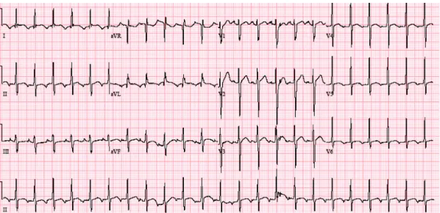
Figure 1: 12 lead ECG on presentation. Narrow complex tachycardia with HR of 139 and nonspecific T wave abnormalities.
Figure 2: 12 lead ECG. P wave become obvious as the heart rate decrease from 139 to 115. P waves are inverted in lead II and AVL, while the P waves were upright in III and AVF, suggesting the possibility of ectopic atrial tachycardia, most likely originating from the left side.
the criteria for noncompaction cardiomyopathy (Figure 4). Left atrium was moderately dilated (49 mm) with normal right atrium (38 mm), top normal left ventricle (diastolic 54 mm, systolic 62 mm).
Electrophysiology study showed sustained atrial tachy-cardia originating on the ridge anterior to the left sided pulmonary veins.
A successful radiofrequency catheter ablation was per-formed at this site without any complications. Patient was started on aspirin, beta-blockers, and ACE inhibitors and anticoagulated with Coumadin. ICD implantation was planned as outpatient for primary prevention of ventricular tachyarrhythmias. After the procedure his AT was abolished completely. Patient’s heart rate improved and on 12 lead ECG done he had positive P waves in lead II and AVF (which were previously negative) (Figure 3).
3. Discussion
Cardiac rhythm disturbances occur frequently in LVNC. The electrocardiographic findings are usually abnormal, but no characteristic changes are defined. The abnormalities mostly reported are branch blocks, ST-T wave changes, AV blocks, left ventricle hypertrophy, and arrhythmias [1]. Ventricular arrhythmias are the most common rhythm disturbance seen
[2,3]. The intertrabecular recesses appear to be a favorable substrate for ventricular arrhythmias. There are several case series and reports describing ventricular ectopic beats, ven-tricular tachycardia and/or venven-tricular fibrillations, and sud-den cardiac deaths in these patients [1]. Atrial arrhythmia usually seen is atrial fibrillation with reported incidence of 5% [4] to 29% [3].
The differentiation of narrow complex tachycardias (NCT) is a commonly encountered diagnostic dilemma. In this case the patient presented with an NCT, which initially appeared to be sinus tachycardia. Sinus tachycardia generally occurs in the setting of underlying physiologic stress or phar-macologic influence, namely, fever, infection, pain, hyperthy-roidism, anxiety, stimulant drug use, or hypotension, among many others. Other causes of NCTs include atrial fibrillation, atrial flutter, AV nodal reentrant tachycardia, atrioventricular reentrant tachycardia, sinus node reentrant tachycardia, uni-focal atrial tachycardias, or multiuni-focal atrial tachycardia.
Figure 3: 12 lead ECG after ablation of atrial tachycardia showed sinus bradycardia with sinus arrhythmia (heart rate of 53 bpm). The P waves are now positive in lead II and AVF.
(a) (b)
(c) (d)
Figure 4: Cardiac MRI showing extensive apical and mid cavity obliteration of the papillary muscles and large trabeculations and intertrabeculation recesses in the left ventricle (a): three chamber image; (b): two chamber image; (c) short axis image; (d) four chamber image.
disease or enhanced automaticity, if there is no underlying heart disease. We believe that LVNC with atrial involvement was likely the cause of AT in our patient.
There were no reported cases of supraventricular tachy-cardia in a followup of 238 patients with LVNC for 4 years
4 Case Reports in Medicine
common in pediatric patients, and orthodromic reentrant tachycardia was reported in some of those patients [7]. One patient with AV nodal reentrant tachycardia [8] and another case of atrial flutter with extreme atrial enlargement in the setting of LVNC have been reported [9]. One case of regular sustained likely macroentry atrial tachycardia (AT) was identified [10]. However, focal atrial tachycardia is rarely described in the literature in this patient population. There is one published case of focal AT in a patient with LVNC [11], which could well represent reentrant tachycardia [10].
The patient described in this case had a focal atrial tachy-cardia arising from the ridge anterior to the left sided pul-monary veins, which was successfully treated with radiofre-quency catheter ablation. The focal AT was probably related to the moderately enlarged left atrium. Our report suggests that although rare, involvement of left atrial myocardium is possibile in LVNC, and these patients may present with focal atrial tachycardia.
Disclosure
This paper has not been submitted elsewhere, is not under review, or published previously. All the authors are aware of and approve the paper being submitted to this journal.
Conflict of Interests
There is no conflict of interests in the paper, including finan-cial, consultant, institutional, and other relationships that might lead to bias or a conflict of interests.
References
[1] R. J. Sarma, A. Chana, and U. Elkayam, “Left ventricular non-compaction,”Progress in Cardiovascular Diseases, vol. 52, no. 4, pp. 264–273, 2010.
[2] T. K. Chin, J. K. Perloff, R. G. Williams, K. Jue, and R. Mohrmann, “Isolated noncompaction of left ventricular myo-cardium. A study of eight cases,”Circulation, vol. 82, no. 2, pp. 507–513, 1990.
[3] M. Ritter, E. Oechslin, G. S¨utsch, C. Attenhofer, J. Schneider, and R. Jenni, “Isolated noncompaction of the myocardium in adults,”Mayo Clinic Proceedings, vol. 72, no. 1, pp. 26–31, 1997. [4] C. St¨ollberger, J. Finsterer, and G. Blazek, “Left ventricular
hypertrabeculation/noncompaction and association with addi-tional cardiac abnormalities and neuromuscular disorders,”
American Journal of Cardiology, vol. 90, no. 8, pp. 899–902, 2002.
[5] G. Fazio, G. Corrado, S. Novo et al., “Supraventricular arrhyth-mias in noncompaction of left ventricle: is this a frequent com-plication?”International Journal of Cardiology, vol. 127, no. 2, pp. 255–256, 2008.
[6] F. Ichida, Y. Hamamichi, T. Miyawaki et al., “Clinical features of isolated noncompaction of the ventricular myocardium: long-term clinical course, hemodynamic properties, and genetic background,”Journal of the American College of Cardiology, vol. 34, no. 1, pp. 233–240, 1999.
[7] J. Fichet, A. Legras, A. Bernard, and D. Babuty, “Aborted sudden cardiac death revealing isolated noncompaction of the left ventricle in a patient with Wolff-Parkinson-White syndrome,”
Pacing and Clinical Electrophysiology, vol. 30, no. 3, pp. 444–447, 2007.
[8] S. G. Enriquez, F. R. Entem, M. Cobo, and J. J. Olalla, “Uncom-mon etiology of syncope in a patient with isolated ventricular noncompaction,”Pacing and Clinical Electrophysiology, vol. 30, no. 4, pp. 577–579, 2007.
[9] J. Suvarna, C. T. Deshmukh, and S. A. Hajela, “Left ventricular noncompaction: a cardiomyopathy often mistaken,” Indian Journal of Medical Sciences, vol. 63, no. 7, pp. 303–307, 2009. [10] S. Seethala, F. Knollman, D. Mcnamara et al., “Exercise-induced
atrial and ventricular tachycardias in a patient with left ventric-ular noncompaction and normal ejection fraction,”Pacing and Clinical Electrophysiology, vol. 34, pp. e94–e97, 2011.
Submit your manuscripts at
http://www.hindawi.com
Stem Cells
International
Hindawi Publishing Corporation
http://www.hindawi.com Volume 2014
Hindawi Publishing Corporation
http://www.hindawi.com Volume 2014
INFLAMMATION
Hindawi Publishing Corporation
http://www.hindawi.com Volume 2014
Behavioural
Neurology
Endocrinology
International Journal ofHindawi Publishing Corporation
http://www.hindawi.com Volume 2014
Hindawi Publishing Corporation
http://www.hindawi.com Volume 2014
Disease Markers
Hindawi Publishing Corporation
http://www.hindawi.com Volume 2014
BioMed
Research International
Oncology
Journal of Hindawi Publishing Corporationhttp://www.hindawi.com Volume 2014
Hindawi Publishing Corporation
http://www.hindawi.com Volume 2014
Oxidative Medicine and Cellular Longevity
Hindawi Publishing Corporation
http://www.hindawi.com Volume 2014
PPAR Research
The Scientific
World Journal
Hindawi Publishing Corporationhttp://www.hindawi.com Volume 2014
Immunology Research
Hindawi Publishing Corporation
http://www.hindawi.com Volume 2014
Journal of
Obesity
Journal ofHindawi Publishing Corporation
http://www.hindawi.com Volume 2014
Hindawi Publishing Corporation
http://www.hindawi.com Volume 2014
Computational and Mathematical Methods in Medicine
Ophthalmology
Journal ofHindawi Publishing Corporation
http://www.hindawi.com Volume 2014
Diabetes Research
Journal ofHindawi Publishing Corporation
http://www.hindawi.com Volume 2014
Hindawi Publishing Corporation
http://www.hindawi.com Volume 2014
Research and Treatment
AIDS
Hindawi Publishing Corporation
http://www.hindawi.com Volume 2014
Gastroenterology Research and Practice
Hindawi Publishing Corporation
http://www.hindawi.com Volume 2014
Parkinson’s
Disease
Evidence-Based Complementary and Alternative Medicine
Volume 2014 Hindawi Publishing Corporation