_____________________________________________________________________________________________________
*Corresponding author: E-mail: appiatee1@gmail.com;
Stroke Secondary to a Parapharyngeal Pleomorphic
Adenoma – Case Report
Peter Appiah-Thompson
1*, Nana Andoh Hanson
2, Juliana Kwakye Arthur
2,
Grace Amoo-Quaye
2and Theophilus Adjeso
31
Department of Surgery, Ear, Nose and Throat (ENT) Unit, School of Medical Sciences, Cape Coast Teaching Hospital, University of Cape Coast, Cape Coast, Ghana. 2Department of Surgery,Ear, Nose and Throat Unit, Cape Coast Teaching Hospital,
Cape Coast, Ghana. 3
DepartmentofEar, Nose and Throat, School of Medicine and Health Sciences, Tamale Teaching Hospital, University for Development Studies, Tamale, Ghana.
Authors’ contributions
This work was carried out in collaboration among all authors. Author PAT designed the report and authors PAT, NAH and JKA wrote the first draft of the manuscript. Authors PAT and TA managed the literature searches. All authors contributed to the design and write – up of this case report. Authors PAT, NAH and JKA formed the operating team for the surgery. All authors read and approved the final manuscript.
Article Information
Editor(s): (1)Dr. José Francisco de Sales Chagas, Professor, Sao Leopoldo Mandic Medical School, Brasil. Reviewers: (1) Muhammad Ghafoor Ali, Pakistan. (2)Michael Bordonaro, Geisinger Commonwealth School of Medicine, USA. (3)Francesca Gorini, National Research Council, Italy. Complete Peer review History:https://sdiarticle4.com/review-history/51779
Received 29 July 2019 Accepted 03 October 2019 Published 15 October 2019
ABSTRACT
Aims: Parapharyngeal Space (PPS) tumours are rare and may present as painless masses only becoming evident because of their mass effect. The diagnosis is more difficult when they do not present with any obvious clinical features. This case report seeks to highlight this type of tumour and the possibility of transoral resection when the lesion is small.
Presentation of Case: We present the case of a parapharyngeal tumour whose mass effect led to a cerebrovascular accident. The lesion was excised piecemeal transorally and the patient is now walking with an improved quality of life. Repeat CT scan showed a normal brain parenchyma and neck.
Discussion: PPS tumours can lead to many consequences thus a high index of suspicion is needed to diagnose and treat them. The type and location of the tumour should guide the type of approach employed.
Conclusion: PPS tumours of the prestyloid area of small size may be excised transorally by gentle and careful dissection.
Keywords: Parapharyngeal space; pharyngomaxillary space; cerebrovascular accident; pleomorphic adenoma.
1. INTRODUCTION
Generally, parapharyngeal space (PPS) tumours are less than 1% of all head and neck tumours [1]. Most PPS tumours are benign (70-80%). The malignant ones are fewer. The post - styloid compartment lesions tend to be of neurogenic origin whereas the prestyloid ones arise from the minor salivary glands in the lateral pharyngeal wall or extensions of tumours of the deep lobe of the parotid [2]. Among the benign tumours of the
PPS, the pleomorphic adenoma is the
commonest [1]. We present a case of a left
parapharyngeal mass which caused a
cerebrovascular accident (CVA) and was excised transorally with resolution of the CVA. We believe that the left parapharyngeal tumour in our case was compressing on the ipsilateral carotid sheath and therefore by inference on the walls of the internal carotid artery thus limiting the blood supply to the left half of the brain. This led to the left brainstem infarct with the resultant right hemiparesis. The occluded vessel recanalized after the tumour was excised with resolution of
the hemiparesis. The other mechanism of occlusion of the artery could have been as a result of an encasement of the vessel by the tumour [3,4]. That was not likely in this case.
2. PRESENTATION OF CASE
A 66-year-old known hypertensive and diabetic man who had been compliant with his medications, had been apparently well until he developed a CVA with right hemiparesis. This made him unable to walk. He was seen at a hospital in another region a month prior to presentation at our hospital, where a computed tomography (CT) scan of the brain and neck was requested. A left acute brainstem infarct was noted on the CT scan. A left parapharyngeal mass was also incidentally identified on the CT scan and the patient thus referred to the ENT Unit of our hospital for further management. The left parapharyngeal lesion on the CT scan as shown in Fig. 1 measured 6.1 x 4.2 cm with hyperdense areas. Patient came to our clinic in a wheelchair.
Prior to the onset of the CVA, patient did not experience any symptoms such as odynophagia, dysphagia, change in voice, throat pain nor any feeling of a mass in the throat. He also had no
nasal congestion, nasal bleeding, nasal
discharge or any neck pain.
On examination, he was afebrile (temperature
35.9°C), not pale, anicteric and fairly hydrated.
No bipedal oedema, finger clubbing, or cyanosis was present. BP of 149/89 mmHg, pulse of 78 beats/min, regular of good volume and random blood sugar of 7.7 mmo1/L were recorded.
On examination, no cranial nerve palsies, no drooling, halitosis, or oral sores were found. There was a left soft palatal bulge with anterior displacement of the left tonsillar region. There was no bleeding or ulceration in the oral cavity or in the oropharynx. No abnormalities were found
in the chest and abdomen. Power in the limbs on the right were 3/5 and in the left limbs were 5/5.
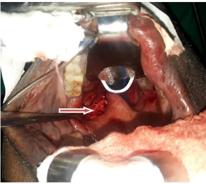
The patient was then prepared and excision of the parapharyngeal mass done transorally using a vertical incision made on the mass, on the lateral wall of the oropharynx upward into the nasopharynx after opening the mouth with a Boyle-Davies mouth gag as shown in Fig. 2 below. A yellowish fibrocartilaginous mass of about 7x7 cm was identified.
The mass was then excised piecemeal with meticulous blunt dissection of attached soft tissues. Histology of the sample was reported as
follows: sections showed benign tumour
composed of a mixture of glandular epithelial as well as mucinous, cartilaginous and hyalinised stromal elements. Features were that of pleomorphic adenoma.
Two years post excision of the lesion, patient is much better, walks without using a wheelchair and has an improved quality of life. Repeat CT scan of the brain and neck showed a normal
brain parenchyma and neck without a
parapharyngeal tumour.
3. DISCUSSION
The PPS or the pharyngomaxillary space is on the lateral aspect of the neck with its medial wall being the buccopharyngeal membrane of the pharynx covering the constrictor muscles and the lateral wall being bounded by the medial pterygoid muscle, mandible and the deep lobe of the parotid gland. Posteriorly lies the prevertebral fascia. The space is pyramidal in shape with the base at the base of skull and the apex at the hyoid bone. It is divided into two compartments by the styloid process and its attached muscles. Namely, the prestyloid/anterior and the
post-styloid/posterior compartments. The main
components of the prestyloid region are minor salivary glands, upper deep cervical lymph nodes and the deep lobe of the parotid gland whereas the post-styloid region contains the carotid sheath, sympathetic chain and cranial nerves IX to XII [5].
PPS tumours make up about 0.5% of all head and neck tumours. The prestyloid tumours are mostly of salivary gland origin whilst the poststyloid tumours tend to be malignant. The prestyloid tumous mostly arise from the minor salivary glands or from the deep lobe of the parotid gland. The tumours from the minor salivary glands are rare. Pleomorphic adenoma makes up about 80-90% of the prestyloid salivary gland tumours [1].
The common symptoms of PPS tumours include a painless mass, oral fullness, dysphagia, dysphonia, trismus and sometimes sore throat. The mass effect of these tumours may include CVA as being discussed in this report. Our patient is very happy now with an improved quality of life [6].
The diagnostic work-up of these lesions include
haematological, serological, fine needle
aspiration cytology (FNAC) and imaging i.e. CT Scan and magnetic resonance imaging (MRI). MRI is more superior [7]. Imaging helps to know the extent of the tumour, type and relations to the surrounding structures. In our case, a CT scan was done as the request was part of the work up for the type of stroke. The PPS tumour was only
diagnosed incidentally. FNAC was not done in our case. The neck palpation was negative as no masses were obvious on the neck.
The head and neck surgeon and the
otorhinolaryngologist have a difficult task in treating these lesions due to the deep location in the neck with relations to important and critical vessels and nerves in the carotid sheath. Excising the tumours can lead to haemorrhage, dehiscence, damage to the vagus or other nerves and fistulae formation [2]. The excision
through external cervical approach with
mandibular split osteotomy is thus more popular. Some surgeons have still used the transcervical approach without splitting the mandible. The transoral approach has been recommended for lesions less than 3cm in diameter due to the limited exposure, possibility of tumour spillage and injury to nerves and vessels [8,9]. The transoral approach was used in this case because this approach gave the most easily accessible route to the tumour as the lesion was not palpable on the neck. Dissection also caused no significant bleeding and the lesion easily peeled off the surrounding structures. The transoral approach though not popular among some surgeons has the advantages of causing less trauma to the surrounding vessels and cranial nerves, giving better cosmesis as no scars are left on the neck and preserves the superficial lobe of the parotid [9]. These complications which are averted in the transoral
approach are common in the external
approaches [10]. The endoscope-assisted
transoral approach is the new method being used. This is said to be less traumatic with fewer complications [11].
Where readily available and affordable carotid angiography and magnetic resonance imaging could be done to diagnose the vessel occlusion. This patient also stood the risk of having a stroke apart from the PPS tumour because he was diabetic and hypertensive [12]. However, these were not likely to have caused his stroke since he was compliant with his medications.
4. CONCLUSION
and careful dissection. Also, in the workup of stroke patients, neck tumours must be ruled out.
CONSENT
Informed written consent was obtained from this patient for the publication of this article.
ETHICAL APPROVAL
As per international standard written ethical approval has been collected and preserved by the author(s).
ACKNOWLEDGEMENTS
We wish express our most sincere gratitude to the ENT nurses of CCTH (Mrs Esther Nketia and her team) for helping take care of this patient.
COMPETING INTERESTS
Authors have declared that no competing interests exist.
REFERENCES
1. Akin I, Karagoz T, Mutlu M, Sahan M,
Onder E, Case report pleomorphic
adenoma of the parapharyngeal space. Case Reports in Otolaryngology. 2014;4. [Article 168401]
Available:http://dx.doi.org/10.1155/2014/16 8401
2. Sergi B, Limongelli A, Scarano E, Fetoni
AR, Paludetti G. Giant deep lobe parotid gland pleomorphic adenoma involving the parapharyngeal space. Report of three cases and review of the diagnostic and
therapeutic approaches, Acta
Otorhinolaryngologica Italica. 2008;28(5): 261–265. View at Google Scholar • View at Scopus.
3. Haaso AN, Bentson JR, Wilson GH,
Vignaud J. Neuroradiology of the
sphenoidal region. Radiology. 1975;114: 619-627.
4. Momose KJ, New PFJ. Nonatheromatous
stenosis and occlusion of the internal carotid artery and its branches. Am J Roentgenol Radium Ther Nucl Med. 1973; 118:550-566
5. Dhingra PL. Diseases of the ear, nose and
throat. 4th Edition. Elsevier. 2006;248.
6. Watkinson JC, Gaze MN, Wilson JA. Stell
and Maran’s head and neck surgery, 4th Edition. Butterworth Heinemann. 2000; 193.
7. Lloyd GA, Phelps PD. The demonstration
of tumours of the parapharyngeal space by
magnetic resonance imaging. British
Journal of Radiology. 1986;59(703):675– 683. View at Publisher • View at Google Scholar • View at Scopus.
8. Bozza F, Vigili MG, Ruscito P, Marzetti A,
Marzettt F. Surgical management of parapharyngeal space tumours: Results of
10-year follow-up. Acta
Otorhinolaryngologica Italica. 2009;29(1): 10–15. View at Google Scholar • View at Scopus.
9. Kovacic M, Rudic M, Kranjcec Z. Transoral
excision of a parapharyngeal space Tumor: Case report, Coll. Antropol. 2012; 36(2):193–195.
10. Casale M, Capuano F, Sabatino L, Pace
P, Oliveto G, et al. A safe transoral surgical approach to parapharyngeal tumor arising from deep lobe of parotid gland. SAGE Open Medical Case Reports. 2016; 4:1–4.
11. Iseri M, Ozturk M, Kara A, Ucar S, Aydin
O, Keskin G. Endoscope-assisted
transoral approach to parapharyngeal space tumors. head & neck; 2014. View at Publisher • View at Google Scholar.
12. Matsuo R, Kamouchi M, Inoue T, Okada Y,
Ibayashi S, Cerebral infarction due to carotid occlusion caused by cervical Vagal Neurilemmoma – Report of a case. Stroke. 2002;33(5):1428-1431.
_________________________________________________________________________________
© 2019 Appiah-Thompson et al.; This is an Open Access article distributed under the terms of the Creative Commons Attribution License (http://creativecommons.org/licenses/by/4.0), which permits unrestricted use, distribution, and reproduction in any medium, provided the original work is properly cited.
Peer-review history: