Gingival Unit Graft Versus Free Gingival Graft for Treatment of
Gingival Recession: A Randomized Controlled Clinical Trial
Niloofar Jenabian 1, Mohadese Yazdanpanah Bahabadi 2, Ali Bijani 3, Morteza Rahimi Rad 4
1 Associate Professor, Oral Health Research Center, Department of the Periodontology, School of Dentistry, Babol University of Medical
Sciences, Babol, Iran
2 Assistant Professor, Department of the Periodontology, School of Dentistry, Kashan University of Medical Sciences, Kashan, Iran 3
Social Determinants of Health Research Center, Health Research Institute, Babol University of Medical Sciences, Babol, Iran
4
Periodontist, Department of Periodontology, School of Dentistry, Golestan University of Medical Sciences, Gorgan, Iran
Abstract
Objectives: Gingival recession can lead to root exposure and discomfort for patients. There are various techniques for root coverage. The aim of this study was to compare the use of gingival unit graft (palatal graft including the marginal gingiva and papillae) with free gingival graft for treatment of localized gingival recession.
Materials and Methods: In this randomized controlled clinical trial, 18 bilateral localized recessions of Miller class I and II were treated in nine systemically healthy patients. Recessions were randomly treated with gingival unit graft in one side and conventional free gingival graft in the other side. Clinical parameters including clinical attachment level, keratinized tissue width, probing depth and vertical recession depth (VRD) were recorded at baseline and at one, three and six months after surgery. The healing index and patient satisfaction were also evaluated. One-way and two-way repeated measures ANOVA and paired t-test were used for statistical analyses.
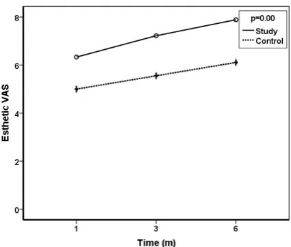
Results: Both techniques caused significant improvement in clinical parameters. Gingival unit graft produced higher satisfaction esthetically (P=0.050, 0.024 and 0.024, respectively at the three time points), higher healing index (P<0.001), higher root coverage percentage at one month after surgery (34.04%, P=0.011) and greater reduction of recession width three months after surgery (P=0.007) but the reduction in VRD at this side was not significantly greater.
Conclusions: Gingival unit graft might be an acceptable modality in Miller Class I/II recession defects. This technique may have advantages over free gingival graft such as significantly superior clinical and esthetic results.
Keywords: Gingival Recession; Transplantation, Autologous; Tooth Root; Transplants
Journal of Dentistry, Tehran University of Medical Sciences, Tehran, Iran (2016; Vol. 13, No. 3)
Corresponding author:
M. Yazdanpanah Bahabadi,
Department of the
Periodontology, School of Dentistry, Kashan University of Medical Sciences, Kashan, Iran
mohadese.yazdanpanah@yahoo. com
Received: 2 February 2016 Accepted: 26 April 2016
INTRODUCTION
Marginal gingival recession refers to the location of the gingival margin apical to the cemento-enamel junction, which results in exposure of root surfaces and loss of attached gingiva [1]. The prevalence of gingival recession varies between 20% and 100% among adults [2-6]. Gingival recession may increase tooth hypersen- sitivity [7] and cause pain, root caries, unesthetic appearance of the gums, periodontal attachment loss and tooth loss [8], and make oral hygiene and plaque control difficult [9]. These clinical problems can be resolved by root coverage treatments, including free gingival graft [10],
pedicle graft [11], connective tissue graft [12], coronally positioned flap [13] and guided tissue regeneration [14]. Although most of these methods result in significant clinical improve-ment [6,15], selection of the method of choice depends on many factors, including factors related to the defect, patient and technique. Therefore, treatment of gingival recession is still a challenge and researchers are working to find new more efficient surgical techniques for this purpose [16].
clinical conditions of grafts including submerg-ing the graft [18,19], "butt joint" adaptation of the graft to the adjacent tissues at the recipient site, root planing with the aim of reducing its prominence, and special suturing techniques to improve adaptation of the graft and its blood supply at the recipient site. The only donor site modification that can promote success in root coverage with non-submerged grafts is to increase graft thickness to maintain its viability on the avascular root surface [10,16,17,20,21]. Use of site-specific donor tissue may increase graft survival at the recipient site, which does not have optimal blood perfusion.
Allen and Cohen [22] used a palatal graft contai-ning marginal gingiva as a free gingival graft without increasing its thickness. They used this technique based on the data in a case report [23]. Gingiva has a unique structure and charac-teristics [24,25]. The vascular plexus of the gingiva is rich of horizontal anastomoses, which perfuse the marginal zone but do not extend to the interproximal area. Marginal and interdental gingival tissues can be used to benefit from better blood perfusion of the donor and recipient sites and, therefore, improve the chances for graft survival [26]. The vascular characteristics of the graft are probably important for rapid anasto-mosis of the capillaries of the recipient site with the injured vessels of the graft [16].
Gingival unit graft should be harvested from an area, which is not esthetically important [26]. There are only a few case studies and just one clinical trial in this respect [22,26]. Kuru and Yildirim [16] compared the gingival unit graft and the conventional free gingival graft in patients with Miller class I/II gingival recession. Reduction in vertical recession and attachment and keratinized tissue gain were significantly higher in gingival unit group. Other human studies have shown that in clinically healthy gingiva, there are significantly different vascular distributions in marginal, attached and inter-dental gingiva [16]. The involvement of marginal
gingiva and papillary tissue in the graft can improve defect coverage, healing process and color adaptation. The aim of the present study was to compare the use of gingival unit graft with the conventional free gingival graft (palatal graft) for treatment of localized gingival recession.
MATERIALS AND METHODS
In this randomized controlled clinical trial with split-mouth design, 18 localized recessions of Miller class I and II were treated in nine systemi-cally healthy patients. Bilateral recessions with a vertical depth of≥2 mm on the buccal aspects of mandibular premolars and incisors were selected. The exclusion criteria were (I) smoking, (II) pregnancy, (III) root surface restorations, (IV) endodontic treatment and (V) poor oral hygiene (O’Leary plaque score >20%). All details of the two treatment modalities were explained to the participants and written informed consent was obtained from them.
The study protocol was approved by the Ethical Committee of Babol University of Medical Sciences and registered at http://www.irct.ir (registration number: IRCT 201403061760N33; date registered: April 27, 2014).
Study design
measurements and randomization were done by another clinician.
Clinical assessments
The clinical parameters were measured at baseline and at one, three and six months after the surgical procedures to the nearest 0.5 millimeter using a University of Michigan "O" probe with Williams marking. The measured clinical para-meters were as follows:
- Keratinized tissue width (KTW): Distance between the most apical part of the gingival margin and mucogingival junction.
- Clinical attachment level (CAL): Distance between the cementoenamel junction (CEJ) and bottom of the pocket
- Probing depth (PD): Distance between the most apical part of the gingival margin and bottom of the pocket at three points of mesial, midbuccal and distal.
- Vertical recession depth (VRD): Distance
between the CEJ and the most apical part of the gingival margin.
- Recession width (RW): Width of exposed root 1mm apical to the CEJ [27].
On the 10th postoperative day (suture removal session), the patients were asked about post-surgical pain using a 10-point visual analog scale (VAS) in which, 0 indicated no pain and 10 represented the worst pain experienced.
In addition, patients were questioned about esthetics at one, three and six months after surgery. Patient satisfaction was assessed using a 10-point VAS (0 indicated "dissatisfied" and 10 indicated "fully satisfied").
The Landry’s healing index was assessed according to Jankovic et al, [28] at 10 days and one month after the surgery (Table 1).
The coverage percentage was calculated using the following formula: [(baseline VRD–new VRD)/baseline VRD]×100 at one, three and six
Table 1: Healing index used in the present study
Score Characteristics
1: Very poor
Tissue color: ≥50% of gingiva red Response to palpation: bleeding Granulation tissue: present
Incision margin: not epithelialized, with loss of epithelium beyond incision margin Suppuration present
2: Poor
Tissue color: ≥50% of gingiva red Response to palpation: bleeding Granulation tissue: present
Incision margin: not epithelialized, with connective tissue exposed
3: Good
Tissue color: ≥25% and <50% of gingiva red Response to palpation: no bleeding
Granulation tissue: none
Incision margin: no connective tissue exposed
4: Very good
Tissue color: <25% of gingiva red Response to palpation: no bleeding Granulation tissue: none
Incision margin: no connective tissue exposed
5: Excellent
Tissue color: all tissues pink Response to palpation: no bleeding Granulation tissue: none
Fig. 1: Surgical procedure and follow-up (A) Initial clinical appearance at the free gingival graft side, (B) Free gingival graft, (C) Suturing, (D) After six months, (E) Initial clinical appearance at the gingival unit graft side, (F) Gingival unit graft, (G) Suturing, (H) After six months
months after surgery.
The CAL was measured at the midpalatal area of the relevant premolar tooth at baseline and at three and six months after gingival unit graft harvesting for clinical evaluation of donor site healing.
Surgical procedures Recipient site
After achieving local anesthesia in both groups, the preparation of the recipient site was begun by making two vertical incisions extending to the adjacent teeth and about 3 to 4 mm beyond the mucogingival junction, as well as a horizontal incision at the mucogingival junction. A sharp split-thickness flap was reflected, and the surfaces between these incisions were de-epithel-ialized (Figs. 1A and 1E).
At the gingival unit side, the interdental papillae were removed; but, at the conventional graft side, the butt-joint incisions were made on the papillary tissue at the level of the CEJ. The exposed surface of the root was planed with hand instruments and rinsed with sterile saline.
Donor site
At the gingival unit side, the palatal tissue, including the marginal and interdental gingiva, was harvested from the palatal aspect of the maxillary premolars (Fig. 1F). On the other side (conventional graft), the palatal tissue was
harvested from the same area on the other side but about 2 mm away from the gingival margin (Fig. 1B). For the grafts at both sides, care was taken to obtain a thickness of about 1 mm. Next, the grafts were contoured, adapted and sutured at the level of the CEJ (Figs. 1C and 1G). Both the donor and recipient sites were covered with periodontal dressing. The sutures were removed after 10 days.
Postsurgical care
Patients were advised not to brush their teeth at the surgical site, avoid chewing hard food, and rinse once daily with 0.2% chlorhexidine diglu-conate mouthwash for three weeks. Systemic antibiotic (500 mg amoxicillin, tid) was pres-cribed for one week. After 10 days, the patients were instructed to resume gentle brushing, directed coronally, at the operated sites. Post-surgical recalls were scheduled every other week during the first month and at one, three and six months after the surgery (Figs. 1D and 1H). Statistical analyses
Statistical analyses were performed using SPSS version 21 (SPSS Inc., IL, USA). Quantitative data of midbuccal measurements of the recession sites were recorded as mean±standard deviation. The data were analyzed using repeated measures two-way ANOVA and Tukey’s post hoc test for
intragroup comparisons. Paired t-test was used
for intergroup comparisons.
B C D
A
RESULTS
Nine patients eligible for this study were treated in a split-mouth design. All patients completed the follow-up sessions. Healing was uneventful and there were no complications.
No significant differences in pre-surgical para-meters were found between the two sides. Paired
t-test found no significant difference between the
two groups at each time point. The gingival unit graft produced significantly higher esthetic satisfaction at one, three and six months after the surgery (P=0.050, P=0.024 and P=0.024, respectively). Higher healing index (4.44±0.88 at the gingival unit graft side and 3.44±1.01 at the free gingival graft side, P<0.001) and lower level of pain (3.33±1.50 for the gingival unit graft side and 5.22±1.64 for the free gingival graft side, P=0.020) were noted at suture removal session. Higher root coverage percentage was obtained one month after surgery (30.04±12.41 at the gingival unit graft side and 20.24±16.78 at the free gingival graft side, P=0.011). Reduction of recession width was observed three months after surgery (P=0.007; Fig. 2)
The mean percentage of root coverage at three months after surgery was 44.04±18.78 (range: 16.66 to 75) and 26.33±24.28 (range: 0 to 66.66) in gingival unit graft side and free gingival graft side, respectively and this difference was not significant (P=0.110). The mean percentage of root coverage at six months after the surgery was 60.52±21.22 (range: 28.57 to 100) and 45.52±21.94 (range: 0 to 66.66) in gingival unit graft side and free gingival graft side, respectively and this difference was not significant (P=0.062, Table 2).
Complete defect coverage, defined as gingival margins at the level of the CEJ, was found in one of nine patients (11%) after six months in unit graft side; whereas, none of the patients showed complete coverage in the other side.
No complications were observed at the gingival unit donor site, and healing was uneventful. The CAL of the relevant maxillary premolar at the
gingival unit donor site was 2.22±1.52, 2.00±1.54 and 2.00±1.54 at baseline, three months and six months after surgery, respec-tively and there was no recession or attachment loss in this area but significant attachment gain was observed after three months (P=0.00). There were no significant changes in midbuccal PD at each side. A significant reduction in VRD was observed after surgery at each side and the coronal movement of the gingival margin at the gingival unit side was greater than that in the other side; this difference was not significant between the two sides (P>0.05, Table 2).
DISCUSSION
This randomized controlled clinical trial lasted for six months and evaluated the treatment of localized gingival recessions (Miller class I/II) with an alternative technique (gingival unit graft) in which, palatal grafts include the marginal gingiva and the papillary tissue to use a site-specific vascular configuration.
Gingival arterioles are oriented in an apico-coronal direction. Capillaries in the marginal gingiva form repetitive networks and several small vessels form loops extending towards the marginal gingiva. Additionally, it has been demonstrated that predominant gingival vessels decrease in size and increase in number as they extend coronally [16]. Thus, in this modified
Table 2: Clinical parameters at baseline and at one, three and six months
P-value2
Six months Three months
One month Baseline
Parameters
<0.001 5.05±1.01d
5.33±1.03c
5.94±1.07b
2.44±1.52a
Study
KTW (mm) Control 2.16±1.47a 5.38±1.43b 4.83±1.52c 4.38±1.36d <0.001 Group*Time= 0.730
Time= <0.001 Group= 0.273
P-value1
<0.001 2.66±1.56c
3.50±1.14b
3.72±0.83b
5.33±1.85a
Study
CAL (mm) Control 5.05±1.66a 3.72±1.00bc 3.72±1.12b 3.00±1.17c <0.001 Group*Time= 0.499
Time= <0.001 Group= 0.904
P-value1
<0.001 1.83±1.47c
2.38±1.29b
2.72±1.09b
4.11±1.63a
Study
VRD (mm) Control 3.72±1.46a 2.83±0.93b 2.61±1.08b 2.00±1.11c <0.001 Group*Time= 0.315
Time= <0.001 Group= 0.960
P-value1
<0.001 1.94±0.72c
2.11±0.82bc
2.50±0.82b
3.00±1.19a
Study
RW (mm) Control 3.16±1.54a 2.50±1.17b 2.66±1.19b 2.44±1.21b 0.011 Group*Time= 0.177
Time= <0.001 Group= 0.205
P-value1
0.356 0.83±0.25a
1.11±0.33a
1.00±0.50a
1.22±083a
Study
PD (mm) Control 1.44±0.28a 0.77±0.08a 1.11±0.11a 1.00±0.08a 0.124 Group*Time= 0.136
Time= 0.168 Group= 0.503
P-value1
In each row, the same superscripted letters show non-significant difference; 1: Repeated measures two-way ANOVA; 2: Repeated measures one-way ANOVA; KTW: Keratinized tissue width; CAL: Clinical attachment level; VRD: Vertical recession depth; RW: Recession width; PD: Probing
technique, the size and number of vessels and vascular configuration of donor tissue would better match those of the recipient site.
In the current study, gingival unit side produced significantly greater esthetic satisfaction at one, three and six months, higher healing score, lower post-surgical pain score, higher root coverage percentage at one month and greater reduction in recession width at three months. These results indicate that this modified technique may improve these periodontal parameters.
Proper plaque control, root surface biocompa-tibility, careful surgical manipulation and tissue thickness have been accepted as important factors affecting the outcome of graft procedures and should be controlled [16].
In the current study, one inclusion criterion was good oral hygiene during the study period. The exposed root surfaces were planed carefully with hand instruments to ensure root surface biocom-patibility. Sutures were made without tension
over the graft to avoid graft displacement or impairment of blood supply. Special care was taken to obtain approximately 1mm thick grafts. Search of the literature yielded one previous comparative controlled clinical trial comparing gingival unit graft and conventional free gingival graft, which did not have a split-mouth design (eight patients in gingival unit graft, nine patients in free gingival graft) [16]. In the current study, in order to eliminate the inter-individual variabi-lity of the treatment effect, a split-mouth design was used. In the afore-mentioned previously published study [16], 50% of the sites in gingival unit group showed complete defect coverage at eight months, but none of the patients in the other group showed complete coverage.
defects. The results showed that gingival unit grafts provided better defect coverage than free gingival grafts [29].
In our study, 11% of the gingival unit sides showed complete coverage at six months. Similar to the above-mentioned comparative study [16], none of the free gingival graft sides showed complete coverage. The mean coverage percen-tage at the gingival unit side was 60.52% at six months compared to 45.52% at the free gingival graft side. The lower degree of coverage in the current study compared to the other study may be due to greater VRD and width at baseline. Probing depth did not change significantly, but keratinized tissue gain was significant in our study. These findings were similar to the findings of the afore-mentioned study [16]. Although reduction of VRD was significant at both sides and greater reduction was observed at the gingival unit side, this difference was not significant. The greatest reduction in VRD was observed during the first month, but then showed another significant reduction during three to six months. Migration of gingival margin in the coronal direction was probably because of creeping attachment. This phenomenon was first described by Goldman [30]. Creeping attachment may be observed one to 12 months after surgery and gives an average coverage of about 1mm in narrow recessions. The current study was limited to six months and a longer follow-up period is needed to draw definite conclusions regarding this phenomenon.
In the current study, esthetic satisfaction was assessed by asking patients their subjective opinion regarding the esthetic outcome. Signifi-cantly higher scores were obtained at one, three and six months after surgery for the gingival unit graft side compared to the free gingival graft side. Kuru and Yildirim [16] showed similar results regarding esthetic satisfaction. These findings, in addition to better root coverage, reveal that gingival unit graft can be used as a modification of free gingival graft, particularly in
areas where esthetics is important. Clinical healing at the gingival unit donor site was uneventful and without complications. It seems that some complications, such as necrosis of the primary palatal flap, which is commonly seen in subepithelial connective tissue grafts, do not occur in gingival unit grafts [31]. The recession of the donor site following denudation of the palatal bone, which is probably seen in laterally positioned flaps, does not seem to occur in this technique [32].
In the current study, care was taken to avoid significant changes in the location of the attachments. Even if the attachment apparatus was slightly injured, new attachment has always been observed to form quickly [33] without any problems.
Significant attachment gain was observed at three months, which was probably because of the formation of new attachments in this area. One of the benefits of this technique is its feasibility, even in presence of a thin palatal fibromucosa [16].
CONCLUSION
The present study showed that the use of gingival unit graft could be an acceptable treatment for Miller Class I/II recession defects. This tech-nique may have advantages over the conven-tional free gingival graft technique such as significantly superior clinical and esthetic outcomes.
Additional studies with longer follow-up periods are required. More studies are needed to support the biological aspect of this technique by evaluating the blood flow and vasculature of the gingival unit graft.
REFERENCES
1- American Academy of Periodontology. Glossary of Periodontol Terms, 4th ed. Chicago: American Academy of Periodontolgy; 2001;44.
years of age and older in the United States, 1988-1994. J Periodontol. 1999 Jan;70(1):30-43.
3- Loe H, Anerud A, Boysen H. The natural history of periodontal disease in man: prevalence, severity, and extent of gingival recession. J Periodontol. 1992 Jun;63(6):489-95.
4- Arowojolu MO. Gingival recession at the University College Hospital, Ibadan--prevalence and effect of some aetiological factors. Afr J Med Med Sci. 2000 Sep-Dec;29(3-4):259-63.
5- Thomson WM, Hashim R, Pack AR. The prevalence and intraoral distribution of periodontal attachment loss in a birth cohort of 26-year-olds. J Periodontol. 2000 Dec;71(12):1840-5.
6- Greenwell H, Fiorellini J, Giannobile W, Offenbacher S, Salkin L, Townsend C, et al. Oral reconstructive and corrective considerations in periodontal therapy. J Periodontol. 2005 Sep;76(9): 1588-600.
7- Rees JS, Addy M. A cross-sectional study of dentine hypersensitivity. J Clin Periodontol. 2002 Nov;29(11):997-1003.
8- Oliver RC, Brown LJ, Loe H. Periodontal diseases in the United States population. J Periodontol. 1998 Feb;69(2):269-78.
9- Goutoudi P, Koidis PT, Konstantinidis A. Gingival recession: a cross-sectional clinical investigation. Eur J Prosthodont Restor Dent. 1997 Jun;5(2):57-61. 10- Holbrook T, Ochsenbein C. Complete coverage of the denuded root surface with a one-stage gingival graft. Int J Periodontics Restorative Dent. 1983;3(3):8-27.
11- Pfeifer JS, Heller R. Histologic evaluation of full and partial thickness lateral repositioned flaps: a pilot study. J Periodontol. 1971 Jun;42(6):331-3.
12- Langer B, Langer L. Subepithelial connective tissue graft technique for root coverage. J Periodontol. 1985 Dec;56(12):715-20.
13- Allen EP, Miller PD Jr. Coronal positioning of existing gingiva: short term results in the treatment of shallow marginal tissue recession. J Periodontol. 1989 Jun;60(6):316-9.
14- Prato GP, Clauser C, Magnani C, Cortellini P. Resorbable membrane in the treatment of human
buccal recession: a nine-case report. Int J Periodon-tics Restorative Dent. 1995 Jun;15(3):258-67. 15- Kassab MM, Badawi H, Dentino AR. Treatment of gingival recession. Dent Clin North Am. 2010 Jan;54(1):129-40.
16- Kuru B, Yildirim S. Treatment of localized gingival recessions using gingival unit grafts: a randomized controlled clinical trial. J Periodontol. 2013 Jan;84(1):41-50.
17- Miller Jr PD. Root coverage using the free soft tissue autograft following citric acid application. III. A successful and predictable procedure in areas of deep-wide recession. Int J Periodontics Restorative Dent. 1985;5(2):14.
18- Miller PD Jr. Root coverage grafting for regeneration and aesthetics. Periodontol 2000. 1993 Feb;1:118-27.
19- Borghetti A, Gardella JP. Thick gingival autograft for the coverage of gingival recession: a clinical evaluation. Int J Periodontics Restorative Dent. 1990;10(3):216-29.
20- Miller PD Jr. Root coverage with the free gingival graft. Factors associated with incomplete coverage. J Periodontol. 1987 Oct;58(10):674-81.
21- Camargo PM, Melnick PR, Kenney EB. The use of free gingival grafts for aesthetic purposes. Periodontol 2000. 2001;27:72-96.
22- Allen AL, Cohen DW. King and Pennel's free graft series: a defining moment revisited. Compend Contin Educ Dent. 2003 Sep;24(9):698-700, 702, 704-6.
23- Pennel BM, Tabor JC, King KO, Towner JD, Fritz BD, Higgason JD. Free masticatory mucosa graft. J Periodontol. 1969 Mar;40(3):162-6.
24- Schroeder HE, Listgarten MA. The gingival tissues: the architecture of periodontal protection. Periodontol 2000. 1997 Feb;13:91-120.
25- Folke LE, Stallard RE. Periodontal microcirculation as revealed by plastic microspheres. J Periodontal Res. 1967;2(1):53-63.
27- Naik AR, Ramesh AV, Dwarkanath CD, Naik MS, Chinnappa AB. Use of autologous platelet rich plasma to treat gingival recession in esthetic periodontal surgery. J Indian Soc Periodontol. 2013 May;17(3):345-53.
28- Jankovic SM, Zoran AM, Lekovic MC, Bozidar DS, Kenneyy BE. The Use of platelet-rich plasma in combination with connective tissue grafts following treatment of gingival recessions. Periodontal pract today. 2007 Jan;4(1):63–71.
29- Yildirim S, Kuru B. Gingival unit transfer using in the Miller III recession defect treatment. World J Clin Cases. 2015 Feb 16;3(2):199-203.
30- Goldman HM CD. Periodontal therapy. 5th ed.
St. Louis: C. V. Mosby Co.; 1973.
31- Zucchelli G, Amore C, Sforza NM, Montebugnoli L, De Sanctis M. Bilaminar techniques for the treatment of recession-type defects. A comparative clinical study. J Clin Periodontol. 2003 Oct;30(10):862-70.
32- Guinard EA, Caffesse RG. Treatment of localized gingival recessions. Part III. Comparison of results obtained with lateral sliding and coronally repositioned flaps. J Periodontol. 1978 Sep;49(9): 457-61.