Effect of Lactic Acid on Microleakage of Class V Low-Shrinkage
Composite Restorations
Sedigheh Sadat Hashemikamangar 1, Seyed Jalal Pourhashemi 2, Zohre Nekooimehr 3, Mehrzad Gholampur Dehaki 4,
Mohamad Javad Kharazifard 5
1 Assistant Professor, Department of Operative Dentistry, Dental School, Tehran University of Medical Sciences, International Campus, Tehran, Iran 2 Associate Professor, Department of Pediatric Dentistry, Dental School, Tehran University of Medical Sciences, International Campus, Tehran, Iran 3 Dentist, Tehran University of Medical Sciences, International Campus, Dental School, Tehran, Iran
4 Assistant Professor, Internal Medicine and Endocrinology, Medical Faculty, AJA University of Medical Sciences, Tehran, Iran 5 Statistical Consultant, Dental Research Center, Dentistry Research Institute, Tehran University of Medical Sciences, Tehran, Iran
Abstract
Objectives: To assess the effect of lactic acid (LA) on microleakage of silorane-based composite restorations and methacrylate-based composites with self-etch and etch-and-rinse bonding systems.
Materials and Methods: Class V cavities were prepared in 120 extracted human teeth, divided into four groups and restored as follows: 1. Silorane-based composite+P90 adhesive system (P90); 2. Filtek Z250+SE Bond (Z250SE); 3. Filtek Z350+SE Bond (Z350SE) and 4. Filtek Z250+Single Bond (Z250SB). Half of the samples in each group were immersed in LA and the other half in distilled water (DW) for seven days. Degree of microleakage was determined by dye penetration. Data were analyzed using Kruskal Wallis and Mann Whitney-U tests (type 1 error was considered 0.05 for primary and 0.017 for post-hoc tests). Results: No significant difference was found in microleakage between LA and DW groups. The difference among groups in gingival margin microleakage was significant (P<0.05). The highest degree of microleakage was seen in Z250SB; which was significantly higher than Z250SE (DW: P=0.012 and LA: P=0.002) and Z350SE (DW: P=0.002 and LA: P=0.014). Microleakage was not significantly different between Z250SE and Z350SE (DW: P=0.683 and LA: P=0.533). The degree of microleakage of P90 in both media was lower than Z250SB and higher than that of Z250SE and Z350SE; but these differences were not significant. Conclusions: Immersion in LA has no effect on microleakage of class V composite restorations regardless of the type of composite and adhesive system. At gingival margins, the highest microleakage occurred in Z250SB followed by P90 and self-etch groups. Keywords: Silorane Composite Resin; Dental Leakage; Lactic Acid
Journal of Dentistry, Tehran University of Medical Sciences, Tehran, Iran (2016; Vol. 13, No. 4)
Corresponding author:
M. Gholampour Dehaki, AJA University of Medical Sciences, Tehran, Iran mehr_ghol@yahoo.com Received: 10 February 2016 Accepted: 5 June 2016
INTRODUCTION
Presence of dental biofilm is fundamental for development of caries. Dental biofilm contains acidogenic bacteria [1] and high concentrations of lactic acid (LA), acetic acid and propionic acid [2,3]. It has been documented that presence of dental biofilm is not related to oral hygiene or technique of plaque removal by the patient [4]. Oral biofilm has the potential of producing organic acids in every individual [5].
Tooth-colored restorative materials, and
particularly composite resins are highly popular in contemporary dentistry [6,7]. However, drawbacks such as stress due to polymerization
shrinkage compromise their clinical success. Stress accumulation in the cavity walls restored with composite resin causes gap and subsequent microleakage [7]. The gap at the tooth-restoration interface allows passage of bacteria
and ions leading to consequent tooth
hypersensitivity, pulp irritation and marginal discoloration [8,9]. There are ways to decrease
polymerization shrinkage stress including
resin matrix and producing low-shrinkage composites like silorane-based composites is one recent solution to decrease polymerization shrinkage [7]. The manufacturers claim that silorane composite has two main advantages: first, its low polymerization shrinkage due to the ring-opening reaction of oxirane molecule and second its increased hydrophobicity due to the presence of siloxane molecule [9,14,15].
Many clinical and laboratory studies have demonstrated that the adhesive interface is the weakest area in adhesive restorations [16-18]. The mechanism of action of the currently used bonding systems (both self-etch and etch and rinse) is based on the formation of a hybrid layer [19,20]. Previous studies showed that hybrid layer is very susceptible to hydrolysis and thus, weakens the dentin-adhesive interface [21,22]. Silva et al, [5] showed that acids present in oral biofilm may affect the bond strength of adhesive systems to human dentin. Lactic acid is a carboxylic acid with –OH and –COOH functional groups in its formulation. It is highly likely that these functional groups form hydrogen bonds with the polar end of methacrylate monomers present in the bonding agent matrix such as –OH in Bis-GMA, –O- in TEGDMA and Bis-EMA and N-H in UDMA, causing greater softening of the matrix [23].
There is controversy regarding the marginal adaptation of silorane composites versus methacrylate-based composites before and after seven days of water storage [24-26]. Low-shrinkage composites may have less destructive effect during polymerization on the adhesive and the cavity walls [15,24]. Thus, the adhesive layer might remain intact. According to this theory and the hydrophobicity of silorane composites, the degradation pattern of silorane composites due to organic acids in biofilm might be different from
that of methacrylate-based composites.
Therefore, this study aimed to assess the effect of LA (as a main organic acid in dental biofilm) on
microleakage of Class V silorane-based
composite restorations compared to restorations with two methacrylate-based (nanofilled and
microhybrid) composites following the
application of self-etch and etch and rinse bonding systems.
MATERIALS AND METHODS
Preparation of specimens:A total of 120 human molar and premolar teeth extracted within the past three months prior to the experiment were collected. The teeth were free from caries, cracks or restorations and had been extracted for orthodontic or periodontal reasons. To remain hydrated, the teeth were stored in distilled water (DW). The collected teeth were rinsed with water and the tissue residues, and debris were removed by a curette and then, an ultrasonic scaler. Teeth surfaces were then polished by low-speed handpiece, a prophy brush and pumice paste. The specimens were disinfected by immersion in 0.5% chloramine T solution for one week followed by storage in DW at 4°C in a refrigerator. The teeth were randomly divided into four groups of 30 (A-D).
Cavity preparation: Standard class V cavities were prepared on the buccal surfaces of all teeth measuring 1.5 in depth, 2 mm occlusogingivally and 4 mm mesiodistally using a high speed handpiece and 008 diamond fissure bur (Stoddard, Garden City, England) under water spray in such a way that the cavities extended to 1mm below the cementoenamel junction. The bur was replaced for every five teeth. The teeth were stored in DW during the experiment.
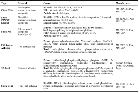
Material application: The teeth received the following restorations (Table 1 summarizes the characteristics of the materials used in this study):
manufacturer’s instructions: The primer was applied to the cavity walls (both enamel and dentin) using a microbrush and agitated for 15 seconds. The primer layer was gently air dried and cured with a LED light curing unit (Valo, Ultradent Products Inc., South Jordan, UT, USA)
with a light intensity of 1000 mW/cm2 for 10
seconds.
Using a new microbrush, bonding agent was applied to the cavity walls and after gentle air drying, it was cured for 10 seconds. P90 composite resin was applied incrementally to the cavity in three increments. The first increment was applied on the occlusal margin and the axial wall, the second increment was applied over the previous layer to the middle third of the axial wall and the third layer was applied over the previous ones to the outer third of the gingival margin obliquely. Each layer was cured for 10 seconds.
Finishing and polishing procedures were carried out using a fine flame diamond bur and flexible coarse, medium, fine and super fine polishing discs (Soflex, 3M ESPE. St. Paul, MN, USA), respectively. The samples were post cured for 20 seconds.
Group B: Teeth in this group were restored with Z250 composite resin (3M ESPE. St. Paul, MN, USA) and SE bond (Kuraray Noritake Dental Inc., Osaka, Japan). Primer was applied to enamel and dentin cavity surfaces by a microbrush and agitated for 20 seconds followed by gentle air-drying. The bonding agent was then applied to cavity surfaces using a microbrush, gently air dried and light cured for 10 seconds using a LED light-curing unit. Cavities were then restored with Z250 composite resin according to the protocol in group A.
Group C: Procedures were performed as in group B except that the cavities were filled with Z350
Table 1. Materials and their Composition
Type Material Content Manufacturer
Filtek Z250
Microhybrid methacrylate-based composite
Bis-GMA, Bis-EMA, UDMA, TEGDMA
Filler: zirconia, silica, 78wt%, 60v%
Particle size: 0.01-3.5 m
3M ESPE, St. Paul, MN, USA
Filtek Z350XT Enamel
Nanofilled methacrylate-based composite
Bis-GMA, UDMA, Bis-EMA, silica, zirconia, nanoparticles (20µm) and nanoagglomerates )0.4-0.6 µm)
Enamel shade: 78.5wt%, 63.3v%
3M-ESPE. St. Paul MN, USA
Filtek P90
Silorane-based composite (microhybrid)
Matrix: 3,4 Epoxycyclohexyl ethyl cyclo poly-methyl siloxane, bis-3,4 epoxycyclohexyl-ethyl-phenyl-methyl silane
Filler: Silanized, quartz, yttrium fluoride 76wt%– 55v%,
Particle size: 0.04-1.7m
3M ESPE, St. Paul MN, USA
P90 System
Adhesive Two step self-etch
Primer: phosphorylated-methacrylate, Vitrebond copolymer Bis-GMA, HEMA, water, ethanol, Silane-treated silica filler, camphorquinone stabilizer
Bond: hydrophobic dimethacrylate, phosphorylated-methacrylate, TEGDMA, Silane-treated silica filler, initiator, stabilizer
3M ESPE, St. Paul, MN, USA
SE Bond Self- etch adhesive
Primer: 10-Methacryloyloxydecyldihydrogen phosphate (MDP), 2-Hydroxyethyl methacrylate, hydrophilic dimethacrylate, dl-camphorquinone, accelerators, water
Bond: 10- Methacryloyloxydecyl dihydrogen phosphate (MDP), bisphenol a diglycidyl methacrylate (Bis-GMA), 2-Hydroxyethyl methacrylate (HEMA), hydrophobic dimethacrylate, dl-Camphorquinone, accelerators, silanized colloidal silica, surface treated sodium fluoride
Kuraray Noritake Dental Inc., Osaka, Japan
Single Bond Total- etch adhesive
Bis-GMA, HEMA, dimethacrylate, ethanol, water, novel photoinitiator system, methacrylate functional copolymer of polyacrylic, polyitaconic acids
composite resin (3M ESPE. St. Paul, MN, USA) and SE bond.
Group D: Teeth in this group were restored with Z250 composite resin (3M ESPE. St. Paul, MN, USA) and Single Bond (3M ESPE. St. Paul, MN, USA). The cavity was first etched with 37% phosphoric acid (3M ESPE. St. Paul, MN, USA). Enamel was etched for 20 seconds and dentin for 15 seconds. The cavity was then rinsed for 20 seconds and gently air-dried. The adhesive was then applied by an applicator according to the manufacturer’s instructions, agitated for 15 seconds and gently air dried for five seconds. The second layer of adhesive was applied to the cavity walls and light cured for 10 seconds using a LED light curing unit with a light intensity of
1000 mW/cm2. Then, half of the teeth in each
group were immersed in LA (pH = 4, 0.01M) and the other half in DW (pH=7) as the control group and stored in an incubator at 37°C for seven days.
Dye penetration technique: Before the
immersion of teeth in dye solution, the apices, root surfaces and the furcation area were sealed with sticky wax. All teeth surfaces except for the restoration area and 1 mm around the restoration margins were sealed with two coats of nail varnish. The teeth were immersed in 2% basic
fuchsin (Sigma Aldrich, Taufkirchen
Germany) in DW and stored in an incubator at 37°C for 24 hours. The samples were rinsed, dried and mounted in clear polyester acrylic resin and longitudinally sectioned in buccolingual direction by a saw with a blade thickness of 0.82mm in a cutting machine (Mecatome, T201A, Persi, France). Gingival and occlusal margin microleakage was assessed under a stereomicroscope (SMZ 800, Nikon, Tokyo, Japan) at x40 magnification and scored as follows:
0: No dye penetration
1: Dye penetration extending up to half the gingival/occlusal wall
2: Dye penetration extending to more than half the gingival/occlusal wall
3: Dye penetration extending into the axial wall and pulp
Statistical analysis: Data were analyzed using SPSS 18. Non-parametric Kruskal Wallis test was used for the analysis of microleakage (ordinal variable). The Mann Whitney U test was used to compare the degree of microleakage at each margin and for each composite in different media. Also, the Kruskal Wallis test was applied to compare the four groups of composites based on the margin and the media. The type of post hoc test was Mann-Whitney U with Bonferroni adjustment. (type 1 error was considered 0.05 for primary and 0.017 for post-hoc tests). Dunn procedure with P-value adjustment was used for pairwise comparisons. Confidence level was set at 95% (α= 0.05).
RESULTS
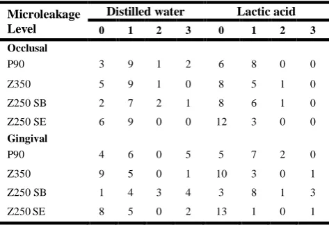
Table 2 shows the frequency of degrees of microleakage in the studied groups.
Table 2: Microleakage values at both occlusal and gingival margins in the studied groups
Microleakage Level
Distilled water Lactic acid
0 1 2 3 0 1 2 3 Occlusal
P90 3 9 1 2 6 8 0 0 Z350 5 9 1 0 8 5 1 0 Z250 SB 2 7 2 1 8 6 1 0 Z250 SE 6 9 0 0 12 3 0 0
Gingival
P90 4 6 0 5 5 7 2 0 Z350 9 5 0 1 10 3 0 1 Z250 SB 1 4 3 4 3 8 1 3 Z250SE 8 5 0 2 13 1 0 1
gingival margin (P<0.05). Pairwise comparisons were made using Dunn procedure with P value adjustment. The highest degree of microleakage was seen in Z250+SB group, being significantly higher than that of Z250+SE (P=0.012 in DW and P=0.002 in LA) and Z350+SE (P=0.002 in DW and P=0.014 in LA). The degree of microleakage was not significantly different in Z250+SE and Z350+SE groups (P=0.683 in DW and P=0.533 in LA). The microleakage of P90 in both media was less than that of Z250+SB and more than that of Z250+SE and Z350+SE; but none of the differences were statistically significant (P>0.017). Microleakage was not different in occlusal and gingival margins (P>0.05) in all groups except for Z250SB (P=0.039 in DW and P=0.008 in LA).
DISCUSSION
In the current study, seven days of immersion in LA did not cause a significant change in microleakage compared to immersion in distilled water. Silva et al, [5] showed that seven days of immersion in LA decreased the bond strength of composite restorations with Clearfil SE Bond and Adper Single Bond compared to artificial saliva. However, chemical agents such as the acids present in dentinal fluid, saliva, dental biofilm, foods and drinks can impact on the tooth-restoration interface as well as the hybrid layer and cause variable patterns of degradation of collagen fibrils and resin [21,27]. In addition, LA is a carboxylic acid with –OH and –COOH functional groups in its formulation. It is highly likely that these functional groups form hydrogen bonds with the polar end of methacrylate monomers present in the bonding agent matrix such as –OH in Bis-GMA, –O- in TEGDMA and Bis-EMA and N-H in UDMA, causing greater softening of the matrix [23].
The current study aimed to assess the early effect of immersion in LA on degree of microleakage and thus, immersion was only performed for seven days and no significant diffidence was
noted in microleakage following immersion of teeth in LA and DW. It might be related to short-term storage. However, long-short-term immersion may have destructive effects on the hybrid layer and resin and cause degradation of tooth-restoration interface. Further studies with longer storage time and thermocycling are required to better elucidate this phenomenon.
The results revealed no significant difference in microleakage of groups restored with silorane-based composite and Z250 bonded with SE bond. In a study by Kermanshah et al, [28] no difference was reported in microleakage of cavities restored with a silorane-based composite and SE bond. In contrast, Al-Boni and Raja [14] demonstrated that microleakage of silorane after thermocycling was less than that of Z250 bonded with self-etch adhesive.
In the current study, the highest degree of microleakage in gingival margin was seen in Z250+SB group. In a study by Hooshmand et al,
[29] the microleakage of silorane-based
composites was reported to be less than that of specimens bonded with Exite (an etch and rinse adhesive).
Single Bond is a two-step, etch and rinse adhesive system. This adhesive system is susceptible to nano-leakage due to inadequate impregnation of adhesive at the resin-dentin interface after polymerization [30]. On the other hand, higher content of hydrophilic monomers in two-step etch and rinse adhesives (compared to
three-step systems) [31] allows greater
penetration following polymerization, facilitates water sorption and increases leakage [32]. However, all adhesive systems have shown some degrees of incomplete polymerization and subsequent microleakage [33-35]; this is especially true about simplified systems like one-step self-etch and two-one-step etch and rinse systems due to higher hydrophilic monomer content [33-35]. SE-Bond is a two-step, self-etch adhesive. In
self-etch adhesives, acidic co-monomers
time. Complete infiltration of adhesive into the substrate is necessary to achieve a stable bond [36]. On the other hand, in mild (pH=2) two-step self-etch adhesives, phosphate or carboxylate groups present in monomers form a chemical bond with the residual hydroxyapatite crystals in dentin collagen network, reinforcing long-term stability [37,38]. In the current study, the degree of microleakage of silorane group was somewhere between that of Z250+SB and the remaining two groups; but none of these
differences were statistically significant.
Silorane composite resins are polymerized via a cationic ring-opening mechanism [7-9]. These new monomers are formed by the reaction of oxirane and siloxane molecules and thus the name silorane [7,8,39, 40]. Silorane primer has a pH of 2.7 and according to the manufacturer’s claim, it causes mild etching of the tooth surface and provides a strong, stable bond [41,42]. Moreover, Mine et al, [43] demonstrated that silorane primer forms a chemical bond with hydroxyapatite crystals. On the other hand, P90 primer and bonding agent are supplied in separate bottles and each one is photo-cured separately after application. Santini and Miletic [44] reported the presence of an intermediate layer with 1µm thickness between the silorane primer and bonding agent using micro-Raman spectroscopy. This area might be the weakest area involved in the mechanism of failure of silorane restorations; further investigations are warranted in this respect.
In the current study, the degree of microleakage of Z350+SE was similar to that of Z250+SE. But, Sharma et al, [45] indicated higher microleakage of Z350 compared to that of Z250 after thermocycling. In our study, similar results were obtained for samples restored with composites in conjunction with SE adhesive.
Z350XT is a nanofilled composite with high filler content. It undergoes less linear shrinkage than microhybrid composites due to smaller monomers and higher filler volume [46]. Low
shrinkage stress may improve marginal fit [24]. On the other hand, Filtek Z250 composite has shown acceptable results with regard to marginal fit [45,47].
CONCLUSION
Immersion in LA had no effect on microleakage of class V composite restorations regardless of the type of composite and adhesive system. At gingival margins, the highest microleakage was seen in Z250SB followed by P90 and self-etch groups.
REFERENCES
1- Roberson T, Heymann HO, Swift Jr EJ.
Sturdevant's art and science of operative dentistry: 6th ed., Missuri, Elsevier Pub. Co., 2013:44-6.
2- Borgström MK, Edwardsson S, Sullivan Å,
Svensäter G. Dental plaque mass and acid production activity of the microbiota on teeth. Eur J Oral Sci. 2000 Oct;108(5):412-7.
3- Distler W, Kröncke A. The acid pattern in human
dental plaque. J Dent Res. 1983 Feb;62(2):87-91.
4- Namiot D, Leszczyńska K, Namiot Z, Chilewicz
M, Bucki R, Kemona A. The occurrence of Helicobacter pylori antigens in dental plaque; an association with oral health status and oral hygiene practices. Adv Med Sci. 2010;55(2):167-71.
5- Silva EM, Almeida GS, Poskus LT, Guimaraes JG.
Influence of organic acids present in the oral biofilm on the microtensile bond strength of adhesive systems to human dentin. J Biomed Mater Res B Appl Biomater. 2012 Apr;100(3):735-41.
6- Mujdeci A, Gokay O. Effect of bleaching agents
on the microhardness of tooth-colored restorative materials. J Prosthet Dent. 2006 Apr;95(4):286-9.
7- D'Alpino PH, Bechtold J, Santos PJ, Alonso RC,
Di Hipolito V, Silikas N, et al. Methacrylate- and silorane-based composite restorations: Hardness, depth of cure and interfacial gap formation as a function of the energy dose. Dent Mater. 2011 Nov;27(11):1162-9.
8- Bagis YH, Baltacioglu IH, Kahyaogullari S.
silorane-based resin composite in wide Class II MOD cavities. Oper Dent. 2009 Sep-Oct;34(5):578-85.
9- Weinmann W, Thalacker C, Guggenberger R.
Siloranes in dental composites. Dent Mater. 2005 Jan;21(1):68-74.
10- Mehl A, Hickel R, Kunzelmann KH. Physical properties and gap formation of light-cured
composites with and without
'softstart-polymerization'. J Dent. 1997 May-Jul;25(3-4):321-30.
11- Braga RR, Ferracane JL, Condon JR.
Polymerization contraction stress in dual-cure cements and its effect on interfacial integrity of bonded inlays. J Dent. 2002 Sep-Nov;30(7-8):333-40.
12- Choi KK, Condon JR, Ferracane JL. The effects of adhesive thickness on polymerization contraction stress of composite. J Dent Res. 2000 Mar;79(3):812-7.
13- Lutz E, Krejci I, Oldenburg T. Elimination of polymerization stresses at the margins of posterior composite resin restorations: A new restorative technique. Quintessence Int. 1986 Dec;17(12):777. 14- Al-Boni R, Raja OM. Microleakage evaluation of silorane based composite versus methacrylate based composite. J Conserv Dent. 2010 Jul;13(3):152-5. 15- Hashemikamangar SS, Ghavam M, Mahinfar N, Kharazi Fard MJ. Effect of 30% hydrogen peroxide on marginal integrity of silorane-based versus methacrylate-based composite restorations. J Dent (Tehran). 2014 Sep;11(5):545-53.
16- Sano H, Yoshikawa T, Pereira P, Kanemura N, Morigamui M, Tagami J, et al. Long-term durability of dentin bonds made with a self-etching primer, in vivo. J Dent Res. 1999 Apr;78(4):906-11.
17- Frankenberger R, Krämer N, Lohbauer U, Nikolaenko SA, Reich SM. Marginal integrity: Is the
clinical performance of bonded restorations
predictable in vitro? J Adhes Dent. 2007;9 Suppl 1:107-16.
18- Hashimoto M, Ohno H, Kaga M, Endo K, Sano H, Oguchi H. In vivo degradation of resin-dentin bonds in humans over 1 to 3 years. J Dent Res. 2000 Jun;79(6):1385-91.
19- Waidyasekera K, Nikaido T, Weerasinghe DS, Ichinose S, Tagami J. Reinforcement of dentin in self-etch adhesive technology: A new concept. J Dent. 2009 Aug;37(8):604-9.
20- Albaladejo A, Osorio R, Toledano M, Ferrari M. Hybrid layers of etch-and-rinse versus self-etching adhesive systems. Med Oral Patol Oral Cir Bucal. 2010 Jan 1;15(1):e112-8.
21- Hashimoto M, Ohno H, Sano H, Kaga M, Oguchi H. Degradation patterns of different adhesives and bonding procedures. J Biomed Mater Res B Appl Biomater. 2003 Jul 15;66(1):324-30.
22- Armstrong S, Vargas M, Chung I, Pashley D, Campbell J, Laffoon J, et al. Resin-dentin interfacial ultrastructure and microtensile dentin bond strength after five-year water storage. Oper Dent. 2004 Nov;29(6):705-12.
23- da Silva EM, Goncalves L, Guimaraes JG, Poskus LT, Fellows CE. The diffusion kinetics of a nanofilled and a midifilled resin composite immersed in distilled water, artificial saliva, and lactic acid. Clin Oral Investig. 2011 Jun;15(3):393-401.
24- Schmidt M, Kirkevang LL, Horsted-Bindslev P, Poulsen S. Marginal adaptation of a low-shrinkage silorane-based composite: 1-year randomized clinical trial. Clin Oral Investig. 2011 Apr;15(2):291-5. 25- Santos PJ, Silva MS, Alonso RC, D'Alpino PH. Hydrolytic degradation of silorane-and methacrylate-based composite restorations: Evaluation of push-out strength and marginal adaptation. Acta Odontol Scand. 2013 Sep;71(5):1273-9.
26- Mahmoud SH, Al-Wakeel Eel S. Marginal adaptation of ormocer-, silorane-, and methacrylate-based composite restorative systems bonded to dentin cavities after water storage. Quintessence Int. 2011 Nov-Dec;42(10):e131-9.
27- Liu Y, Tjäderhane L, Breschi L, Mazzoni A, Li N, Mao J, et al. Limitations in bonding to dentin and experimental strategies to prevent bond degradation. J Dent Res. 2011 Aug;90(8):953-68.
Dent Assoc Iran 2013; 25(2): 91-8.
29- Hooshmand T, Tabari N, Keshvad A. Marginal leakage and microhardness evaluation of low-shrinkage resin-based restorative materials. Gen Dent. 2013 Jan-Feb;61(1):46-50.
30- Tay FR, Pashley DH, Yoshiyama M. Two modes of nanoleakage expression in single-step adhesives. J Dent Res. 2002 Jul;81(7):472-6.
31- Tay FR, Pashley DH. Have dentin adhesives become too hydrophilic? J Can Dent Assoc. 2003 Dec;69(11):726-31.
32- Tay FR, Frankenberger R, Krejci I, Bouillaguet S, Pashley DH, Carvalho RM, et al. Single-bottle adhesives behave as permeable membranes after polymerization. I. In vivo evidence. J Dent. 2004 Nov;32(8):611-21.
33- Breschi L, Cadenaro M, Antoniolli F, Sauro S, Biasotto M, Prati C, et al. Polymerization kinetics of dental adhesives cured with LED: correlation between extent of conversion and permeability. Dent Mater. 2007 Sep;23(9):1066-72.
34- Cadenaro M, Antoniolli F, Sauro S, Tay FR, Di Lenarda R, Prati C, et al. Degree of conversion and permeability of dental adhesives. Eur J Oral Sci. 2005 Dec;113(6):525-30.
35- Cadenaro M, Breschi L, Antoniolli F, Navarra CO, Mazzoni A, Tay FR, et al. Degree of conversion of resin blends in relation to ethanol content and hydrophilicity. Dent Mater. 2008 Sep;24(9):1194-200.
36- Summitt JB, Robbins JW. Fundamentals of operative dentistry. 4th ed., Chicago, Quintessence Pub. Co.; 2013:228-33.
37- De Munck J, Van Landuyt K, Peumans M, Poitevin A, Lambrechts P, Braem M, et al. A critical review of the durability of adhesion to tooth tissue: methods and results. J Dent Res. 2005 Feb;84(2):118-32.
38- Van Meerbeek B, Yoshihara K, Yoshida Y, Mine A, De Munck J, Van Landuyt KL. State of the art of self-etch adhesives. Dent Mater. 2011
Jan;27(1):17-28.
39- Unterbrink GL, Liebenberg WH. Flowable resin composites as" filled adhesives": literature review and clinical recommendations. Quintessence Int. 1999 Apr;30(4):249.
40- Yu H, Wegehaupt FJ, Wiegand A, Roos M, Attin T, Buchalla W. Erosion and abrasion of tooth-colored restorative materials and human enamel. J Dent. 2009 Dec;37(12):913-22.
41- Krifka S, Federlin M, Hiller KA, Schmalz G. Microleakage of silorane- and methacrylate-based class V composite restorations. Clin Oral Investig. 2012 Aug;16(4):1117-24.
42- Hegde MN, Vyapaka P, Shetty S. A comparative evaluation of microleakage of three different newer direct composite resins using a self etching primer in class V cavities: An in vitro study. J Conserv Dent 2009 Oct;12(4):160.
43- Mine A, De Munck J, Van Ende A, Cardoso MV, Kuboki T, Yoshida Y, et al. TEM characterization of a silorane composite bonded to enamel/dentin. Dent Mater. 2010 Jun;26(6):524-32.
44- Santini A, Miletic V. Comparison of the hybrid layer formed by Silorane adhesive, one-step self-etch and etch and rinse systems using confocal micro-Raman spectroscopy and SEM. J Dent. 2008 Sep;36(9):683-91.
45- Sharma RD, Sharma J, Rani A. Comparative
evaluation of marginal adaptation between
nanocomposites and microhybrid composites
exposed to two light cure units. Indian J Dent Res. 2011 May-Jun;22(3):495.
46- Pereira RA, Araujo PA, Castañeda-Espinosa JC, Mondelli RF. Comparative analysis of the shrinkage stress of composite resins. J Appl Oral Sci. 2008 Jan-Feb;16(1):30-4.