www.Genes&Cancer.com Genes & Cancer, Vol. 9 (5-6), May 2018
Tumor metabolism regulating chemosensitivity in ovarian
cancer
Chae Young Han
1, David A. Patten
3,4, Richard B. Richardson
3, Mary-Ellen Harper
4,
and Benjamin K. Tsang
1,21 Department of Obstetrics and Gynecology and Cellular and Molecular Medicine, University of Ottawa, and Chronic Disease
Program, Ottawa Hospital Research Institute, Ottawa, Ontario, Canada
2 State Key Laboratory of Quality Research in Chinese Medicine, Macau Institute for Applied Research in Medicine and Health,
Macau University of Science and Technology, Avenida Wai Long, Taipa, Macao, China
3 Canadian Nuclear Laboratories (CNL), Radiobiology and Health Branch, Chalk River Laboratories, Chalk River, Ontario,
Canada
4 Department of Biochemistry, Microbiology and Immunology, Faculty of Medicine, University of Ottawa, Ottawa, Canada
Correspondence to: Benjamin K. Tsang, email: btsang@ohri.ca
Keywords: ovarian cancer, chemoresistance, tumor metabolism, hexokinase 2, p53
Received: June 12, 2018 Accepted: August 14, 2018 Published: August 25, 2018
Copyright: Han et al. This is an open-access article distributed under the terms of the Creative Commons Attribution License 3.0 (CC BY 3.0), which permits unrestricted use, distribution, and reproduction in any medium, provided the original author and source are credited.
ABSTRACT
Elevated metabolism is a key hallmark of multiple cancers, serving to fulfill high anabolic demands. Ovarian cancer (OVCA) is the fifth leading cause of cancer deaths in women with a high mortality rate (45%). Chemoresistance is a major hurdle for OVCA treatment. Although substantial evidence suggests that metabolic reprogramming contributes to anti-apoptosis and the metastasis of multiple cancers, the link between tumor metabolism and chemoresistance in OVCA remains unknown. While clinical trials targeting metabolic reprogramming alone have been met with limited success, the synergistic effect of inhibiting tumor-specific metabolism with traditional chemotherapy warrants further examination, particularly in OVCA. This review summarizes the role of key glycolytic enzymes and other metabolic synthesis pathways in the progression of cancer and chemoresistance in OVCA. Within this context, mitochondrial dynamics (fission, fusion and cristae structure) are addressed regarding their roles in controlling metabolism and apoptosis, closely associated with chemosensitivity. The roles of multiple key oncogenes (Akt, HIF-1α) and tumor suppressors (p53, PTEN) in metabolic regulation are also described. Next, this review summarizes recent research of metabolism and future direction. Finally, we examine clinical drugs and inhibitors to target glycolytic metabolism, as well as the rationale for such strategies as potential therapeutics to overcome chemoresistant OVCA.
I. INTRODUCTION
Ovarian cancer (OVCA) is the fifth
leading cause
of cancer deaths in women and has a high mortality rate
(30-50%) [1, 2
]. Late diagnosis and the development of
chemoresistance are major hurdles to successful therapy.
Due to non-specific clinical symptoms and the absence of
early biomarkers, OVCA is usually diagnosed in advanced
stages. Epithelial OVCA accounts for more than 90% of
incidences and has the highest mortality rates [
3]. Within
epithelial OVCA, high-grade serous is most frequently
found (70%) among different sub-types (high-grade,
low-grade, endometroid, clear cell, and mucinous) [
2].
The standard treatment of OVCA is debulking surgery
followed by chemotherapy, but 70% of patients in
advanced stage experience chemoresistance or relapse
within 15 months following treatment [
4].
multifactorial, partly due to defects in apoptosis,
dysregulation of an oncogene, defects in tumor
suppressors, and increased metabolism [
5, 6
]. The master
tumor suppressor p53 is responsible for apoptosis of
cancer cells and is critical to the chemoresponsiveness of
OVCA [
7
, 8].
TP53 mutations occur in 70% cases of OVCA and
in more than 90% cases in high-grade serous epithelial
OVCA [
9], and is closely associated with chemoresistance
[10
]. Understanding in-depth metabolic and molecular
mechanisms are required in the pursuit for effective
therapeutic targets to overcome chemoresistant OVCA.
Metabolic reprogramming enables cancer cells to fulfill
their high proliferation and survival potentials [
11].
Since Otto Warburg proposed a high rate of aerobic
glycolysis in cancer cells (Warburg effect) in 1923 [
12,
13
], the unique metabolic character of cancer cells has
been intensely investigated as a target for cancer therapy.
The Warburg effect specifically proposes that glycolysis
is a main metabolic pathway for ATP generation and
oxidative phosphorylation (OXPHOS) is impaired in
cancer cells; however, recent research has demonstrated
that both pathways are relatively elevated in cancer cells
compared to normal cells [
11, 14
]. Moreover, it may be
that the ability of cancer cells to switch between energy
substrates and metabolic pathways (termed bioenergetic
flexibility) is associated with poor prognosis, including
metastasis [
15, 16
]. Glycolysis works as precursor
pathway, since its metabolites and products are required
for downstream pathways including: the tricarboxylic acid
(TCA) cycle and OXPHOS; pentose phosphate pathway
(PPP) for ribonucleotide synthesis and Nicotinamide
adenine dinucleotide phosphate (NADPH); glycosylation
and gluconeogenesis; amino acid biosynthesis, and fatty
acid synthesis [
11,
17]. In addition to glycolysis, the PPP
pathway is highly elevated in cancer [
18,
19
], providing
anabolic substrates for cancer growth and reductive
intermediates (e.g., NADPH) for glutathione (GSH)
synthesis and protection from oxidative damage.
Fatty acid metabolism is similarly altered in
cancer. Unsaturated lipids are increased in OVCA [20,
21
], which induces stemness, whereas lipid desaturation
impairs cancer stemness and tumor initiation [
21].
Cellular glycogen accumulation is observed as a feature
of clear cell, a sub-type of epithelial OVCA, which
frequently develops chemoresistance [
22
]. Also, glycogen
accumulation is elevated in hypoxic conditions, a core
component of the solid tumor microenvironment [
22, 23].
These findings suggest that specific metabolic phenotypes
enhance chemoresistance in OVCA.
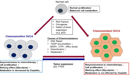
However, it remains unclear whether metabolic
reprogramming in cancer is associated with
chemoresistance and what therapeutic approaches
could modulate metabolic phenotypes associated with
chemoresistance of OVCA (Figure 1). In this review,
we discuss: 1) the cellular and molecular mechanisms
involved in metabolic reprogramming; 2) the influence
of metabolism on apoptosis and the related functions
of mitochondria; 3) whether and how oncogenes/
tumor suppressors regulate the role of key glycolysis
enzymes; 4) future strategies for targeting metabolism
in cancer treatment, including combining such therapies
with traditional chemotherapeutics; and lastly, 5) the
importance of tumor microenvironment and potential
novel metabolic strategies.
II. THE ROLE OF GLYCOLYTIC
METABOLISM IN CHEMORESISTANCE
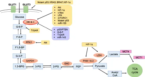
Accelerated glycolysis is a key hallmark of
tumorigenesis [
12, 24, 25
]. Glycolysis is the first metabolic
pathway converting glucose to pyruvate. Beyond their
metabolic functions, key glycolytic enzymes have been
shown to enhance the survival and progression of tumors
associated with drug resistance (Figure 2). Metabolic
adaptation of excessive glycolysis in cancer is mediated
by hyperactive glycolytic enzymes, and this is primarily
due to dysregulation of tumor suppressors or activation of
oncogenes [26].
Glucose transporters
Glucose transporters (GLUTs) are a large group of
membrane transport proteins that facilitate the transport
of glucose over a plasma membrane [27
]. There are 14
identified GLUTs (SCL2A1 to SCL2A14). Among them,
GLUT1 has been identified as a hypoxic marker and plays
the critical role in the Warburg effect [
28
]. In cancer cells,
GLUT1 is highly expressed and facilitates metastasis of
tumor and poor prognosis in multiple cancers including
lung, stomach, breast, and kidney [
28,
29]. High level of
GLUT1 expression is observed in membrane of high-grade
serous OVCA, whereas GLUT1 expression is negatively
correlated with fasting glucose concentration, explaining
its glucose uptalking [
30
]. In OVCA, by modulating
GLUT1 membrane trafficking, phytochemicals such as
resveratrol decreases glycolysis and induces apoptosis
[31].
Hexokinase II
bound to the outer mitochondrial membrane (OMM), and
localized within mitochondria [
33
]. Both HKI and HKII
are inhibited by G-6-P, its catalytic product via feedback
inhibition [
13
]. HKII, a predominant isoform in insulin
sensitive tissues (adipose, skeletal, and cardiac muscles),
is highly expressed in multiple tumors [
34]. Recent
research demonstrates that HKII is highly associated
with tumorigenesis and cancer cell survival [
25, 35
]. In
mouse models, deletion of HKII significantly decreased
tumor burden and prolonged survival, suggesting that
HKII is a critical factor involved in tumor progression
[36
]. Inhibition of HKII restored normal glycolysis and
OXPHOS pathway as well as mitochondrial biogenesis in
glioblastoma [37]. HKII depletion also increased intrinsic
apoptosis by increased mitochondrial permeability in
chemoresistant brain cancer cells [37]. Lysophosphatidic
acid (LPA), a lipid growth factor and G protein-coupled
receptor (GPCR) ligand are significantly increased in
OVCA, triggering the activation of hypoxia inducible
factor-1α (HIF-1α) and inducing GLUT1 and HKII,
thus shifting cells towards a glycolytic metabolism [
38].
Zhang et al. have demonstrated that HKII contributes
to chemoresistance by enhancing CDDP-induced
extracellular signal-regulated kinases (ERK) 1/2
phosphorylation and autophagy [39
].
Phosphofructokinase 1 (PFK1)
Phosphofructokinase (PFK) 1 is responsible for
converting fructose-6-phosphate (F-6-P) to fructose 1,6
bisphosphate (F-1,6-BP), the second irreversible step in
glycolysis [
40, 41
]. Somatic mutation of PFK and altered
structure promote altered glycolytic flux in cancer [
42].
The most potent allosteric activator of PFK is
fructose-2,6-bisphosphate (F-2,6-BP) and its synthesis and
degradation depend on the 6-phosphofructo-2-kinase/
fructose-2,6-bisphosphatases (PFKFB) [
43
]. PFKFBs
phosphorylate F-6-P to F-2,6-BP, which in turn activates
PFK1, forwarding glycolytic flux to lactate. Among the
five PFKFB isoenzymes (PFKFB 1-4, and TIGAR),
PFKFB3 has the highest kinase/phosphate activity ratio,
leading to elevated F-2,6-BPase and resulting in high
glycolysis rate. PFKFB3 promotes cell cycle progression
and suppresses apoptosis by facilitating cyclin dependent
kinase (Cdk)-induced degradation of p27, a key apoptotic
activator [40
]. Its product, F-2,6-BP is shown to be highly
expressed in ovarian and breast cancers [
43
]. Conversely,
PFKFB4 depletion increased apoptosis and the production
of reactive oxygen species (ROS) in mitotically arrested
cells. It also significantly enhanced mitotic cell death in
OVCA with paclitaxel treatment [
44].
Pyruvate kinase
Pyruvate kinase (PK) catalyzes the last irreversible
reaction of glycolysis, converting phosphoenolpyruvate
to pyruvate. Two distinct PK genes, [L/R
(in liver and
red blood cells) and
PKM2
(in muscle)] encode four
PK isoforms: PKL, PKR, PKM1, and PKM2. Among
them, PKM2 is allosterically regulated by various
metabolites [
45, 46
]. The activity of PKM2 enables cancer
cells to adapt to altered tumor metabolic conditions.
Phosphorylated PKM2 dimers stimulate a high rate of
nucleotide and amino acid biosynthesis, while indirectly
sustaining the Warburg effect [
46
]. Depletion of PKM2 in
a breast cancer mouse xenograft model showed a reversal
of the Warburg effect and inhibits tumor growth [47
].
High expression of PKM2 is found in malignant OVCA,
suggesting its late stage detection role of poor prognosis
[48
]. The association of PKM2 with tumorigenesis
depends on the type of cancer, as the suppression of
PKM2 did not show anti-tumor activity in breast and
colon cancer, but did in leukemia [49
, 50].
Lactate dehydrogenase
Lactate dehydrogenase (LDH) catalyzes the
interconversion of pyruvate and lactate, and of NADH
and NAD+. Four LDH isozymes exist (LDH-A, LDH-B,
LDH-C, and LDH-D), with LDH-A being predominantly
expressed in the majority of tissues. Elevated levels of
LDH-A are associated with multiple solid tumors and
poor prognosis of cancer [
51
]. As LDH-A promotes the
reduction of pyruvate to lactate for NADH production,
whereas LDH-B favors the reverse reaction; LDH-A is a
key enzyme involved in the Warburg effect. Depletion of
LDH-A in tumor cells and in mice reduces tumor growth
[52
]. Recent research demonstrates that LDH supports
enhanced rates of oxidative metabolism in tumors e.g.,
TCA cycle activity [
53, 54
]. Moreover, excessive activity
of LDH in glycolysis can lead to local acidification of the
tumor microenvironment, a favorable condition for tumor
invasion and metastasis [
55, 56
]. High level of LDH is
observed in serum and peritoneal fluid from OVCA
patients compared with that of benign tumor, suggesting
its possible role as potential prognostic biomarker [57
, 58].
Lactate, the end product of LDH, a key gluconeogenic
precursor, is moved intracellularly and intercellularly
(e.g., Lactate shuttle)
via monocarboxylate transporters
MCT1 and MCT4, proposed by San-Millan et. al [59
].
This notion is also supported by the evidence that high
expression of MCT1 promotes the CDDP resistance by
antagonizing the effect of first apoptosis of signal receptor
(Fas) in epithelial OVCA [60
]. High levels of lactate, as
key elements for energy source and tumorigenesis, have
been observed along with subsequent active glycolysis in
many cancers [
61].
III. THE INFLUENCE OF ELEVATED
GLYCOLYSIS ON
MITOCHONDRIAL-MEDIATED APOPTOSIS IN CANCER
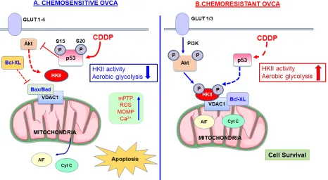
A. The role of mitochondrial HKII for cell
survival
Intrinsic (mitochondria-mediated) apoptosis is
initiated by the loss of mitochondrial membrane potential,
increased mitochondrial outer membrane permeabilization
(MOMP), and the subsequent release of mitochondrial
death proteins including cytochrome c [
62
]. In addition
to its role in glycolysis, HKII is involved in protecting
mitochondria and the suppression of intrinsic apoptosis
[63
] (Figure 3), whereas its depletion of HKII enhances
apoptosis and sensitivity to external stimuli. Ectopic
expression of HKII protects lung cancer cells and renal
epithelial cells from oxidative injury or cell death [
64, 65].
Depletion of HKII in human HCC cells enforces metabolic
pathway toward OXPHOS through maintaining TCA flux,
thus facilitating cell death and inhibiting tumorigenesis
[66].
In line with its role in regulating MOMP,
HKI and HKII can bind to voltage-dependent anion
channel 1 (VDAC1), a likely component of the
mitochondrial permeability transition pore (mPTP)
[25,
67]. Approximately 80% of HKII is localized with
mitochondria in cancer cells [
33
]. Accumulating studies
showed that HKII bound to mitochondria (mito-HKII)
enhances tight coupling of glucose phosphorylation to
mitochondrial ATP generation [
68,
69
]. DeWaal et al.
showed that ectopic expression of mitochondrial
binding-deficient HKII failed to restore cell proliferation and
tumorigenesis, emphasizing pivotal role of mito-HKII
[66]. Mitochondrial Ca
2+overload and ROS induce the
opening of mPTP and consequent rupture. Mito-HKII
provides cellular protection against this Ca
2+overload and
mPTP opening [
35].
The interaction of HKII with Bcl-2 members also
affects apoptotic processes, particularly the execution
of intrinsic apoptosis. Some anti-apoptotic molecules of
Bcl-2 family (e.g., Bcl-2 and Bcl-xL) stay in the outer
mitochondrial membrane (OMM), whereas pro-apoptotic
members (Bad, Bax, and Bid) are translocated from the
cytosol to the OMM upon initiation of apoptotic signaling
[70, 71]. In mitochondrial-mediated cell death, activated
Bax/Bak forms a pore at the OMM, induces MOMP,
and results in the release of apoptotic factors such as
cytochrome c from the inter-membrane space [71], which
then accelerates the opening of VDAC [72]. Conversely,
anti-apoptotic Bcl-2 family members, such as Bcl-xL
interact with VDAC, inhibiting cytochrome c release by
modulating OMM integrity or MOMP [70
,
73
].
Mito-HKII inhibits binding of Bax to Bcl-2 through competitive
inhibition and antagonizes truncated Bid (tBid)-mediated
apoptosis [
74
,
75
]. Upon cell stress (e.g., glucose
restriction or treatment with Metformin, Clotrimazole,
Flavonoid-429, 2-DG, or 3-Bromopyruvate) HKII is
detached from VDAC [76-79], inducing dysregulation
of mitochondria by opening the mPTP, increasing the
MOMP, and subsequent inducing of
mitochondrial-mediated apoptosis [74
, 80
]. HKII dissociation from
mitochondria using an artificial peptide also was shown
to induce apoptosis without changing mitochondrial
membrane potential, ROS production, or OXPHOS [78
].
This suggests that detachment of HKII from mitochondria
is critical for intrinsic apoptosis. However, the molecular
mechanism of HKII detachment remains to be elucidated.
Cristae structural alterations and changes in the
ultrastructure of the inner mitochondrial membrane
compartments also significantly contribute to apoptotic
signaling [81, 82
]. Optic Atrophy 1 (OPA1) is a
pro-fusion dynamin-related protein in the inner mitochondrial
membrane that regulates cristae structure. OPA1 associates
with itself to form oligomers, which act to maintain
narrow cristae and narrow cristae junctions. However,
Bax/Bak activation can disrupt OPA1 oligomerization,
thereby widening cristae, and aiding in the release of
pro-apoptotic factors and ultimately inducing apoptosis [
82].
We observed that CDDP induced OPA1 processing and
intact OPA1 is associated with chemoresistance [
83, 84].
However, it remains unknown if and how mito-HKII is
involved in Bax - OPA1 interaction and how this affects
OPA1 oligomerization and cristae structure [
81].
B. The role of mitochondrial fission/fusion in
metabolic reprogramming of cancer
Mitochondria are highly dynamic organelles
regulated by fission and fusion events and, as described
above, cristae undergo important ultrastructural changes.
Indeed, mitochondrial dynamism plays essential roles
in fundamental cellular processes and metabolism, and
changes in mitochondrial dynamics have been linked
to cancer [
89
,
90]. Mitochondrial fusion is regulated by
the dynamin GTPases, Mitofusin 1 and 2 (Mfn1, Mfn2)
and OPA1, while mitochondrial fission is regulated
by dynamin-related protein (Drp1) and recruitment/
activation factors (Mff, Fis1, MiD49, and Mid50) [91
].
Mitochondrial fusion is additionally correlated with
altered cristae structure, enhanced assembly of respiratory
super-complexes and ATP synthase assembly, often
shifting cells from glycolysis to OXPHOS [92
,
93
]. The
role of mitochondrial fission/fusion in the regulation
of apoptosis and cell survival remains controversial.
In OVCA cells, we observed significant higher level of
mitochondrial fusion in chemoresistant cells than in their
chemosensitive counterparts [
83,
94
]. CDDP increased
significant mitochondrial fission in chemosensitive cells,
but not in chemoresistant cells. Piceatannol, a metabolite
of functional food compound in red wine, sensitized
OVCA cells to CDDP
in vitro
and
in vivo, by inducing
dephosphorylation of mitochondrial fusion protein,
Drp1 at site of Ser
637, and promoting mitochondrial
fission and apoptosis [95
]. This indicates that higher
proportion of tubular network in mitochondria harboring
chemoresistance cells are closely related to anti-apoptosis
and cell survival.
Few studies have reported the mitochondrial
dynamics and energy metabolism in OVCA. Generally,
through either enhanced mitochondrial fission, or
decreased mitochondrial fusion, cancer cells have
increased mitochondrial fragmentation, which has
a functional role to promote glycolysis for energy
production over OXPHOS [96]. Pro-survival factor,
Survivin (BIRC5) overexpression leads to mitochondrial
fragmentation through increased mitochondrial fission,
shifting energy production toward glycolysis [97
,
98
].
Also, the inhibition of glycolysis attenuates the
anti-apoptotic action of Survivin. However, cancer cells
can adapt their mitochondrial structure in response to
metabolic challenges similar to non-cancerous cells
[
96
,
99
, 100
]. These reports demonstrate that cancer
cells adapt to stressful energy conditions by modulating
mitochondrial morphologies and functions generally
associated with increased glycolysis.
C. Mitochondrial biogenesis and glycolysis
metabolism
To maintain energy metabolism and function upon
external stress, mitochondrial autophagy (mitophagy) and
mitochondrial biogenesis are cooperatively required to
regulate cellular homeostasis and survival [
101
]. Intact
mitochondrial function and mitochondrial biogenesis are
indeed required for survival of cancer cells [
102]. Cancer
cells respond to changes in glucose and other nutrient
sources by altering mitochondrial biogenesis [
101, 103].
Also, the key oncogenic transcription factor c-MYC
(v-myc avian myelocytomatosis viral oncogene homolog)
has been shown to facilitate glycolysis and mitochondrial
biogenesis with associated increases in ATP production
[104, 105].
D. ROS and glycolytic metabolism
Aberrant high levels of ROS generation are
often associated with increase in antioxidant defenses
in cancer cells [106
]. ROS are potentially damaging
byproducts of mitochondrial oxidative metabolism, but
also function as second messengers in the transduction
of extracellular signals that control cellular proliferation
and cell cycle progression [107]. As a master regulator of
ROS and mitochondrial energy metabolism, peroxisome
proliferator-activated receptor gamma co-activator 1-alpha
(PGC-1α) is partly responsible for this detoxification
[15
]. Through stimulating bioenergetic potential,
PGC-1α promotes migration and invasion of breast cancer
and facilitates chemoresistance [
16]. Other transcription
factors, nuclear respiratory factors (Nrf1 and Nrf2),
the estrogen-related receptors (ERR-α, -β and -γ) and
the nuclear factor erythroid 2-related factor 2 (Nrf2/
NFE2L2) act as master regulators of anti-oxidants [
108].
In spheroid OVCA cell cultures, ROS induced high
expression of PGC-1α, a phenomenon associated with
metastasis, stemness, and chemoresistance [109
]. Also,
Nrf2 is overexpressed in clear cell ovarian carcinoma
exhibiting high glycolytic phenotype, and high nuclear
expression of Nrf2 is associated with poor prognosis of
OVCA patients [110, 111
]. GSH, an important antioxidant,
is critical for modulating apoptosis. In OVCA, GSH is
inversely associated with chemoresistance and levels
of GSH are 15-50 fold higher in chemosensitive cells
than chemoresistant cells [
112
]. Mitochondrial GSH
protects cells from ROS during mitochondrial respiration
[113, 114
]. Moreover, NADPH produced by the PPP is
required for the generation of reduced GSH [
115
]. Hence,
GSH functions as metabolic link between PPP and ROS
balance.
IV. REGULATORY MECHANISMS
OF GLYCOLYTIC METABOLISM IN
OVARIAN CANCER
A. Aberrant activation of glycolysis by oncogenes
Accumulating evidence suggests that multiple tumor
suppressor/oncogenes affect glucose metabolism [
11,
116
]. The phosphoinositide-3 kinase (PI3K)/Akt axis is
a key oncogenic cell signaling pathway in cell survival
and tumor progression,and may promote glycolytic
reprogramming. For example, acute insulin treatment
leads to the trafficking of glucose transporter in a PI3K
/Akt-dependent manner [117]. PI3K is a phospholipid
kinase that phosphorylates the 3′ hydroxyl (OH) group
of the inositol ring of phosphoinositide lipids [
118
]. PI3K
phosphorylates the membrane lipid phosphatidylinositol
4,5-bisphosphate (PIP
2) to form phosphatidylinositol
3,4,5-triphosphate (PIP
3), whereas this process is regulated
by tumor suppressors phosphatase and tensin homolog
(PTEN) to dephosphorylate PIP
3[
119]. Akt is serine/
threonine kinase and is highly upregulated in cancer cells
[120
]. Akt is activated by its recruitment to the plasma
membrane by PIP
3, followed by phosphorylation of Thr
308and Ser
473by PDK1 [
121].
Akt promotes glycolysis, suppresses apoptosis, and
elicits cell survival
via multiple mechanisms [
34, 122,
123
]. PI3K pathway activation or mutational changes in
genetic and function were common in OVCA [
124
]. Effect
of inhibition of Akt depends on genetic heterogeneity
of cancer. Inhibition of Akt1 selectively caused cancer
growth in subset of OVCA cells lines, but not applied to
all of OVCA subset, due to high expression of other Akt
isoforms [
125, 126
]. Akt enhances mitochondrial HKII
activity and in turn OMM stability, thereby increasing its
anti-apoptotic action. Akt directly phosphorylates HKII
at the consensus binding site of (RARQKT*) Thr
473.
FV-429 inhibits glycolysis by attenuating Akt-mediated
phosphorylation of HKII, resulting in apoptotic induction
[
77]. As oncogenes, KRAS and BRAF activate Akt and
mutation of KRAS/BRAF enhance the expression and
plasma membrane trafficking of GLUT1 [129-131
]. Still,
it remains unclear how the interplay of Akt/p53 affects the
HKII-mediated glycolysis and other metabolic processes
in cancer.
c-MYC is an oncogene and transcription factor that
regulates multiple genes involved in cell proliferation,
metabolism, and apoptosis [
132]. Constitutive activation
of c-MYC is frequently found in human cancer [
133].
In OVCA, protein expression of c-MYC is higher in
chemosresistant cells compared with its counterpart
sensitive cells [134, 135
]. High c-MYC expression is also
associated with decreased overall survival and disease free
survival rate [135
]. c-MYC contributes to chemoresistance
of cancer in part by controlling metabolism [
136
]. c-MYC
binds and activates the promoters of key metabolic
enzymes including GLUT1, HKII, PFK, and enolase 1
(ENO1), leading to an activation of the glycolysis pathway
[
137-139]. In addition, c-MYC cooperates with HIF-1α
to promote HKII and PDKI, again shifting metabolic
pathway to glycolysis [
136].
mTOR is a serine/threonine kinase involved
in promoting energy metabolism, cell growth, and
proliferation. Elevated mTOR suppresses induction of
autophagy and frequently leads to the development of
tumors [
34, 116
]. mTOR is composed of two distinct
functional complexes, mTOR complex 1 and 2 (mTORC1
and mTORC2), with their respective defining components
Raptor and Rictor [51
]. Akt activates mTOR, promoting
anabolic metabolism and fatty acid synthesis, whereas
AMPK represses mTOR, leading to catabolic energy
production and fatty acid oxidation. In response to
glucose deprivation, HKII can bind to mTORC1 by its
TOS motif, decreasing TORC1 activity and positively
regulate autophagy [
140]. Under glucose depletion,
inhibition of HKII is attenuated, while overexpression of
HKII elevates glucose deprivation-induced autophagy [
34,
140
]. This indicates that cells increase their adaptability
and survival by mTOR-HKII interaction under varied
energy conditions.
MicroRNAs (miRNAs) are small, non-coding
RNAs and post-transcriptional inhibitory regulators of
gene expression. As a tumor suppressor miRNA,
143 downregulates HKII, whereas oncogenic
miR-155 upregulates HKII expression [
141
]. Overall, key
oncogenes promote glycolysis metabolism and cell
survival, whereas tumor suppressors regulate it in OVCA,
incurring its responsiveness to chemotherapy.
B. Regulation of glycolysis by p53
p53, key tumor suppressor is involved in the
regulation of cellular proliferation, apoptosis, and
metabolism [
51]. p53 elicits its apoptotic action
via
transcription-dependent and -independent mechanisms
[8
]. Our lab has demonstrated that activation of functional
p53 signaling is required for apoptosis in response to
chemotherapy such as CDDP [
6, 142
]. In addition to its
fundamental role in apoptosis, p53 is shown to negatively
regulate glycolysis [
143
], as its mutation may lead to
the reliance on glycolysis of cancer cells [9
]. Moreover,
co-deletion of p53/PTEN in prostate cancer can further
increase HKII levels. PTEN deletion increase HKII
mRNA translation
via activation of the
Akt-mTOR-4EBP1 pathway, whereas p53 loss enhances HKII
mRNA stability by inhibiting miR-143 biogenesis [
144].
p53 plays a critical role in maintaining mitochondrial
integrity and function, potentially through modulating
SCO2 (the synthesis of cytochrome c oxidase protein)
and cytochrome oxidase II (COX II) [
145, 146]. SCO2
is required for the OXPHOS, mitochondrial respiration,
and the assembly of the mitochondrial complex with COX
II. Therefore, depletion of p53 impairs mitochondrial
structure and respiration, shifting cells toward glycolytic
metabolism. p53 can also regulate the protein TP53
inducible glycolysis and apoptosis regulator (TIGAR)
[147]. TIGAR has a dual function in regulating glycolysis
to maintain homeostatic balance system in the cell. TIGAR
leads to the accumulated production of G-6-P, preventing
glycolysis. However, under hypoxic conditions, TIGAR
is translocated to mitochondria and binds to HKII,
decreasing ROS, promoting glycolysis, and providing cell
protection [
147
, 148
]. Mutant p53 promotes the plasma
membrane trafficking of GLUT1
via
Rho-associated
protein kinase (ROCK) signalling [149
]. Despite p53’s
regulatory role in metabolism, detailed molecular and
temporal mechanisms remain to be further elucidated.
V. TARGETING METABOLISM FOR
CANCER THERAPY
A. Current status of therapeutic approaches in
targeting metabolism
For the numerous aforementioned reasons, targeting
glucose metabolism has been considered as a promising
therapeutic strategy. However, side effects and barriers
to effective treatments have led to several unsuccessful
clinical trials (Table. 1).
PPARα agonist, fenofibrate (FF) leads to cancer cell
death by inducing endoplasmic reticulum stress [
151].
Despite these promising results, 2-DG clinical trials were
stopped in phase I, since 2-DG resulted in intolerable
hypoglycemia and reduced white blood cell counts in
leukemia patients [152]. Also, it is toxic when given
concurrently with radio therapy in glioma patients [
153].
With regards to safety and efficacy, minimal concentration
of 2-DG with use of concomitant chemotherapeutic agent
or other glucose analog inhibitors with better specificity
could be considered.
Given that HKII has variable isoforms and HKI is
also highly expressed in normal cells, target specificity is
of ultimate importance for safety and efficacy. Other drugs
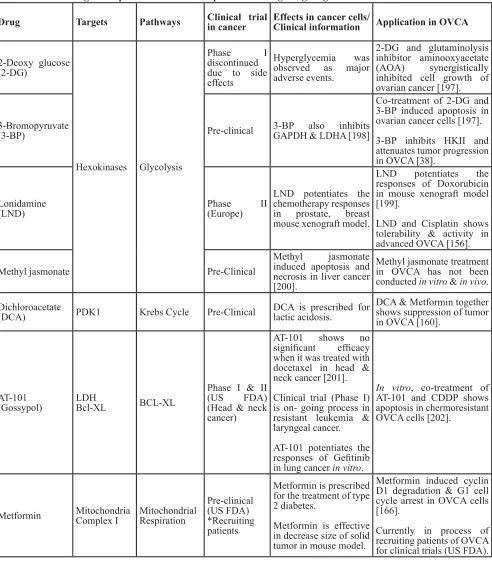
Table 1: Current drug development & clinical trial process for drugs targeting metabolism
Drug
Targets
Pathways
Clinical trial
in cancer
Effects in cancer cells/
Clinical information
Application in OVCA
2-Deoxy glucose
(2-DG)
Hexokinases Glycolysis
Phase
I
discontinued
due to side
effects
Hyperglycemia
was
observed as major
adverse events.
2-DG and glutaminolysis
inhibitor aminooxyacetate
(AOA)
synergistically
inhibited cell growth of
ovarian cancer [
197].
3-Bromopyruvate
(3-BP)
Pre-clinical
3-BP also inhibits
GAPDH & LDHA [19
8]
Co-treatment of 2-DG and
3-BP induced apoptosis in
ovarian cancer cells [
197].
3-BP inhibits HKII and
attenuates tumor progression
in OVCA [38].
Lonidamine
(LND)
Phase
(Europe)
II
LND potentiates the
chemotherapy responses
in prostate, breast
mouse xenograft model.
LND potentiates the
responses of Doxorubicin
in mouse xenograft model
[199].
LND and Cisplatin shows
tolerability & activity in
advanced OVCA [156].
Methyl jasmonate
Pre-Clinical
Methyl
jasmonate
induced apoptosis and
necrosis in liver cancer
[200].
Methyl jasmonate treatment
in OVCA has not been
conducted
in vitro & in vivo
.
Dichloroacetate
(DCA)
PDK1
Krebs Cycle
Pre-Clinical
DCA is prescribed for
lactic acidosis.
DCA & Metformin together
shows suppression of tumor
in OVCA [160].
AT-101
(Gossypol)
LDH
Bcl-XL
BCL-XL
Phase I & II
(US FDA)
(Head & neck
cancer)
AT-101 shows no
significant
efficacy
when it was treated with
docetaxel in head &
neck cancer [
201].
Clinical trial (Phase I)
is on- going process in
resistant leukemia &
laryngeal cancer.
AT-101 potentiates the
responses of Gefitinib
in lung cancer
in vitro
.
In vitro, co-treatment of
AT-101 and CDDP shows
apoptosis in chermoresistant
OVCA cells [202].
Metformin
Mitochondria
Complex I
Mitochondrial
Respiration
Pre-clinical
(US FDA)
*Recruiting
patients
Metformin is prescribed
for the treatment of type
2 diabetes.
Metformin is effective
in decrease size of solid
tumor in mouse model.
Metformin induced cyclin
D1 degradation & G1 cell
cycle arrest in OVCA cells
[166].
such as 3-Bromo pyruvate and methyl jasmonate, which
are known to specifically detach HKII from VDAC of
mitochondria, showed anti-neoplastic effects in vitro
and
in vivo mouse tumor models [
154, 155]. These drugs have
been shown to facilitate mitochondria-mediated apoptosis,
whereas their targets cover a broad spectrum, including
inhibition of glyceraldehyde -3-phosphate dehydrogenase
(GAPDH) and LDH-A.
LND (
indazole-3-carboxylic acid), inhibits HKII
and aerobic glycolysis in hypoxic conditions. Since its
combined therapy with temozolomide showed anti-tumor
effects in brain cancer, clinical trials for combinational
approaches are underway [
63
]. In advanced OVCA, LND
has been shown to be active and tolerable in phase II
clinical trial when combined with CDDP and Paclitaxel
[156
]. Though the results are promising, the possible
application of LND in treating chemoresistant OVCA
requires further investigation.
3-(3-Pyrinidyl)-1-(4-Pyrinidyl)-2-propane-1-one
(3-PO) inhibits PKFB3 family of enzymes which regulate
F-2,6-BP and the activity of PFK1, a rate-limiting step of
glycolysis. 3-PO shows a suppressive effect in glycolytic
flux and growth of cancer cells in vitro
and
in vivo
[
157
].
3-PO and its derivatives are currently in clinical trials for
their efficacy and safety.
Dichloroacetate (DCA) was originally used for
hereditary lactic acidosis [
158,
159]; however, DCA
is also considered as an anti-cancer drug that targets
metabolism. DCA inhibits PDK1, thereby stimulating
the activity of PDH, and shifting cells from glycolysis
and lactate product to mitochondrial respiration. DCA
treatment induced apoptosis in endometrioid cancer [
158].
Combined treatment with DCA and Metformin showed
a synergistic effect in the suppression of OVCA [
160].
Also, DCA treatment with the anti-angiogenesis agent,
bevacizumab (Avastin)
, enhanced anti-tumor effects
in brain cancer through reversing hypoxic adaptation
[161
]. Considering that bevacizumab was recently
approved (2016) for recurrent platinum-sensitive OVCA,
concomitant use of bevacizumab with DCA may broaden
its scope to chemoresistant OVCA [
161
]. Clinically, this
drug has recently shown successful efficacy as durable
remission for four years in glioblastoma and resistant
non-Hodgkin lymphoma, but further clinical studies need to be
conducted in other solid tumors in combinational therapy
[162, 163].
In addition, repurposing Metformin, the first-line
medication for type 2 diabetes, has gained immense
interest as potential cancer treatments [
164
]. Metformin
inhibits mitochondrial complex I and targeting
mitochondrial Bcl-2 family of anti-apoptotic proteins
in cancers. Metformin has also been shown to directly
inhibit HKI and HKII by mimicking of its product,
G-6-P as a competitive inhibitor [
165
]. Accumulating
evidence supports its anti-tumor activity [159
, 160, 166,
167]. Metformin antagonizes proliferation of cancer
cells by suppressing flow of glucose- and
glutamine-derived metabolic intermediates, leading to decreased
citrate production and lipid biosynthesis [
168]. Recent
research demonstrates that its anti-tumor activity is
increased when Metformin is used along with p53
stabilizers [167]. Currently, Metformin and combined
chemotherapy (Cisplatin and Carboplatin) are in initial
phase (recruiting patients) of clinical trial in multiple solid
tumors, including OVCA [
164
], worldwide (USA, UK,
Netherland, Australia, and Norway) [
16,
169
].
AT-101 (Gossypol) is used as a Bcl-xL inhibitor
and is also shown to target lactate dehydrogenase. It is
currently in phase I clinical trial as combined therapy
with other apoptosis-inducing chemotherapy agents
(Docetaxel, Cisplatin, and Carboplatin) in relapsed
leukemia and laryngeal cancer. Though Imatinib (Gleevec)
has never been in clinical trial covering the spectrum of
targeting metabolism, it has recently been showed to also
inhibit glycolysis activity [159]. Still, implementation
of pre-approved drug or broadening the indications
of current drugs targeting metabolism is an on-going
process. Based on the outcome of previous clinical
trials, specifically targeting metabolism seems justified,
but the promising synergistic anti-tumor effects with
conventional chemotherapy seem promising. As adjuvant
chemotherapy, these strategies should be implemented to
maximize synergistic effect to current chemotherapeutic
agents such as Paclitaxel and Carboplatin.
In addition to chemical inhibitors of tumor
metabolism, restriction of nutrition has been considered as
a plausible option. The ketogenic diet (low carbohydrate,
high fat and adequate protein) under certain settings may
suppress tumor growth and increase chemosensitivity
[
170
,
171]. The combination of a ketogenic diet with
hyper-oxygen conditions also showed anti-tumor effects
in mice [170]. However, chemotherapy frequently
results in muscle wasting and cachexia, and thus patients
undergoing chemotherapy are recommended to consume
elevated calories and protein. In tumor bearing mice,
high caloric food consumption increased survival, even
in the absence of chemotherapy [172]. Still, dietary
interventions in tandem with traditional therapeutics are
under development and possible side effects, such as
hypoglycemia, should be considered seriously [173].
B. Challenges & Strategies for targeting tumor
metabolism
adaptive responses by another metabolic pathway, possibly
resulting in side effects. Therefore, for the use of these
strategies, we need to proactively investigate systematic
effects of these drugs, including their toxicology and
potential side effects in the kidneys or other organs.
VI. TUMOR MICROENVIRONMENT &
IMMUNOMETABOLISM
A. The influence of tumor microenvironment on
glycolytic metabolism
Due to rapid growth and altered metabolism, the
solid tumor contains areas of hypoxia (low oxygen),
increased acidification (due to high rates of glycolysis)
and energy substrate limitation (due to increased distance
from supporting blood vessels). Tumor cells adapt
and thrive in these seemingly hostile environments by
activating multiple cell survival pathways. HIFs are
master transcription factors required for metabolic and
survival adaptations to hypoxia [174]. Active
HIF-1 is a heterodimer of an oxygen sensitive HIF-HIF-1α and
constitutively expressed HIF-1β. HIF-1α stability in
hypoxia allows for the transcription of many
HIF-dependent genes, including multiple glycolytic genes
[
175
,
176]. For example, HIF-1α promotes the expression
of pyruvate dehydrogenase kinase (PDK) 2 and PDK4
which inactivates PDH. PDH is responsible for converting
pyruvate to acetyl-CoA, which is used in TCA cycle [
138].
However, excessive PDK inactivates PDH and results in
the suppression of TCA cycle and OXPHOS activities,
shifting the generation of ATP toward glycolysis [177
].
In non-small cell lung cancer, aberrant expression of
HIF-1α stimulates HKII expression, contributing to elevated
glycolysis [178]. In turn, PKM2 function as an upstream
effector and binds to HIF-1α, promoting glycolysis
metabolism and tumorigenesis [179]. HIF-1α cooperates
with c-Myc, an oncogenic transcription factor, and
induces key metabolic genes, HK2 and PDK1 to promote
glycolysis [
136-138
]. HIF-1α increases the expression of
GLUT1, glucose uptake, and glucose phosphorylation
[146, 180].
B. Reverse Warburg effect
The Warburg effect in the tumor microenvironment
has recently been investigated from a different perspective,
called reverse Warburg effect [
181
]. In this alternative
model, epithelial cancer cells obtain energy from adjacent
stromal fibroblasts, facilitating tumor growth [
181].
These fibroblasts lose their stromal characteristics and are
transformed into wound healing cells as a feeding source
for cancer cells. In this transformed stroma, the absence
of caveolin (marker for stroma) and high glycolytic
enzyme expression is evident [
181
]. However, the specific
conditions in which reverse Warburg effect occurs and
how it affects tumorigenesis remain to be elucidated.
C. IL-6 (Cytokine)/ CXCL14 effect on glycolysis
and tumor microenvironment
In the tumor microenvironment, immunometabolism
and cytokines cooperate and play critical roles in
modulating the functions of immune cells and cancer cells
[182, 183
]. Cytokines are a family of secreted proteins
that stimulate chemotaxis and cell growth. Specific
proinflammatory cytokines are sometimes associated with
cancer progression [184
]. In OVCA patients, high level
of the pro-inflammatory cytokine interleukin-6 (IL-6) is
present in the serum and ascites, which is associated with
poor clinical outcomes [
184, 185
]. Increased IL-6 and its
receptor IL-6R induce invasion and metastasis of OVCA
through activating the down-stream JAK-STAT3 pathway
[186
]. In a colon cancer mouse model, IL-6 treatment
stimulated key glycolytic genes, including PFKFB3 and
aerobic glycolysis, whereas the anti-IL-6R antibody
decreased glycolysis [187], suggesting an important role
of IL-6 in cellular metabolism.
Small cytokine, CXCL14 is a key driver for
promoting metastasis of cancer-associated fibroblast
(CAF) cells in breast and prostate cancer [
188,
189
].
CXCL14 is elevated in tumor stromal cells and promotes
tumor cell proliferation and metastasis [
188
]. Clinically,
an elevated level of CXCL14 is also correlated with poor
prognosis [
190]. In OVCA, CXCL14 in CAF mediates
delivery of long non-coding RNA, LINC00092, a driver
of metastasis [191]. Mechanistically, CAFs with high
CXCL-14 promotes LINC00092 in OVCA. LINC00092
binds to PFKFB2, promoting cancer metastasis [191
].
Chemokine growth-regulated oncogene (Gro)1 functions
as an inducer of senescence in fibroblast, leading to
malignant transformation of ovarian epithelial cells [192
].
This suggests that Gro1 may be involved in the reverse
Warburg effect, with adjacent fibroblasts providing
nutrients to the cancer cells. Precisely how multiple
cytokines interplay in the tumor microenvironment and
regulate cellular metabolism remains to be determined.
Infiltrating T cells compete with tumor cells for
energy substrates as both cell types consume copious
energy. Incidentally, glucose deprivation of CD8
+T cells in
the tumor microenvironment decreases its cytotoxic action
on tumor cells [
11,
193]. Conversely, CD8
+T cells abolish
this effect by altering GSH and cysteine metabolism in
fibroblasts, suggesting a critical role of CD8
+T cells in
D. Influence of exosome delivery on tumor
metabolism
Exosomes are nano-sized vesicles derived from
endocytic compartments and released by multiple cell
types, including cancer cells. Accumulating evidence
suggests that purified exosomes contain functional miRNA
and small RNA, and long non-coding RNA [195
]. Still, it
remains unclear whether and how exosomes affect tumor
microenvironment and tumor metabolism.
Nonetheless, using exosomes, cancer
cell-derived miR-122 has been transferred to normal cells
in premetastatic niches, thereby suppressing glucose
uptake and utilization by down-regulating PK. Using
glucose source derived from these niche cells, cancer
cells facilitate the massive energy needs during metastatic
growth [196]. This metabolic programming of cancer
facilitates metastasis to other organs. Hence, cancer
cell-derived extracellular miR-122 can reprogram systemic
energy metabolism to facilitate disease progression and
metastasis of breast cancer [196
].
VII. SUMMARY & FUTURE
DIRECTIONS
Elevated metabolism is a key characteristic of
multiple cancers. Many key glycolytic enzymes, including
HKII, PFK, and PKM2 are elevated in tumor cells, and are
involved in anti-apoptotic and cell survival mechanisms
associated with chemoresistance. Considering that these
enzymes are regulated by oncogenes (e.g., Akt, mTOR)
and tumor suppressors (e.g., p53), it is likely that defective
control of tumor suppressors may lead to dysregulated
metabolism and growth of cancer cells. In addition,
the tumor microenvironment contributes to elevated
metabolism in cancer cells. In this context, targeting
dysregulated metabolism may be an effective strategy
to suppress tumor growth. However, recent clinical
trials have demonstrated that targeting metabolism alone
is not sufficient for cancer therapy. Instead, adjuvant
chemotherapy in combination with targeting tumor
metabolism may be the optimal therapeutic strategy,
especially in overcoming chemoresistant OVCA. With
continued emphasis on the design of specific inhibitors of
tumor metabolism, the application of combined therapies
is likely to improve anti-cancer strategies.
ACKNOWLEDGMENTS
This work was supported by grants from the
Canadian Institute of Health Research [CIHR; to BKT
(MOP 126144) and to MEH (FDN 143278)]. RBR, as
an adjunct professor, is aided by the library facilities
of McGill University. CYH is a recipient of doctoral
admission scholarship from the University of Ottawa.
DAP is a recipient of postdoctoral fellowship from
Canadian Nuclear Laboratories.
CONFLICTS OF INTEREST
The authors declare no conflicts of interest exists.
REFERENCES
1. Kurman RJ, Shih Ie M. Pathogenesis of ovarian cancer: lessons from morphology and molecular biology and their clinical implications. Int J Gynecol Pathol. 2008; 27: 151-60. doi: 10.1097/PGP.0b013e318161e4f5.
2. Vang R, Shih Ie M, Kurman RJ. Ovarian low-grade and high-grade serous carcinoma: pathogenesis, clinicopathologic and molecular biologic features, and diagnostic problems. Adv Anat Pathol. 2009; 16: 267-82. doi: 10.1097/PAP.0b013e3181b4fffa.
3. Rosen DG, Yang G, Liu G, Mercado-Uribe I, Chang B, Xiao XS, Zheng J, Xue FX, Liu J. Ovarian cancer: pathology, biology, and disease models. Front Biosci (Landmark Ed). 2009; 14: 2089-102.
4. Hennessy BT, Coleman RL, Markman M. Ovarian cancer. Lancet. 2009; 374: 1371-82. doi: 10.1016/S0140-6736(09)61338-6.
5. Galluzzi L, Senovilla L, Vitale I, Michels J, Martins I, Kepp O, Castedo M, Kroemer G. Molecular mechanisms of cisplatin resistance. Oncogene. 2012; 31: 1869-83. doi: 10.1038/onc.2011.384.
6. Fraser M, Bai T, Tsang BK. Akt promotes cisplatin resistance in human ovarian cancer cells through inhibition of p53 phosphorylation and nuclear function. Int J Cancer. 2008; 122: 534-46. doi: 10.1002/ijc.23086.
7. Moll UM, Wolff S, Speidel D, Deppert W. Transcription-independent pro-apoptotic functions of p53. Curr Opin Cell Biol. 2005; 17: 631-6. doi: 10.1016/j.ceb.2005.09.007. 8. Vousden KH, Lu X. Live or let die: the cell’s response
to p53. Nat Rev Cancer. 2002; 2: 594-604. doi: 10.1038/ nrc864.
9. Richardson RB. p53 mutations associated with aging-related rise in cancer incidence rates. Cell Cycle. 2013; 12: 2468-78. doi: 10.4161/cc.25494.
10. Marabese M, Marchini S, Marrazzo E, Mariani P, Cattaneo D, Fossati R, Compagnoni A, Signorelli M, Moll UM, Codegoni AM, Broggini M. Expression levels of p53 and p73 isoforms in stage I and stage III ovarian cancer. Eur J Cancer. 2008; 44: 131-41. doi: 10.1016/j.ejca.2007.10.011. 11. Hay N. Reprogramming glucose metabolism in cancer: can
it be exploited for cancer therapy? Nat Rev Cancer. 2016; 16: 635-49. doi: 10.1038/nrc.2016.77.
12. Warburg O. On the origin of cancer cells. Science. 1956; 123: 309-14.
requirements of cell proliferation. Science. 2009; 324: 1029-33. doi: 10.1126/science.1160809.
14. Pfeiffer T, Morley A. An evolutionary perspective on the Crabtree effect. Front Mol Biosci. 2014; 1: 17. doi: 10.3389/fmolb.2014.00017.
15. St-Pierre J, Drori S, Uldry M, Silvaggi JM, Rhee J, Jager S, Handschin C, Zheng K, Lin J, Yang W, Simon DK, Bachoo R, Spiegelman BM. Suppression of reactive oxygen species and neurodegeneration by the PGC-1 transcriptional coactivators. Cell. 2006; 127: 397-408. doi: 10.1016/j. cell.2006.09.024.
16. Andrzejewski S, Klimcakova E, Johnson RM, Tabaries S, Annis MG, McGuirk S, Northey JJ, Chenard V, Sriram U, Papadopoli DJ, Siegel PM, St-Pierre J. PGC-1alpha Promotes Breast Cancer Metastasis and Confers Bioenergetic Flexibility against Metabolic Drugs. Cell Metab. 2017; 26: 778-87 e5. doi: 10.1016/j. cmet.2017.09.006.
17. Pedersen PL. Warburg, me and Hexokinase 2: Multiple discoveries of key molecular events underlying one of cancers’ most common phenotypes, the “Warburg Effect”, i.e., elevated glycolysis in the presence of oxygen. J Bioenerg Biomembr. 2007; 39: 211-22. doi: 10.1007/ s10863-007-9094-x.
18. Zheng W, Feng Q, Liu J, Guo Y, Gao L, Li R, Xu M, Yan G, Yin Z, Zhang S, Liu S, Shan C. Inhibition of 6-phosphogluconate Dehydrogenase Reverses Cisplatin Resistance in Ovarian and Lung Cancer. Front Pharmacol. 2017; 8: 421. doi: 10.3389/fphar.2017.00421.
19. Catanzaro D, Gaude E, Orso G, Giordano C, Guzzo G, Rasola A, Ragazzi E, Caparrotta L, Frezza C, Montopoli M. Inhibition of glucose-6-phosphate dehydrogenase sensitizes cisplatin-resistant cells to death. Oncotarget. 2015; 6: 30102-14. doi: 10.18632/oncotarget.4945.
20. Zhang C, Zhang D, Cheng JX. Coherent Raman Scattering Microscopy in Biology and Medicine. Annu Rev Biomed Eng. 2015; 17: 415-45. doi: 10.1146/annurev-bioeng-071114-040554.
21. Li J, Condello S, Thomes-Pepin J, Ma X, Xia Y, Hurley TD, Matei D, Cheng JX. Lipid Desaturation Is a Metabolic Marker and Therapeutic Target of Ovarian Cancer Stem Cells. Cell Stem Cell. 2017; 20: 303-14 e5. doi: 10.1016/j. stem.2016.11.004.
22. Iida Y, Aoki K, Asakura T, Ueda K, Yanaihara N, Takakura S, Yamada K, Okamoto A, Tanaka T, Ohkawa K. Hypoxia promotes glycogen synthesis and accumulation in human ovarian clear cell carcinoma. Int J Oncol. 2012; 40: 2122-30. doi: 10.3892/ijo.2012.1406.
23. Uekuri C, Shigetomi H, Ono S, Sasaki Y, Matsuura M, Kobayashi H. Toward an understanding of the pathophysiology of clear cell carcinoma of the ovary (Review). Oncol Lett. 2013; 6: 1163-73. doi: 10.3892/ ol.2013.1550.
24. Mathupala SP, Rempel A, Pedersen PL. Glucose catabolism
in cancer cells: identification and characterization of a marked activation response of the type II hexokinase gene to hypoxic conditions. J Biol Chem. 2001; 276: 43407-12. doi: 10.1074/jbc.M108181200.
25. Mathupala SP, Ko YH, Pedersen PL. Hexokinase II: cancer’s double-edged sword acting as both facilitator and gatekeeper of malignancy when bound to mitochondria. Oncogene. 2006; 25: 4777-86. doi: 10.1038/sj.onc.1209603. 26. Annibaldi A, Widmann C. Glucose metabolism in cancer
cells. Curr Opin Clin Nutr Metab Care. 2010; 13: 466-70. doi: 10.1097/MCO.0b013e32833a5577.
27. Thorens B, Mueckler M. Glucose transporters in the 21st Century. Am J Physiol Endocrinol Metab. 2010; 298: E141-5. doi: 10.1152/ajpendo.00712.2009.
28. Younes M, Brown RW, Stephenson M, Gondo M, Cagle PT. Overexpression of Glut1 and Glut3 in stage I nonsmall cell lung carcinoma is associated with poor survival. Cancer. 1997; 80: 1046-51.
29. Lidgren A, Bergh A, Grankvist K, Rasmuson T, Ljungberg B. Glucose transporter-1 expression in renal cell carcinoma and its correlation with hypoxia inducible factor-1 alpha. BJU Int. 2008; 101: 480-4. doi: 10.1111/j.1464-410X.2007.07238.x.
30. Pizzuti L, Sergi D, Mandoj C, Antoniani B, Sperati F, Chirico A, Di Lauro L, Valle M, Garofalo A, Vizza E, Corrado G, Tomao F, Rinaldi M, et al. GLUT 1 receptor expression and circulating levels of fasting glucose in high grade serous ovarian cancer. J Cell Physiol. 2018; 233: 1396-401. doi: 10.1002/jcp.26023.
31. Gwak H, Haegeman G, Tsang BK, Song YS. Cancer-specific interruption of glucose metabolism by resveratrol is mediated through inhibition of Akt/GLUT1 axis in ovarian cancer cells. Mol Carcinog. 2014. doi: 10.1002/mc.22227. 32. Guo C, Ludvik AE, Arlotto ME, Hayes MG, Armstrong
LL, Scholtens DM, Brown CD, Newgard CB, Becker TC, Layden BT, Lowe WL, Reddy TE. Coordinated regulatory variation associated with gestational hyperglycaemia regulates expression of the novel hexokinase HKDC1. Nat Commun. 2015; 6: 6069. doi: 10.1038/ncomms7069. 33. Kabir F, Wilson JE. Mitochondrial hexokinase in brain:
coexistence of forms differing in sensitivity to solubilization by glucose-6-phosphate on the same mitochondria. Arch Biochem Biophys. 1994; 310: 410-6. doi: 10.1006/ abbi.1994.1186.
34. Roberts DJ, Miyamoto S. Hexokinase II integrates energy metabolism and cellular protection: Akting on mitochondria and TORCing to autophagy. Cell Death Differ. 2014. doi: 10.1038/cdd.2014.173.
36. Patra KC, Wang Q, Bhaskar PT, Miller L, Wang Z, Wheaton W, Chandel N, Laakso M, Muller WJ, Allen EL, Jha AK, Smolen GA, Clasquin MF, et al. Hexokinase 2 is required for tumor initiation and maintenance and its systemic deletion is therapeutic in mouse models of cancer. Cancer Cell. 2013; 24: 213-28. doi: 10.1016/j.ccr.2013.06.014. 37. Wolf A, Agnihotri S, Micallef J, Mukherjee J, Sabha N,
Cairns R, Hawkins C, Guha A. Hexokinase 2 is a key mediator of aerobic glycolysis and promotes tumor growth in human glioblastoma multiforme. J Exp Med. 2011; 208: 313-26. doi: 10.1084/jem.20101470.
38. Ha JH, Radhakrishnan R, Jayaraman M, Yan M, Ward JD, Fung KM, Moxley K, Sood AK, Isidoro C, Mukherjee P, Song YS, Dhanasekaran DN. LPA Induces Metabolic Reprogramming in Ovarian Cancer
via
a Pseudohypoxic Response. Cancer Res. 2018; 78: 1923-34. doi: 10.1158/0008-5472.CAN-17-1624.39. Zhang XY, Zhang M, Cong Q, Zhang MX, Zhang MY, Lu YY, Xu CJ. Hexokinase 2 confers resistance to cisplatin in ovarian cancer cells by enhancing cisplatin-induced autophagy. Int J Biochem Cell Biol. 2018; 95: 9-16. doi: 10.1016/j.biocel.2017.12.010.
40. Yalcin A, Clem BF, Imbert-Fernandez Y, Ozcan SC, Peker S, O’Neal J, Klarer AC, Clem AL, Telang S, Chesney J. 6-Phosphofructo-2-kinase (PFKFB3) promotes cell cycle progression and suppresses apoptosis
via
Cdk1-mediated phosphorylation of p27. Cell Death Dis. 2014; 5: e1337. doi: 10.1038/cddis.2014.292.41. Costa Leite T, Da Silva D, Guimaraes Coelho R, Zancan P, Sola-Penna M. Lactate favours the dissociation of skeletal muscle 6-phosphofructo-1-kinase tetramers down-regulating the enzyme and muscle glycolysis. Biochem J. 2007; 408: 123-30. doi: 10.1042/BJ20070687.
42. Webb BA, Forouhar F, Szu FE, Seetharaman J, Tong L, Barber DL. Structures of human phosphofructokinase-1 and atomic basis of cancer-associated mutations. Nature. 2015; 523: 111-4. doi: 10.1038/nature14405.
43. Atsumi T, Chesney J, Metz C, Leng L, Donnelly S, Makita Z, Mitchell R, Bucala R. High expression of inducible 6-phosphofructo-2-kinase/fructose-2,6-bisphosphatase (iPFK-2; PFKFB3) in human cancers. Cancer Res. 2002; 62: 5881-7.
44. Taylor C, Mannion D, Miranda F, Karaminejadranjbar M, Herrero-Gonzalez S, Hellner K, Zheng Y, Bartholomeusz G, Bast RC, Jr., Ahmed AA. Loss of PFKFB4 induces cell death in mitotically arrested ovarian cancer cells. Oncotarget. 2017; 8: 17960-80. doi: 10.18632/ oncotarget.14910.
45. Israelsen WJ, Vander Heiden MG. Pyruvate kinase: Function, regulation and role in cancer. Semin Cell Dev Biol. 2015; 43: 43-51. doi: 10.1016/j.semcdb.2015.08.004. 46. Gao X, Wang H, Yang JJ, Liu X, Liu ZR. Pyruvate kinase
M2 regulates gene transcription by acting as a protein kinase. Mol Cell. 2012; 45: 598-609. doi: 10.1016/j. molcel.2012.01.001.
47. Christofk HR, Vander Heiden MG, Harris MH, Ramanathan A, Gerszten RE, Wei R, Fleming MD, Schreiber SL, Cantley LC. The M2 splice isoform of pyruvate kinase is important for cancer metabolism and tumour growth. Nature. 2008; 452: 230-3. doi: 10.1038/nature06734. 48. Chao TK, Huang TS, Liao YP, Huang RL, Su PH, Shen
HY, Lai HC, Wang YC. Pyruvate kinase M2 is a poor prognostic marker of and a therapeutic target in ovarian cancer. PLoS One. 2017; 12: e0182166. doi: 10.1371/ journal.pone.0182166.
49. Wang YH, Israelsen WJ, Lee D, Yu VWC, Jeanson NT, Clish CB, Cantley LC, Vander Heiden MG, Scadden DT. Cell-state-specific metabolic dependency in hematopoiesis and leukemogenesis. Cell. 2014; 158: 1309-23. doi: 10.1016/j.cell.2014.07.048.
50. Kobierzycki C, Piotrowska A, Latkowski K, Zabel M, Nowak-Markwitz E, Spaczynski M, Kedzia W, Pula B, Podhorska-Okolow M, Dziegiel P. Correlation of Pyruvate Kinase M2 Expression with Clinicopathological Data in Ovarian Cancer. Anticancer Res. 2018; 38: 295-300. doi: 10.21873/anticanres.12221.
51. Vousden KH, Ryan KM. p53 and metabolism. Nat Rev Cancer. 2009; 9: 691-700. doi: 10.1038/nrc2715.
52. Xie H, Valera VA, Merino MJ, Amato AM, Signoretti S, Linehan WM, Sukhatme VP, Seth P. LDH-A inhibition, a therapeutic strategy for treatment of hereditary leiomyomatosis and renal cell cancer. Mol Cancer Ther. 2009; 8: 626-35. doi: 10.1158/1535-7163.MCT-08-1049. 53. Feron O. Pyruvate into lactate and back: from the Warburg
effect to symbiotic energy fuel exchange in cancer cells. Radiother Oncol. 2009; 92: 329-33. doi: 10.1016/j. radonc.2009.06.025.
54. Sonveaux P, Vegran F, Schroeder T, Wergin MC, Verrax J, Rabbani ZN, De Saedeleer CJ, Kennedy KM, Diepart C, Jordan BF, Kelley MJ, Gallez B, Wahl ML, et al. Targeting lactate-fueled respiration selectively kills hypoxic tumor cells in mice. J Clin Invest. 2008; 118: 3930-42. doi: 10.1172/JCI36843.
55. Gatenby RA, Gillies RJ. Why do cancers have high aerobic glycolysis? Nat Rev Cancer. 2004; 4: 891-9. doi: 10.1038/ nrc1478.
56. Gatenby RA, Gawlinski ET, Gmitro AF, Kaylor B, Gillies RJ. Acid-mediated tumor invasion: a multidisciplinary study. Cancer Res. 2006; 66: 5216-23. doi: 10.1158/0008-5472.CAN-05-4193.
57. Schneider D, Halperin R, Langer R, Bukovsky I, Herman A. Peritoneal fluid lactate dehydrogenase in ovarian cancer. Gynecol Oncol. 1997; 66: 399-404. doi: 10.1006/ gyno.1997.4792.
58. Boran N, Kayikcioglu F, Yalvac S, Tulunay G, Ekinci U, Kose MF. Significance of serum and peritoneal fluid lactate dehydrogenase levels in ovarian cancer. Gynecol Obstet Invest. 2000; 49: 272-4. doi: 10.1159/000010258.