Journal of Engineering Sciences, Volume 6, Issue 2 (2019), pp. C 1–C 5 C 1 JOURNAL OF ENGINEERING SCIENCES
ЖУРНАЛ ІНЖЕНЕРНИХ НАУК
ЖУРНАЛ ИНЖЕНЕРНЫХ НАУК
Web site: http://jes.sumdu.edu.ua DOI: 10.21272/jes.2019.6(2).c1 Volume 6, Issue 2 (2019)
Biosynthesis of Silver Nanoparticles Extracted Using Proteus
Shameran J. S.1*, Sewgil S. A.2, Awara Kh. S.31
Department of Chemistry, Koya University, D. Mitterrand Blvd., Koya, Iraq;
2
Hawler Medical University, 100 M St.; PO Box 178, Erbil, Iraq;
3
Department of Biology, Koya University, D. Mitterrand Blvd., Koya, Iraq
Article info:
Paper received:
The final version of the paper received: Paper accepted online:
January 29, 2019 April 7, 2019 April 12, 2019
*Corresponding Author’s Address:
[email protected]
Abstract. This study is focused on the evaluation of dependable and eco-friendly methods for the synthesis of metal nanoparticles is a significant step in the area of application of nanotechnology. One of the alternatives to obtain this purpose is to use natural techniques such as biological approach. Here, we examine biosynthesis of metallic na-noparticles using extract Proteus sp. the metal nana-noparticles were successfully synthesized via reduction of silver sul-fate employed extracted cell of bacterium Proteus sp. Nevertheless, the extracellular acts as a reducing agent to con-vert silver ion from its aqueous solution and the synthetic were formed within 2 hrs. On the other hand, scanning electron microscopy (SEM) which describes the surface morphology of bio-reduction of Ag-nanoparticles demon-strated that the spherical nature occurred through the bio-synthesis process and the particles are mostly circular and irregular in shape, UV-visible exhibit a peak at 423 nm corresponding to the plasmon of silver nanoparticle and XRD pattern was taken and presented that all peaks were indexed by hexagonal wurtzite phase (PIXcel 1D). In spite of that, the band gap energy measured (2.93 eV) and suggested strong scattering of the X-ray in the crystalline phase. Finally, we concluded that this study offers the remarkable report that biological synthetic of metal nanoparticle is helpful to avoid the negative influence of physical and chemical process that is inappropriate for medical applica-tions.
Keywords: bandgap, Proteus, bio-reduction, metallic nanoparticle.
1
Introduction
Noble metallic nanoparticles have now become the target of focused study. It is known that the chemical methods use corrosive chemicals to the synthesis of na-noparticles. In addition, the need in this time is the devel-opment of methods for the synthesis of nanoparticles by eco-friendly benign methods. Researchers in this field are eagerly looking into bio-synthesis for non-toxic systems. The biological process of the microorganism and bacte-rium origin have suggested eco-friendly methods for the synthesis of nanoparticles [1, 3]. However, the fabrica-tion, characterizafabrica-tion, and application of biologically synthesized nanomaterials have become a significant section of nanotechnology Bio-motivate techniques ex-tremely lead to the synthesis of nanostructures that are uniform in the shape and size. The demands of bio-synthesis of nanoparticles were started as the chemical and physical processes were been costly [2]. Many re-searches confirmed that the biosynthetic of silver nano-particles using via chemical process produce some un-wanted materials which absorbed on the surface of the
nanoparticles may have hostile effects in medical applica-tions. Thus, many of the latest antibacterial agents devel-oped in the last decades; none of them has been achieved its activity against multi-drug resistant bacteria [5, 7]. Newly, nanotechnology has very remarkable in the phar-maceutical and biomedical field as alternative antimicro-bial agent design in the view of the fact that renovation the occurrence and infective diseases of antibiotic-resistant strains, especially within gram-negative bacteria. Also, there is an increasing concern for silver nanoparti-cles on account of the antimicrobial properties [14]. Sil-ver is a powerful inorganic antimicrobial agent, safe and non-toxic that is capable of killing about 600 types of diseases [11].
2
Literature Review
C 2 MANUFACTURING ENGINEERING: Materials Science amount of dosage [3]. Definitely, in the case of silver
nanoparticles, the broad spectrum antimicrobial activity enhances their use in biomedical applications, food pro-duction, cosmetics, clothing, numerous household prod-ucts, and water and air purification [2, 3, 9]. Synthesis of nanoparticles via biosynthetic process provided non-toxic, eco-friendly and economic through an alternative to the various chemical and physical methods. Microbial such as yeasts, mold fungi, and bacteria are mostly pre-ferred for nanoparticles biosynthetic due to their rapid rate of growth, ease of cultivation and their ability to grow at obtainable conditions of pressure, pH and tem-perature [5]. In a previous study, pointed out that bacteri-al sp. have various ranges of capability to adsorb heavy metals and produce nanoparticles during detoxification methods [6]. Designation of nanoparticles with suitable shape and size disparity is one of the great challenges of current nanotechnology [10].
3
Research Methodology
3.1
Proteus sp.
Proteus is included under the Enterobacteriaceae and is gram-negative, a rod shape, capsulated, motile, non-lactose fermenting, swarm across the surface of blood agar [16, 17]. It is one of the most common bacteria in soil and water containing decaying organic matter of animal origin and usually occurs in large numbers in sewage.
3.2
Experiments
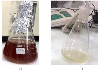
Proteus sp. was obtained at the Department of Medical Microbiology of Koya University. The bacterial stock cultures were maintained on nutrient agar slants at 4 °C. Gram staining technique for the bacterial sample has been conducted to confirm Proteus sp. Fresh bacterial sample inoculated into 200 ml of nutrient broth and incubated in a shaker incubator at 37 °C for 24 hrs. To obtain the bio-mass the culture medium centrifuged at 5 000 rpm for 15 min, then washed many times with double distilled water to obtain a wet amount of the biomass (cells) [22]. The collected cells digested in 100 mL double distilled water for 24 hrs, the biomass was separated via 0.15– 0.21 mm using Durapore membrane and the resulted filtrate was extracted from the cell. The final solution was light yellow and used for the reduction of silver sulfate (0.0006M). The converted time (Ag+ ions to Ag0) was 2 hrs to get brownish colloids. Further, the batch experi-ment carried out in bright condition (Figure 1).
a b
Figure 1 – Colloids of silver nanoparticles and the extracted cells filtrate of Proteus sp.
4
Results
Proteus, both in mixed and pure cultures, has been found to be associated with a variety of pathological con-ditions. Pathogens found mainly in urinary tract infection or commensals found in the normal intestine and sewage [17]. Proteus species are opportunistic pathogens found with varying frequencies in the normal intestinal flora and differ from another group of Enterobacteriaceae in the production of very potent Urease which aids their rapid identification [R2004]. As shown in Figure 2 the bacteria are gram-negative, bacilli shaped [18].
Figure 2 – Gram stain of Proteus sp.
UV–visible spectroscopy measurement was accom-plished by utilizing a double-beam spectrophotometer NORAN operated and scans in the range of 300–700 nm at a resolution of 2.0 nm [15]. The photo-absorption abil-ity of the Ag- nanoparticle was detected by the spectrum as validated in Figure 3. The Ag-nanoparticles exhibited strong absorption at a wavelength of 423 nm.
Figure 3 – Spectrum of Ag colloids measurement
Nevertheless, the band gap energy of the Ag-nanoparticles measured by the following formula:
, 1240
g g
E
(1)
Where, λgis the wavelength (Figure 4), validated that the high ability of Ag-nanoparticles to absorb light by recorded form the measurement of the band gap energy which is Eg = 2.93 eV of the biosynthesis Ag-colloids
Journal of Engineering Sciences, Volume 6, Issue 2 (2019), pp. C 1–C 5 C 3 Figure 4- Band gap plot measurement of Ag-nanoparticles
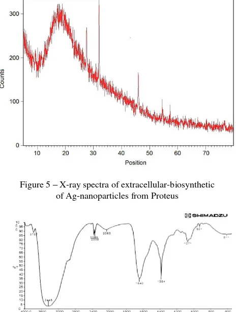
Figure 5 proves the crystalline nature of Ag-nanoparticles using X-ray spectra. However, the diffrac-tion patterns at the values 38.011, 44.113, 45.293 and 54.483 elucidated the reflections of metal silver [19]. Besides with the four peaks above some other unassigned peaks were also observed at 26.540, 30.010, 32.670, 41.713 54.394 and 59.270. Finally, the high intensity of these peaks confirmed strong scattering of the X-ray in the crystalline phase [20].
The FT-IR spectra of Ag-nanoparticles synthesized from extracellular of Proteus are given in Figure 6. The measurements were achieved to characterize the possible bio-molecules responsible for capping and effective stabi-lization of the Ag-nanoparticles biosynthesized by ex-tracted Proteus which indicate peaks at 3 445, тв. 3 787 cm–1 assigned to stretching aldehyde C-H stretch-ing and O-H respectively. The peaks 2 338, 2 063, and 2 359 cm–1 corresponds to C-N stretching of amine [15]. This proposed that the biological molecules might be possibly performed functions of stabilization and for-mation of Ag-nanoparticles in the aqueous media [12, 21].
Surface morphology of biosynthetic of Ag-nanoparticles (Figure 7) clearly demonstrated the pres-ence of nanoparticles in both dispersed and aggregated form. The size diameter of the Ag-nanoparticles has been noticed to lie between 20 to 40 nm and the shapes were indicated as spherical. Similarly, in a size range of 30– 50 nm was reported elsewhere [12, 18]. Also, [22] report-ed that morphology analysis (SEM) of Ag-nanoparticles synthesized from a mushroom revealed the spherical nature of Ag-nanoparticles and size distribution in a range of 40 nm [13].
Figure 5 – X-ray spectra of extracellular-biosynthetic of Ag-nanoparticles from Proteus
Figure 6 – FT-IR spectra of bio-reduction and formation of Ag-nanoparticles
Figure 7 – Images of surface morphology of biosynthetic Ag-nanoparticles in both form
5
Conclusions
The bio-reduction of silver ions has been successful-ly occurred through biosynthetic extracellular microor-ganisms. Here, we demonstrate that materials released from intera-cell and cell wall of Proteus. The expected mechanism for the formation of Ag-nanoparticles includes reduces polysuccerides and
C 4 MANUFACTURING ENGINEERING: Materials Science
References
1. Pliatsuk, L. D., Chernysh, Y. Y., Ablieieva, I. Y., Kozii, I. S., Balintova, M., Matiash, Y. O. (2018). Sulfur utilization in the sys-tems of biological wastewater denitrification. Journal of Engineering Sciences, Vol. 5(1), pp. H7–H15, https://doi.org/10.21272/jes.2018.5(1).h2
2. Ghosh, S., Jagtap, S., More, P., Shete, U. J., Maheshwari, N. O., Rao, S. J., Pal, J. K. (2015). Dioscorea bulbifera mediated syn-thesis of novel Au core Ag shell nanoparticles with potent antibiofilm and antileishmanial activity. Journal of Nanomaterials, Vol. 16(1), pp. 161.
3. Salih, S. J., Smail, A. K. (2016). Synthesis, characterization and evaluation of antibacterial efficacy of zinc oxide nanoparticles.
Pharmaceutical and Biological Evaluations, Vol. 3(3), pp. 327–333.
4. Saravanan, M., Barik, S. K., MubarakAli, D., Prakash, P., Pugazhendhi, A. (2018). Synthesis of silver nanoparticles from Bacil-lus brevis (NCIM 2533) and their antibacterial activity against pathogenic bacteria. Microbial Pathogenesis, Vol. 116, pp. 221– 226.
5. Sarsar, V., Selwal, M. K., Selwal, K. K. (2015). Biofabrication, characterization and antibacterial efficacy of extracellular silver nanoparticles using novel fungal strain of Penicillium atramentosum KM. Journal of Saudi Chemical Society, Vol. 19(6), pp. 682–688.
6. Fariq, A., Khan, T., Yasmin, A. (2017). Microbial synthesis of nanoparticles and their potential applications in biomedicine.
Journal of Applied Biomedicine, Vol. 15(4), pp. 241–248.
7. Balashanmugam, P., Santhosh, S., Giyaullah, H., Balakumaran, M. D., Kalaichelvan, P. T. (2013). Mycosynthesis, characteriza-tion and antibacterial activity of silver nanoparticles from Microporusxanthopus: a macro mushroom. International Journal of Innovative Research in Science, Engineering and Technology, Vol. 2(11), pp. 1–9.
8. Kalpana, D., Lee, Y. S. (2013). Synthesis and characterization of bactericidal silver nanoparticles using cultural filtrate of simu-lated microgravity grown Klebsiella pneumoniae. Enzyme and Microbial Technology, Vol. 52(3), pp. 151–156.
9. Ali, D. M., Sasikala, M., Gunasekaran, M., Thajuddin, N. (2011). Biosynthesis and characterization of silver nanoparticles using marine cyanobacterium, Oscillatoria willei NTDM01. Digest Journal of Nanomaterials and Biostructures, Vol. 6(2), pp. 385– 390.
10. Vahabi, K., Mansoori, G. A., Karimi, S. (2011). Biosynthesis of silver nanoparticles by fungus Trichoderma reesei (a route for large-scale production of AgNPs). Insciences Jornal, Vol. 1(1), pp. 65–79.
11. Hosseini, M. R., Sarvi, M. N. (2015). Recent achievements in the microbial synthesis of semiconductor metal sulfide nanoparti-cles. Materials Science in Semiconductor Processing, Vol. 40, pp. 293–301.
12. Salih, S. J., Rashid, B. Z. (2015). Cranberry stem as an efficient adsorbent and eco-friendly for removal of toxic dyes from in-dustrial wastewater. Physico Studies. International Journal of Pharmaceutical Chemistry, Vol. 5(6), pp. 207–217.
13. Usman, A. R., Kuzyakov, Y., Lorenz, K., Stahr, K. (2006). Remediation of a soil contaminated with heavy metals by immobiliz-ing compounds. Journal of Plant Nutrition and Soil Science, Vol. 169(2), pp. 205–212.
14. Yanovska, H. O., Bolshanina, S. B., Kuznetsov, V. M. (2017). Formation of hydroxyapatite coatings with addition of chitosan from aqueous solutions by thermal substrate method. Journal of Engineering Sciences, Vol. 4(2), 2017. https://doi.org/10.21272/jes.2017.4(2).f1
15. Salih, S. J., Anwer, S. S., Faraj, R. H. (2017). A biosorption of Mercury from wastewater using isolated Aspergillus sp. Modi-fied 1, 10-Phenanthroline: Hill isotherm model. Science Journal of University of Zakho, Vol. 5(4), pp. 288–295.
16. Ray, C. G., Ryan, K. J. (2004). Sherris Medical Microbiology: An Introduction to Infectious Diseases. McGraw-Hill.
17. Mobley, H. L., Belas, R., Lockatell, V., Chippendale, G., Trifillis, A. L., Johnson, D. E., Warren, J. W. (1996). Construction of a flagellum-negative mutant of Proteus mirabilis: effect on internalization by human renal epithelial cells and virulence in a mouse model of ascending urinary tract infection. Infection and Immunity, Vol. 64(12), pp. 5332–5340.
18. Jayaseelan, C., Rahuman, A. A., Kirthi, A. V., Marimuthu, S., Santhoshkumar, T., Bagavan, A., Rao, K. B. (2012). Novel mi-crobial route to synthesize ZnO nanoparticles using Aeromonas hydrophila and their activity against pathogenic bacteria and fungi. Spectrochimica Acta Part A: Molecular and Biomolecular Spectroscopy, Vol. 90, pp. 78–84.
19. Singh, P., Kim, Y. J., Zhang, D., Yang, D. C. (2016). Biological synthesis of nanoparticles from plants and microorganisms.
Trends in Biotechnology, Vol. 34(7), pp. 588–599.
20. Hussain, I., Singh, N. B., Singh, A., Singh, H., Singh, S. C. (2016). Green synthesis of nanoparticles and its potential applica-tion. Biotechnology Letters, Vol. 38(4), pp. 545–560.
21. Agarwal, H., Kumar, S. V., Rajeshkumar, S. (2017). A review on green synthesis of zinc oxide nanoparticles – An eco-friendly approach. Resource-Efficient Technologies, Vol. 3(4), pp. 406–413.
Journal of Engineering Sciences, Volume 6, Issue 2 (2019), pp. C 1–C 5 C 5
УДК 546.57:54.05
Біосинтез наночастинок срібла, екстрагованих із застосуванням
Proteus
ШамеранЙ. С.1, Севґіл С. А.2, Авара Х. С.2
1 Університет м. Койа, б
-р Д. Міттерана, м. Койа, Ірак;
2 Медичний університет ім. Хоулера, вул. 100 М, 178, м. Ербіл, Ірак
Анотація.Запропонованедослідження спрямоване на оцінювання надійних і екологічних методів синтезу металевих наночастинок, що дозволили зробити значний крок у сфері застосування нанотехнологій. Однією з
альтернатив для досягнення поставленої цієї мети є використання природних методів, закрема біологічного підходу. У роботі розглядається біосинтез металевих наночастинок з використанням екстракту Proteus. При
цьому металеві наночастинки успішно синтезувались за допомогою відновлення сульфату срібла, що екстрагується клітками бактерії Proteus. Тим не менш, позаклітинне середовище діє як відновник для
перетворення іонів срібла з його водного розчину, і синтез відбувається впродовж 2 год. З іншого боку,
скануюча електронна мікроскопія, що описує морфологію поверхні біоредукції наночастинок срібла, продемонструвала, що у процесі біосинтезу утворюється сферична форма, а частинки в основному круглої та неправильної форми, видимі в ультрафіолетовому спектрі частинки проявляють пік при довжині хвиль 423 нм, що відповідає плазмону наночастинок срібла,а рентгенографічна картина показала, що всі піки були
визначены гексагональною фазою сульфыда цинку. Незважаючи на це, визначено енергію 2,93еВ, а також
запропоновано сильне рентгенівське випромінювання для кристалічноїфази. У результаті зроблено висновок,
що проведене дослідження підтверджує той факт, що біосинтез металевих наночастинок дозволяє запобігти негативного впливу фізичних і хімічних процесів , що відбуваються у засобах медичного застосування.