COMPARATIVE ANALYSIS OF TARGETING KEY GENES IN STAT PATHWAY IN K562 CELLS
Vinod Rajendran
Sir H N Medical Research Society, Padmashri Gordhanbapa Chowk,
A R T I C L E I N F O
INTRODUCTION
Chronic myeloid leukemia (CML) or chronic myelogenous leukemia is a myeloproliferative neoplasm which has an incidence rate of 1–2 cases per 100000 adults per year. It accounts for approximately 15% of the newly diagnosed cases of leukemia with a 5-year survival rate of 67.6% (American Cancer Society, 2017).
The characteristic feature of CML is the presence of the Philadelphia (Ph) chromosome which results from a reciprocal chromosomal translocation at the t(9;22) position. This aberration has been observed to be present in the bone marrow (BM) in 95% cases of CML (Gong Z et al
translocation t(9;22) occurs when the Abelson (
tyrosine kinase (TK) gene located on chromosome 9, juxtaposes to the (BCR) gene, a breakpoint cluster region located on chromosome 22, leading to an increased TK activi (Quintás-Cardama A and Cortes J, 2009).
Uncontrolled cancer cell proliferation along with improved resistance to apoptosis, and alteration of cell adhesion properties are mechanisms that attribute to BCR
cells and have been implicated in the pathogenesis of CML. The disease is known to progress in three phases, the chronic,
International Journal of Current Advanced Research
ISSN: O: 2319-6475, ISSN: P: 2319-6505,
Available Online at www.journalijcar.org
Volume 7; Issue 9(D); September 2018
DOI: http://dx.doi.org/10.24327/ijcar.2018
Copyright©2018 Vinod Rajendran and Sudha S. Deo
which permits unrestricted use, distribution, and reproduction in any medium, provided the original work is properly cited. Article History:
Received 4th June, 2018
Received in revised form 25th July, 2018 Accepted 18th August, 2018
Published online 28th September, 2018
Key words:
BCR-ABL, STAT5A, STAT5B, BCL-XL, BCL2L1, siRNA, CML, K562, gene silencing, apoptosis.
*Corresponding author: Vinod Rajendran
Sir H N Medical Research Society, Padmashri Gordhanbapa Chowk, Raja Rammohan Roy Road, Mumbai
COMPARATIVE ANALYSIS OF TARGETING KEY GENES IN STAT PATHWAY IN K562 CELLS
Vinod Rajendran and Sudha S. Deo
, Padmashri Gordhanbapa Chowk, Raja Rammohan Roy Road, Mumbai
A B S T R A C T
BCR-ABL is one of the main mutation found in Chronic Myeloid Leukemia (CML). It is
said to activate STAT5 and in turn activate BCL-XL (BCL2L1
silenced these genes separately but a comparative analysis among them has never been done to find out the best target to induce apoptosis.
In the present study, the effects of downregulating BCR
XL in human chronic myeloid leukemia cell line (K562 cells) were investigated through RNA interference (RNAi) and the proliferation inhibition and apoptosis induction were analyzed thereafter. K562 cells were transfected with various concentrations of siRNA and the expressions of aforesaid genes were determined by reverse transcription
polymerase chain reaction (RT-PCR) and Western blot analysis. K562 cell pro and apoptosis were analyzed using MTT and flow cytometry respectively.
RT-PCR and western blotting results post siRNA transfection confirmed the targeted gene suppression and protein reduction in the cell. The cell proliferation assay and apopto assay revealed that silencing BCL-XL had the highest killing effect on K562 cells as compared to knocking down BCR-ABL, STAT5A and
expression profile study validated the formerly known direct dependency of
BCR-ABL via STAT5 in CML.
myeloid leukemia (CML) or chronic myelogenous leukemia is a myeloproliferative neoplasm which has an 2 cases per 100000 adults per year. It accounts for approximately 15% of the newly diagnosed cases ate of 67.6% (American
The characteristic feature of CML is the presence of the Philadelphia (Ph) chromosome which results from a reciprocal chromosomal translocation at the t(9;22) position. This aberration has been observed to be present in the bone marrow et al., 2017). The translocation t(9;22) occurs when the Abelson (ABL1), a tyrosine kinase (TK) gene located on chromosome 9, ) gene, a breakpoint cluster region located on chromosome 22, leading to an increased TK activity
Uncontrolled cancer cell proliferation along with improved resistance to apoptosis, and alteration of cell adhesion BCR-ABL-positive he pathogenesis of CML. The disease is known to progress in three phases, the chronic,
accelerated and blast crisis phases, with increased accumulation of genetic aberrations at every phase (Baccarani M et al., 2012).
BCR-ABL gene have been shown to participate in many pathways like MAPK, RAS,
(Kumar H et al., 2015). One of the important pathways in CML is the JAK/STAT pathway which includes
a downstream component in the pathway. Studies performed in vitro have shown that BCR-ABL can directly instigate the tyrosine phosphorylation along with dim
bypassing JAK2, followed by translocation of STAT5 dimers to the nucleus, wherein they trigger the transcription of pro survival genes (Hantschel O et al
In an earlier study, a correlation was observed between STAT5 levels with TKI resistance and disease progression in patient cancer cells. It was then shown that increased levels of STAT5 caused reduced TKI-mediated cytotoxicity, and decrease of STAT5 levels correlated with enhanced TKI
Although this may suggest that STAT5 inhibition may serve as a potent target for TKI resistance in patients with advanced disease, this strategy may also prove functional for eliminating CML cells residing in the protective BM microenvironment (Warsch W et al., 2011).
B-cell lymphoma (BCL) - 2 protein family members are important regulators in the apoptotic pathway associated with individual components, such as BCL
apoptosis or other factors, and BAX and BAD which can
International Journal of Current Advanced Research
6505, Impact Factor: 6.614
www.journalijcar.org
2018; Page No. 15462-15467
//dx.doi.org/10.24327/ijcar.2018.15467.2823
Vinod Rajendran and Sudha S. Deo. This is an open access article distributed under the Creative Commons Attribution License, which permits unrestricted use, distribution, and reproduction in any medium, provided the original work is properly cited.
Sir H N Medical Research Society, Padmashri Gordhanbapa Mumbai-400004
COMPARATIVE ANALYSIS OF TARGETING KEY GENES IN STAT PATHWAY IN K562 CELLS
Raja Rammohan Roy Road, Mumbai – 400004
is one of the main mutation found in Chronic Myeloid Leukemia (CML). It is
BCL2L1). In the past, researchers have
omparative analysis among them has never been
BCR-ABL, STAT5A, STAT5B and
BCL-in human chronic myeloid leukemia cell lBCL-ine (K562 cells) were BCL-investigated through RNA interference (RNAi) and the proliferation inhibition and apoptosis induction were analyzed thereafter. K562 cells were transfected with various concentrations of siRNA and the expressions of aforesaid genes were determined by reverse transcription - quantitative PCR) and Western blot analysis. K562 cell proliferation and apoptosis were analyzed using MTT and flow cytometry respectively.
PCR and western blotting results post siRNA transfection confirmed the targeted gene suppression and protein reduction in the cell. The cell proliferation assay and apoptosis had the highest killing effect on K562 cells as
and STAT5B. A further all four gene
expression profile study validated the formerly known direct dependency of BCL-XL on
t crisis phases, with increased accumulation of genetic aberrations at every phase (Baccarani
gene have been shown to participate in many , RAF, JUN kinase, MYC etc ., 2015). One of the important pathways in pathway which includes STAT5 gene as a downstream component in the pathway. Studies performed in ABL can directly instigate the tyrosine phosphorylation along with dimerization of STAT5, bypassing JAK2, followed by translocation of STAT5 dimers to the nucleus, wherein they trigger the transcription of
pro-et al., 2012).
In an earlier study, a correlation was observed between STAT5 h TKI resistance and disease progression in patient-cancer cells. It was then shown that increased levels of STAT5
mediated cytotoxicity, and decrease of STAT5 levels correlated with enhanced TKI-mediated killing. st that STAT5 inhibition may serve as a potent target for TKI resistance in patients with advanced disease, this strategy may also prove functional for eliminating CML cells residing in the protective BM microenvironment
2 protein family members are important regulators in the apoptotic pathway associated with individual components, such as BCL-XL which can suppress apoptosis or other factors, and BAX and BAD which can
Research Article
International Journal of Current Advanced Research Vol 7, Issue 9(D), pp 15462-15467, September 2018
promote apoptosis. BCL-XL (BCL2L1) are frequently amplified or overexpressed in numerous tumor types including CML. BCL-2, as well as a second member of this family of anti-apoptotic proteins, BCL-XL, have been suggested as BCR/ABL-regulated effector molecules (Gotlib J., et al., 2013). BCL-XL is one of the target of the signal transducer and activator of transcription 5 (STAT5) (Goff DJ et al., 2013). Furthermore, transfection of Ph+ K562 cells with a dominant-negative isoform of STAT5 led to a decrease in BCL-XL expression and subsequent apoptosis of the cells, suggesting BCL-XL as an important factor in the prevention of programmed cell death in the context of Ph+ leukemias (Horita M et al., 2000).
Tyrosine kinase inhibitor (TKI) therapy are given as a front-line treatment for all the newly diagnosed patients. This includes drugs like Imatinib, Dasatinib, Nilotinib, Ponatinib, etc. Yet, 40% of patients who fail the TKI therapy (Imatinib) are found to harbor a mutation known as T315I where threonine is substituted with isoleucine (deLavallade H, et al., 2008). The incidence of drug resistance has increased over time owing to mutations in kinase and non-kinase domain in CML. Small interfering RNA (siRNA) mediated gene silencing have been shown to be a very powerful tool which is specific and effective and thus has many therapeutic applications. In humans, several siRNAs against members of apoptotic pathway, transcription factors, tyrosine kinase signaling and other signaling pathway are available at our disposal (Landry B et al., 2015). Researchers have silenced BCR-ABL (Valencia-Serna J et al., 2013), STAT5 (Kaymaz BT et al., 2013) and BCL-XL (Bogenberger JM et al., 2014) and seen that K562 cells undergo apoptosis. But the question remains that silencing which of the following target in STAT pathway – BCR-ABL, STAT5A, STAT5B and BCL-XL, will be most potent in killing K562 cells.
In this study, a comparative analysis of silencing BCR-ABL, STAT5A, STAT5B and BCL-XL individually in human chronic myeloid leukemia cell line (K562) were evaluated through interference by respective siRNAs. First, the effect of downregulating these gene expressions in K562 cells were analyzed and the proliferation inhibition and apoptosis induction were confirmed. Secondly, comparisons were made between the efficacy of each siRNA and its ability to induce apoptosis in K562 was investigated through Annexin V / PI staining through flow cytometry. BCL-XL gene silencing proved to be best target when compared with BCR-ABL, STAT5A and STAT5B in killing K562 cells. This study provided an evidence that targeting genes in STAT pathway of CML, other than BCR-ABL, was also very effective in inducing apoptosis in cancer cells.
MATERIALS AND METHODS
Cell culturing: K562 cells were obtained from National Center
for Cell Science (Pune, India). The cells were cultured in Iscove's Modified Dulbecco's Medium (IMDM, Gibco, USA) supplemented with 10% fetal bovine serum (Gibco, USA) and antibiotics penicillin (100 kU/L) and streptomycin (100 mg/L) at 37℃ in a humidified incubator with 5% CO2.
Gene knockdown in K562: ON-TARGETplus siRNAs for
BCR-ABL, STAT5A, STAT5B and BCL-XL were from Dharmacon, USA. BCR-ABL siRNA was synthesized using the following sequence
- 5ʹ GCAGAGUUCAAAAGCCCUUdTdT 3ʹ (Scherr M et al., 2003) and the remaining siRNAs were ordered off the shelf. All of the transfections were performed by using HiPerFect transfection kit (Qaigen, USA) according to the manufacturer's instructions. Briefly, 2 x 10⁵ K562 cells were plated in per well of a 24-well plate in 100 μl serum containing IMDM. Required amount of siRNA was diluted in 100 μl serum free IMDM and 6 μl of HiPerFect transfection reagent was dissolved to the diluted siRNA for 5 minutes at room temperature. The complex (106 µl) was added to the cells, gently shaken and thoroughly incorporated. After 6 hours, 400 μl culture medium containing serum was added to the cells and incubated until further analysis. siRNA transfection efficiency was analyzed by counting positively transfected cells by SiGLO Red (Dharmacon, USA) per 100 cells under fluorescent microscope. The cells transfected with Non-targeting (NT) siRNA (Dharmacon, USA) was used as control.
Reverse Transcription - Quantitative polymerase chain
reaction (RT-qPCR): Total RNA was extracted from the cells
by NucleoSpin® RNA/Protein kit by Macherey-Nagel GmbH & Co. (Germany) and reversed transcribed into cDNA by utilizing High-Capacity cDNA Reverse Transcription Kit (Applied Biosystems, USA) according to the manufacturer's instructions. The primer sequences for qPCR were as follows - BCR-ABL forward, 5'-GTGTGAAACTCCAGACTGTC‑3' and reverse, 5'‑CAAAATCATACAGTGCAACGA‑3' (Xu C et al., 2014), STAT5A forward, 5'-GAAGCTGAACGTG-CACATGAATC‑3' and reverse, 5'‑GTAGGGACAGAGTCT TCA-CCTGG‑3' (Kaymaz BT et al., 2013), STAT5B forward, 5'-AGTTTGATTCTCAGGAA-AGAATGT‑3' and reverse, 5'‑TCCATCAACAGCTTTAGCA- GT‑3' (Kaymaz BT et al., 2013), BCL-XL forward, 5'-TGCATTGTTCCCAT-AGAGTTCCA‑3' and reverse, 5'‑CCTGAATGACCAC-CTAGAGCCTT‑3' (Changchien JJ et al., 2015) and GAPDH forward, 5'-GTCAACGGATTTGGTCGTATTG‑3' and reverse, 5'‑CATGGGTGGAATCA-TATTGGAA‑3' (Wattanapanitch M et al., 2014). Quantitative PCR was performed in StepOnePlus™ Real-Time PCR System (Applied Biosystems, USA) in triplicates using SYBR™ Select Master Mix (Applied Biosystem, USA). The reaction mixture (20µl) was initially set at 50°C for 2 minutes for UDG Activation, then at 95˚C for 2 minutes for AmpliTaq® DNA Polymerase, UP Activation and then subjected to 40 PCR cycles of 95˚C for 3 seconds and 60˚C for 30 seconds. mRNA levels were normalized to GAPDH levels.
MTT test: K562 cells were seeded in 24-well plates at a
density of 2x10⁵ cells/well and transfected with required amount of siRNAs. Cultures were incubated at 37˚C in a fully humidified atmosphere with 5% CO2. After the appropriate incubation, the wells were added with 60 µl of MTT (0.5 mg/ml) and incubated for another 4 hours. Each well was then added with 600 µl of DMSO. The absorbance was quantified at 490nm. Cell proliferation rate was calculated as the percentage of amount of absorbance of treated cells upon absorbance of control cells (cells treated with NT siRNA).
Apoptosis detection assay: The cell apoptotic rate was
minutes and washed with precooled PBS. Cells were then suspended in 1X Binding Buffer at a concentration of 1 x 10 cells/ml. 5 µl of FITC Annexin V and 5 µl PI were added to the cell suspension and incubated in dark at room temperature for 15 minutes. 400 µl of 1X Binding Buffer was added to each tube and analyzed by BD FACSAria II flow cytometry within 1 hour.
Western blot analysis: Total proteins were extracted from the
cells by using NucleoSpin® RNA/Protein kit by Macherey Nagel GmbH & Co. (Germany) following the manufacturer’s protocol. Traditional SDS-PAGE was performed and proteins were transferred to nitrocellulose membranes and then incubated with specific antibodies after blocking with 5% BSA solution. The membranes were washed with Tris
saline and Tween-20 (TBST) and incubated with HRP conjugated second antibody for 1 hour at RT. The blot was developed with SuperSignal™ West Pico PLUS Chemiluminescent Substrate, Thermo Scientific, USA and the signal was exposed to X-ray film. All antibodies were from Thermo Scientific, USA and they were used in the following dilutions – GAPDH (1:5000), BCR-ABL (1:500),
(1:1000), STAT5B (1:1000), BCL-XL (1:250) and Goat anti mouse IgG (H+L) secondary antibody HRP (1:50,000).
Statistical analysis: Results were expressed as mean ±
standard deviation (SD) of atleast three independent experiments performed in triplicate. GraphPad Prism 7 (GraphPad Software, Inc, CA, USA) was used for statistical analysis. The results were compared using students t
one-way ANOVA. Differences between values were considered significant at p<0.05.
RESULTS
Confirmation of siRNA transfection:
successful transfection of siRNA into K562 cells, we used siGLO Red siRNA at a concentration of 50 nM, 100 nM and 200 nM (nmol/l) for 24 hours. The fluorescent microscopy image showed the aggregation of siRNA near the nuclear region in K562 cells (Figure 1).
Figure 1 400X magnification of K562 transfected with 50 nM siGLO Red
siRNA. A. Phase contrast image of K562. B. siGLO siRNA seen as red dots. C. Nuclear region stained in blue by DAPI. D. Merge of all the three images.
The transfection efficiency was estimated by counting successfully transfected cells per 100 cells in the field of view. We achieved an average transfection efficiency of 69 ± 6.5%, 75 ± 5.5% and 85 ± 4.9% for 50 nM, 100 nM and 200 nM siRNA at 24 hours respectively. The average cytotoxicity of HiPerFect was found to be 9 ± 3.5% when compared with non transfected cells for 24 hours. The experiment indicated that as the concentration of siRNA was increased, the transfection efficiency improved proportionately.
Effect of siRNA on K562 cells: To explore the gene silencing
effect of siRNAs in K562 cells, we transfected each siRNA in three concentrations – 50nM, 100nM and 200nM for 24 and 48 hours. After K562 cells were transfected with siRNA, the gene minutes and washed with precooled PBS. Cells were then suspended in 1X Binding Buffer at a concentration of 1 x 10⁶
ells/ml. 5 µl of FITC Annexin V and 5 µl PI were added to the cell suspension and incubated in dark at room temperature for 15 minutes. 400 µl of 1X Binding Buffer was added to each tube and analyzed by BD FACSAria II flow cytometry
Total proteins were extracted from the cells by using NucleoSpin® RNA/Protein kit by Macherey-Nagel GmbH & Co. (Germany) following the manufacturer’s
PAGE was performed and proteins se membranes and then incubated with specific antibodies after blocking with 5% BSA solution. The membranes were washed with Tris‑buffered 20 (TBST) and incubated with HRP-conjugated second antibody for 1 hour at RT. The blot was
with SuperSignal™ West Pico PLUS Chemiluminescent Substrate, Thermo Scientific, USA and the ray film. All antibodies were from Thermo Scientific, USA and they were used in the following ABL (1:500), STAT5A XL (1:250) and Goat anti-mouse IgG (H+L) secondary antibody HRP (1:50,000).
Results were expressed as mean ± standard deviation (SD) of atleast three independent ate. GraphPad Prism 7 (GraphPad Software, Inc, CA, USA) was used for statistical analysis. The results were compared using students t-test and way ANOVA. Differences between values were
To confirm the successful transfection of siRNA into K562 cells, we used siGLO Red siRNA at a concentration of 50 nM, 100 nM and 200 nM (nmol/l) for 24 hours. The fluorescent microscopy image showed the aggregation of siRNA near the nuclear
400X magnification of K562 transfected with 50 nM siGLO Red siRNA. A. Phase contrast image of K562. B. siGLO siRNA seen as red dots. C. Nuclear region stained in blue by DAPI. D. Merge of all the three images.
The transfection efficiency was estimated by counting successfully transfected cells per 100 cells in the field of view. We achieved an average transfection efficiency of 69 ± 6.5%, 75 ± 5.5% and 85 ± 4.9% for 50 nM, 100 nM and 200 nM espectively. The average cytotoxicity of HiPerFect was found to be 9 ± 3.5% when compared with non-transfected cells for 24 hours. The experiment indicated that as the concentration of siRNA was increased, the transfection
To explore the gene silencing effect of siRNAs in K562 cells, we transfected each siRNA in 50nM, 100nM and 200nM for 24 and 48 hours. After K562 cells were transfected with siRNA, the gene
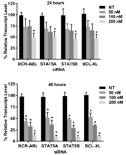
expression was detected through quantitative polymerase chain reaction (qPCR) analysis and western blot analysis. The qPCR results showed that the mRNA level of each gene were lowered in the transfected cells when compared to the non targeting (NT) siRNA transfec
significant amount of silencing observed when all four siRNAs were used only at 200nM for 24 hours individually while at 48 hours all the concentration used were found to be statistically significant when compared to their respect
p<0.05. (Figure 2)
Figure 2 Gene expression after individual gene silencing: K562 was
transfected with 50nM, 100nM and 200nM each of BCR
STAT5B and BCL-XL siRNA for 24 and 48 hours. The relative transcript level of each gene are expressed in % as compared to gene expression of NT siRNA treated K562 cells. GAPDH gene expression was used to normalize the
expression of other target genes. Values are expressed in % ± SD; n=3; * p<0.05 when each value compared with their respe
The western blot results correlated with the RT
Protein expression were seen to be reducing when 200nM of siRNA was used for 24 hours. When siRNA was used for 48 hours, protein reduction was seen in all the samples. The highest protein reduction was seen when 200nM siRNA was used for 48 hours. (Figure 3)
Figure 3 Western Blot analysis after individual gene silencing: K562 was transfected with BCR-ABL siRNA, STAT5A siRNA, STAT5B siRNA and BCL
respectively. Each siRNA was used at 50 nM, 100 nM and 200 nM concentration for 24 and 48 hours. With each sample western blotting was carried out for that specific protein
whose gene was silenced with the respective siRNA. NT1
NT siRNA for 24 hours. NT2 – K562 treated with 200 nM NT siRNA for 48 hours. 1 to 6 – K562 treated with respective siRNAs at mentioned concentration and time points.
ion was detected through quantitative polymerase chain reaction (qPCR) analysis and western blot analysis. The qPCR results showed that the mRNA level of each gene were lowered in the transfected cells when compared to the non-targeting (NT) siRNA transfected controls. There was a significant amount of silencing observed when all four siRNAs were used only at 200nM for 24 hours individually while at 48 hours all the concentration used were found to be statistically significant when compared to their respective controls with
Gene expression after individual gene silencing: K562 was transfected with 50nM, 100nM and 200nM each of BCR-ABL, STAT5A,
XL siRNA for 24 and 48 hours. The relative transcript gene are expressed in % as compared to gene expression of NT
gene expression was used to normalize the expression of other target genes. Values are expressed in % ± SD; n=3; *
p<0.05 when each value compared with their respective Control.
The western blot results correlated with the RT-PCR result. Protein expression were seen to be reducing when 200nM of siRNA was used for 24 hours. When siRNA was used for 48 hours, protein reduction was seen in all the samples. The protein reduction was seen when 200nM siRNA was
Western Blot analysis after individual gene silencing: K562 was transfected ABL siRNA, STAT5A siRNA, STAT5B siRNA and BCL-XL siRNA
was used at 50 nM, 100 nM and 200 nM concentration for 24 and 48 hours. With each sample western blotting was carried out for that specific protein whose gene was silenced with the respective siRNA. NT1 – K562 treated with 200 nM
International Journal of Current Advanced Research Vol 7, Issue 9(D), pp 15462-15467, September 2018
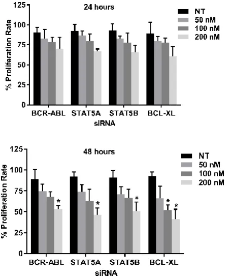
Suppression of tumor cells proliferation: The siRNA
transfected K562 were determined through MTT test to evaluate whether all the four siRNAs inhibits cell proliferation individually. In this study, 50 nM, 100 nM and 200 nM of BCR-ABL, STAT5A, STAT5B and BCL-XL siRNA were transfected into K562 cells for 24 and 48 hours. The results revealed that the cell proliferation rate of the transfected cells were lower than that of the NT siRNA transfected cells (Control). Statistically significant difference was found when BCL-XL siRNA was used at 100nM for 48 hours when compared with the control. All the results were statistically significant when 200nM of each siRNA was used for 48 hrs. Compared with that of NT siRNA transfected cells, the lowest proliferation rate for each siRNA used was observed at 200 nM for 48 hrs. (Figure 4).
Figure 4 Cell proliferation rate after individual gene silencing: MTT assay
was performed after K562 was transfected with 50nM, 100nM and 200nM each of BCR-ABL, STAT5A, STAT5B and BCL-XL siRNA for 24 and 48 hrs. Proliferation rate are expressed in % as compared to the proliferation rate in K562 cells treated with NT siRNA (Control). Values are expressed in % ±
SD; n=3; * p<0.05 when each value compared with their Control.
Apoptosis of K562 cells: In previous experiments, all the four
siRNA’s silencing could inhibit K562 cells proliferation. When each siRNA was used at 200 nM for 48 hours, the best silencing effect and the lowest proliferation rate was achieved. To investigate whether silencing these genes accelerated K562 cell apoptosis, we transfected 200 nM of each siRNA in to K562 individually and compared the rate of apoptosis with NT siRNA (200 nM) transfected cells after 48 hours. Apoptosis was analyzed using Annexin V / PI staining through flow cytometry. Compared to the control, the apoptotic cells increased highest by 51.34 ± 12.4% in BCL-XL silenced cell population and lowest by 39.35 ± 7.2% in STAT5B silenced cell population. All the results were statistically significant when compared with control cells. These results indicated that silencing BCR-ABL, STAT5A, STAT5B and BCL-XL individually induced K562 cells to undergo apoptosis. (Figure 5)
Figure 5 Apoptotic status after individual gene silencing: K562 cells were
assayed through flow cytometry by using Annexin V / PI staining after siRNA transfection for 48 hours. Representative scatter plot for: A – with 200 nM of NT siRNA, B – with 200 nM BCR-ABL siRNA, C – with 200 nM STAT5A siRNA, D – with 200 nM STAT5B siRNA, E – with 200 nM BCL-XL siRNA
has been shown. F - Graphical representation of entire data. Results are expressed in % ± SD; n=3, *p<0.05 when compared with Control.
Gene expression profile of individual gene silencing: From
the above experiments, it became evident that 200 nM of siRNA for 48 hours was better than any concentration of siRNA used for any duration. We wanted to study the effects of each siRNA on the expression of other three genes. For this purpose, 200 nM of each siRNA was transfected in to K562 and gene expression of all four genes were studied after 48 hrs. When BCR-ABL and STAT5A genes were silenced individually, the BCL-XL expression was lowered than the control sample by around 30%. Similarly, when STAT5A, STAT5B and BCL-XL gene was suppressed, the expression of BCR-ABL was lowered as compared to the control by approximately 15 - 25%. Finally, when BCR-ABL siRNA was used, the expression of STAT5A reduced by around 10%. None of the above mentioned differences were statistically significant, except when the target gene expression was determined for each of the siRNA used. (Figure 6).
Figure 6 Gene expression of all targets after individual gene silencing: K562
was transfected with 200nM each of BCR-ABL, STAT5A, STAT5B and BCL-XL siRNA for 48 hours. The relative transcript level of each gene are expressed in % as compared to gene expression of NT siRNA treated K562 cells (Control). GAPDH gene expression was used to normalize the expression
of other target genes. Values are expressed in % ± SD; n=3; * p<0.05 when each value compared with their respective Control.
DISCUSSION
Various downstream pathways are deregulated with the onset of disease including Wnt/β‐catenin pathway, PI3K/AKT/mTOR pathway and JAK/STAT pathway. JAK/STAT pathway plays a role in normal hematopoiesis but is deregulated in variety of myeloproliferative disorders including CML (Arrigoni E et al., 2018).
Research up till now have shown, the role of each of the protein in the JAK/STAT pathway in CML. BCR-ABL shows the constitutive activity of phosphorylating tyrosine residue in various proteins of the cell (Oliver Hantschel. 2012). STAT5A and STAT5B have been shown to function separately in CML. It has been shown that STAT5 along with AKT drives the oncogenesis in CML (Bibi S et al., 2014). It has also been shown that BCL-XL plays a very important role in maintaining the cell survival and evading apoptosis in K562 cells (Yin S et al., 2011).
The siRNA gets entry into the cell within 4 hours of transfection with lipid based delivery system and almost real time when other techniques like electroporation or nucleofection are used. Usually 24 hours is found to be less for any siRNA to show its gene suppression activity. Therefore, in this study when the individual genes were silenced in K562 cells, the gene expression reduced to a greater extent at 48 hours’ time point. When every siRNA was used at 200 nM for 48 hours, the highest suppression rates were seen for STAT5B gene as compared to other three genes. This could be attributed to the resident gene expression in the K562 cells which would be lower than the other three genes in this study. BCR-ABL band at 50nM for 48 hours did not exactly correlate with the mRNA level. The reason being that BCR-ABL oncoprotein has a half-life of approximately 40 hours (Dhut S et al., 1990). Therefore, though the mRNA was silenced the residual protein in the cell was present for longer hours. This could also be the reason for a lower inhibition of cell proliferation rate when BCR-ABL was silenced as compared to when other genes were silenced in this study.
The cell proliferation assay revealed that the most potent siRNA amongst the four used in this study was BCL-XL siRNA. Researchers in past have shown the significant role of BCL-XL in CML and other myeloproliferative disorders (Harb JG et al., 2013). This is the first time, we are showing that in K562 cells, targeting BCL-XL has the highest ability to reduce cell proliferation as compared to targeting BCR-ABL, STAT5A or STAT5B. This result was confirmed by apoptosis assay which clearly showed that more amount of cells underwent apoptosis when BCL-XL gene was silenced.
The gene expression profile study revealed a lot of interesting observations. When BCR-ABL was silenced, there was 10% reduction of STAT5A gene expression. This reduction was not statistically significant. The reason being, BCR-ABL phosphorylates STAT5A and doesn’t have any direct effect on the expression of STAT5A gene expression. Many studies have shown a reduction in Phosphorylated STAT5A (p-STAT5A) when BCR-ABL is blocked using a kinase inhibitor, but the level of STAT5A protein is unaltered (Schaller-Schönitz M et al., 2014). Since silencing BCR-ABL does inhibit the STAT5A phosphorylation which was responsible for the activation of BCL-XL, therefore, in our study we found a remarkable reduction of BCL-XL when BCR-ABL was silenced using siRNA. This directly correlates with the fact that when STAT5A is silenced directly by siRNA, it would also exert
similar suppression effect on the expression of BCL-XL which was evident in our study (Horita M et al., 2000).
Silencing STAT5A, STAT5B and BCL-XL showed a small decrease in BCR-ABL expression. We think that this could be because of some feedback mechanism which might signal the cell to reduce the expression of BCR-ABL gene as its phosphorylation targets are being knocked down. We also believe that if BCR-ABL expression is responsible for the disease progression (Gaiger A et al., 1995), then the reverse is also true. In other words, when K562 cell is undergoing apoptosis, the expression of BCR-ABL should reduce owing to various apoptotic signaling.
One of the limitation of the present study would be the use of only one cell line model K562 to ascertain our hypothesis. Secondly, this study focuses only on one of the many pathways known in CML and there is a lack of in-vivo experimentations. However, this study provided evidence revealing that targeting BCL-XL as against BCR-ABL, STAT5A or STAT5B, is better option to induce apoptosis in K562 cells. For future studies, we might be targeting multiple genes silencing in STAT5 pathway in CML.
Conflicts of Interest
The authors declare no conflicts of interest. Acknowledgements
This project was funded by Sir HN Medical Research Society
Bibliography
American Cancer Society. Cancer Facts & Figures 2017. Atlanta: American Cancer Society.
Arrigoni E, Del Re M, Galimberti S, Restante G, Rofi E, Crucitta S, Baratè C, Petrini M, Danesi R, Di Paolo A. 2018. Concise Review: Chronic Myeloid Leukemia: Stem Cell Niche and Response to Pharmacologic Treatment. Stem Cells Transl Med., 7(3):305-314. Baccarani M, Pileri S, Steegmann JL, Muller M, Soverini S,
Dreyling M; ESMO Guidelines Working Group. 2012. Chronic myeloid leukemia: ESMO Clinical Practice Guidelines for diagnosis, treatment and follow-up. Ann Oncol., 23 Suppl 7:72-7.
Bibi S, Arslanhan MD, Langenfeld F, Jeanningros S, Cerny-Reiterer S, Hadzijusufovic E, Tchertanov L, Moriggl R, Valent P, Arock M. 2014. Co-operating STAT5 and AKT signaling pathways in chronic myeloid leukemia and mastocytosis: possible new targets of therapy. Haematologica, 99(3):417-29.
Bogenberger JM, Kornblau SM, Pierceall WE, Lena R, Chow D, Shi CX, Mantei J, Ahmann G, Gonzales IM, Choudhary A, Valdez R, Camoriano J, Fauble V, Tiedemann RE, Qiu YH, Coombes KR, Cardone M, Braggio E, Yin H, Azorsa DO, Mesa RA, Stewart AK, Tibes R. 2014. BCL-2 family proteins as 5-Azacytidine-sensitizing targets and determinants of response in myeloid malignancies. Leukemia, 28(8):1657-65. Changchien JJ, Chen YJ, Huang CH, Cheng TL, Lin SR,
Chang LS. 2015. Quinacrine induces apoptosis in human leukemia K562 cells via p38 MAPK-elicited BCL2 down-regulation and suppression of ERK / c-Jun-mediated BCL2L1 expression. Toxicol Appl Pharmacol., 284(1):33-41.
International Journal of Current Advanced Research Vol 7, Issue 9(D), pp 15462-15467, September 2018
patients with chronic myeloid leukemia: incidence of sustained responses in an intention-to-treat analysis. J ClinOncol 26: 3358-3363.
Dhut S, Chaplin T, Young BD. 1990. BCR-ABL and BCR proteins: biochemical characterization and localization. Leukemia, 4(11):745-50.
Gaiger A, Henn T, Hörth E, Geissler K, Mitterbauer G, Maier-Dobersberger T, Greinix H, Mannhalter C, Haas OA, Lechner K, Lion T. 1995. Increase of bcr-abl chimeric mRNA expression in tumor cells of patients with chronic myeloid leukemia precedes disease progression. Blood, 86(6):2371-8.
Goff DJ, Court Recart A, Sadarangani A, Chun HJ, Barrett CL, Krajewska M, Leu H, Low-Marchelli J, Ma W, Shih AY, Wei J, Zhai D, Geron I, Pu M, Bao L, Chuang R, Balaian L, Gotlib J, Minden M, Martinelli G, Rusert J, Dao KH, Shazand K, Wentworth P, Smith KM, Jamieson CA, Morris SR, Messer K, Goldstein LS, Hudson TJ, Marra M, Frazer KA, Pellecchia M, Reed JC, Jamieson CH. 2013. A Pan-BCL2 inhibitor renders bone-marrow-resident human leukemia stem cells sensitive to tyrosine kinase inhibition. Cell Stem Cell, 12(3):316-28.
Gong Z, Medeiros LJ, Cortes JE, Zheng L, Khoury JD, Wang W, Tang G, Loghavi S, Luthra R, Yang W, Kantarjian HM, Hu S. 2017. Clinical and prognostic significance of e1a2 BCR-ABL1 transcript subtype in chronic myeloid leukemia. Blood Cancer J., 7(7):e583. Hantschel O, Warsch W, Eckelhart E, Kaupe I, Grebien F,
Wagner KU, Superti-Furga G, Sexl V. 2012. BCR-ABL uncouples canonical JAK2-STAT5 signaling in chronic myeloid leukemia. Nat Chem Biol., 8(3):285-293. Harb JG, Neviani P, Chyla BJ, Ellis JJ, Ferenchak GJ, Oaks
JJ, Walker CJ, Hokland P, Roy DC, Caligiuri MA, Marcucci G, Huettner CS, Perrotti D. 2013. Bcl-xL anti-apoptotic network is dispensable for development and maintenance of CML but is required for disease progression where it represents a new therapeutic target. Leukemia, 27(10): 10.1038
Horita M, Andreu EJ, Benito A, Arbona C, Sanz C, Benet I, Prosper F, Fernandez-Luna JL. 2000. Blockade of the Bcr-Abl kinase activity induces apoptosis of chronic myelogenous leukemia cells by suppressing signal transducer and activator of transcription 5-dependent expression of Bcl-xL. J Exp Med., 191(6):977-84. Kaymaz BT, Selvi N, Gündüz C, Aktan C, Dalmızrak A,
Saydam G, Kosova B. 2013. Repression of STAT3, STAT5A, and STAT5B expressions in chronic myelogenous leukemia cell line K--562 with unmodified or chemically modified siRNAs and induction of apoptosis. Ann Hematol., 92(2):151-62.
Kumar H, Raj U, Gupta S, Tripathi R, Varadwaj PK. 2015. Systemic Review on Chronic Myeloid Leukemia: Therapeutic Targets, Pathways and Inhibitors. J Nucl Med Radiat Ther., 6:257.
Landry B, Valencia-Serna J, Gul-Uludag H, Jiang X, Janowska-Wieczorek A, Brandwein J, Uludag H. 2015. Progress in RNAi-mediated Molecular Therapy of Acute and Chronic Myeloid Leukemia. Mol Ther Nucleic Acids, 12; 4:e240.
Oliver Hantschel. 2012. Structure, Regulation, Signaling, and Targeting of Abl Kinases in Cancer. Genes Cancer, 3(5-6): 436-446.
Quintás-Cardama A, Cortes J. 2009. Molecular biology of bcr-abl1-positive chronic myeloid leukemia. Blood, 113(8):1619-1630.
Schaller-Schönitz M, Barzan D, Williamson AJ, Griffiths JR, Dallmann I, Battmer K, Ganser A, Whetton AD, Scherr M, Eder M. 2014. BCR-ABL Affects STAT5A and STAT5B Differentially. PLoS ONE, 9(5): e97243. Scherr M, Battmer K, Winkler T, Heidenreich O, Ganser A,
Eder M. 2003. Specific inhibition of bcr-abl gene expression by small interfering RNA. Blood, 101(4):1566-9.
Valencia-Serna J, Gul-Uludağ H, Mahdipoor P, Jiang X, Uludağ H. 2013. Investigating siRNA delivery to chronic myeloid leukemia K562 cells with lipophilic polymers for therapeutic BCR-ABL down-regulation. J Control Release., 172(2):495-503.
Warsch W, Kollmann K, Eckelhart E, Fajmann S, Cerny-Reiterer S, Hölbl A, Gleixner KV, Dworzak M, Mayerhofer M, Hoermann G, Herrmann H, Sillaber C, Egger G, Valent P, Moriggl R, Sexl V. 2011. High STAT5 levels mediate imatinib resistance and indicate disease progression in chronic myeloid leukemia. Blood, 117(12):3409–20.
Wattanapanitch M, Klincumhom N, Potirat P, Amornpisutt R, Lorthongpanich C, U-pratya Y, Laowtammathron C, Kheolamai P, Poungvarin N, Issaragrisil S. 2014. Dual Small-Molecule Targeting of SMAD Signaling Stimulates Human Induced Pluripotent Stem Cells toward Neural Lineages. PLoS One, 9(9):e106952. Xu C, Fu H, Gao L, Wang L, Wang W, Li J, Li Y, Dou L,
Gao X, Luo X, Jing Y, Chim CS, Zheng X, Yu L. 2014. BCR-ABL/GATA1/miR-138 mini circuitry contributes to the leukemogenesis of chronic myeloid leukemia. Oncogene, 33(1):44-54.
Yin S, Wang R, Zhou F, Zhang H, Jing Y. 2011. Bcl-xL Is a Dominant Antiapoptotic Protein that Inhibits Homoharringtonine-Induced Apoptosis in Leukemia Cells. Mol Pharmacol., 79(6):1072-83.
How to cite this article:
Vinod Rajendran and Sudha S. Deo.2018, Comparative Analysis of Targeting Key Genes in Stat Pathway in K562 Cells. International Journal of Current Advanced Research, 07(9), pp. 15462-15467.
DOI: http://dx.doi.org/10.24327/ijcar.2018.15467.2823