INTRODUCTION
Metastasis is a leading reason for can-cer mortality and involves a complex, multistep progress by which tumor cells disseminate to distant sites to establish discontinuous secondary colonies (1,2). A large number of factors are validated in the process of metastasis, such as cy-tokines, chemokines, hormones, growth factors, cell adhesion molecules, matrix metalloproteinase, and hypooxygen mi-croenvironment (3-5). However, mecha-nisms of regulating the multistep pro-gression from primary tumor initiation to proliferation in metastatic sites remain poorly elucidated.
Recent studies suggest that phos-phatase of regenerating liver (PRL), a newly identified tyrosine phosphatase family, plays a causative role in tumor metastasis. The PRLs (PRL-1, -2, and -3) are relatively small proteins of ~22 kDa
with at least 75% amino acid sequence similarity, containing the protein tyrosine phosphatase (PTP) active domain (CX5R) and a C-terminal CAAX sequence for prenylation (6). Among these PRLs, PRL-1 is overexpressed in breast, prostate, ovarian, and pancreatic cancers, while PRL-2 can lead to epithelial cell transformation and forms tumors in nude mice (6-8). PRL-3 plays a more no-table role in metastatic cancer cells. Kato et al. (9) indicate that downregulation of endogenous PRL-3 in human DLD-1 cells, treated with PRL-3 small interfer-ing RNA, inhibits the cells’ motility and metastasis formation in liver. Overex-pression of PRL-3 in mouse low-metasta-tic B16 cells and CHO cells not only pro-motes their migration and invasion in vitro, but also enhances their metastatic ability to lung and liver in experimental passive metastatic models, which are
es-tablished by injecting tumor cells into mice via tail vein (10,11). Although these data validate the function of PRL-3 in tumor metastasis, most of them were ob-tained by simulating the partly metasta-tic process from tumor cells to distant tis-sues through the bloodstream. Therefore, the role of PRL-3 in the whole metastatic progress from primary tumor to distant sites is an untilled field.
Accumulating evidence in clinical trials indicates that PRL-3 gene not only is commonly overexpressed in colorectal cancer metastasis (9), liver carcinomas (10), and ovarian tumors, but is also as-sociated with clinic stages of cancer pro-gression (12). Bardelli et al. (13) reported that PRL-3 could be widely detected in metastatic sites from colorectal cancer, such as lymph nodes, liver, brain, and ovary. More recently, PRL-3 was found in endothelial cells besides malignant cells, which suggests that PRL-3 may play a functional role in endothelial compart-ment of tumors, such as promoting an-giogenesis (8). These data support the fact that PRL-3 can be used as an attrac-tive target for innovaattrac-tive anticancer therapeutics.
Melanoma Cells In Vitro and In Vivo
Address correspondence and reprint requests toQiang Xu, State Key Laboratory of Pharmaceutical Biotechnology and the Model Animal Genetics Research Center, School of Life Sciences, Nanjing University, No. 22 Han Kou Road, Nanjing 210093, China. Phone and Fax: 86-25-8359-7620; E-mail: molpharm@163.com
Submitted September 16, 2006; Accepted for publication February 5, 2007.
Feng Qian,
1Yu-Pei Li,
1Xia Sheng,
1Zi-Chao Zhang,
1Ran Song,
1Wei Dong,
1Shao-Xian Cao,
1Zi-Chun Hua,
1Qiang Xu
1,21
State Key Laboratory of Pharmaceutical Biotechnology, School of Life Sciences; 2the Model Animal Genetics Research Center, Nanjing University, Nanjing, China
Phosphatase of regenerating liver-3 (PRL-3) has been proposed to promote the invasion of tumor cells to metastasis sites. How-ever, the effect of PRL-3 on spontaneous metastasis has not been clearly demonstrated, and whether PRL-3 could become a new therapeutic target in malignant tumor is still unknown. In this study, we used PRL-3 siRNA as a molecular medicine to specif-ically reduce the expression of PRL-3 in B16-BL6 cells, a highly metastatic melanoma cell line. In vitro, PRL-3 siRNA significantly in-hibited cell adhesion and migration, but had no effect on cell proliferation. In the spontaneous metastatic tumor model in vivo, PRL-3 siRNA treatment remarkably inhibited the proliferation of primary tumor, prevented tumor cells from invading the draining lymph nodes, and prolonged the life span of mice. Therefore, our results indicate that PRL-3 plays a critical role in promoting the whole process of spontaneous metastasis and tumor growth initiation, and that inhibiting PRL-3 will improve malignant tumor therapy.
RNA interference (RNAi), a sequence-specific, posttranscriptional gene silenc-ing mechanism, is triggered by small in-terfering double-stranded RNA (siRNA) with degradation of mRNA homologous in sequence to the siRNA (14,15). With its efficient and specific ability to downreg-ulate gene expression, RNAi has been widely used for analysis of gene function (16,17) and in vivo treatment of many tumors by injecting naked siRNA or siRNA expression vector (18,19).
The present work, therefore, aims at confirming the role of endogenous PRL-3 in the whole metastatic process of B16-BL6 cells from footpad to draining lymph node in C57BL/6J mice. Further-more, we evaluated the clinical ability of PRL-3 siRNA to prolong the survival time of mice in the spontaneous metasta-sis model.
MATERIALS AND METHODS
Cell Lines
Highly metastatic B16-BL6 cells and 293T cells were maintained in Dulbecco’s modified Eagle’s medium (DMEM) (Life Technologies, Grand Island, NY, USA) supplemented with 10% fetal bovine serum (FBS) (Life Technologies), 100 U/mL penicillin, and 100 μg/mL streptomycin and incubated at 37°C in a humidified atmosphere containing 5% CO2in air.
Animals
C57BL/6J mice (6 to 8 weeks old) were obtained from the Shanghai Laboratory Animal Center (Shanghai, China). Throughout the experiments, mice were maintained with free access to pellet food and water in plastic cages at 21 ± 2°C and kept on a 12-h light-dark cycle. Animal welfare and experimental proce-dures were performed strictly in accor-dance with the care and use of laboratory animals (National Research Council, Washington, DC, USA) and the related ethics regulations of our university. All efforts were made to minimize the ani-mals’ suffering and to reduce the number of animals used.
Construction of Vectors and Transient Transfection in Cells
Myc-PRL-3 was generated by RT-PCR with the following primers: 5′-GCGGAT CCACCATGGAGCAGAAGCTGATCTC CGAGGAGGACCTCGCCCGCATGAAC CGGCCTGCGCCTG-3′and 5′-CTGGTA CCCTACATGACGCAGCATCTGGTC-3′ from muscle total RNA of C57BL/6J mouse. The PCR fragments were cloned into BamHI/KpnI sites of pTARGET (Promega).
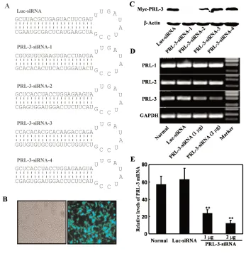
Oligonucleotides encoding four differ-ent PRL-3 siRNAs shown in Figure 1A were commercially synthesized (Invitro-gen, Shanghai, China) and cloned into BamHI/HindIII sites of pRNA-U6.1/ Neo (Genescript). pRNA-U6.1/Neo-Luciferase-siRNA (Genescript) was used as the monitor and a negative control.
Transient transfections were per-formed with polyethylenimine (PEI) liposome complex (kindly provided by Dr. Zi-Chun Hua, Nanjing University). Briefly, 4 μL PEI (1 mg/mL) and plas-mids were diluted in 50 μL MEM (Life Technologies) and mixed, then incubated at room temperature for 20 min before addition of 900 μL MEM. The mixture was spread onto cells. Cells were switched to 10% FBS DMEM after 6 h and incubated at 37°C for another 18 h, then collected and prepared for the experiments.
Semiquantitative RT-PCR and Real-Time PCR
First-strand cDNAs were generated by reverse transcription using oligo(dT) from RNA samples treated by DNase. The specific primers and cycle number for each gene we used are as follows:
PCR products were electrophoresed on a 1.5% agarose gel and visualized by ethidium bromide staining. The gel im-ages were captured and analyzed by the Gel Imaging and Documentation Digi-Doc-It System (v. 1.1.23; UVP, Upland, CA, USA). Real-time quantitative PCR was performed with the ABI Prism 7000 sequence detection system (Applied Biosystems, Foster City, CA, USA) using EvaGreen dye (Biotium, USA), and threshold cycle numbers were obtained using ABI Prism 7000 SDS software v. 1.0. The primer sequences used in this study were the same as above. Condi-tions for amplification were one cycle of 94°C for 5 min followed by 35 cycles of 94°C for 30 s, 60°C for 1 min, and 72°C for 30 s.
Western Blot
The Western blot was performed as described (20). The cells, cotransfected with pTARGET-Myc-PRL-3 (1 μg) and U6.1/Neo-Luc-siRNA or pRNA-U6.1/Neo-PRL3-siRNA-1, -2, -3, or -4 (1μg), were collected and lysed (50 mM Tris, pH 8.0, 150 mM NaCl, 1% NP-40, 0.1% SDS, 5 mM EDTA, 0.1 mM PMSF, 0.15 U/mL aprotinin, 1 μg/mL pepstatin, and 10% glycerol). Anti-Myc (clone 9E10) and anti-actin (Santa Cruz Biotechnol-ogy) antibodies were used for Western blot.
Cell Proliferation Assay
B16-BL6 cells were transiently trans-fected with or without PRL-3 siRNA (1 and 2 μg), or Luc-siRNA (2 μg) for
Product
Gene length Primer Cycle no.
PRL-1 472 bp Forward: 5′-CAACCAATGCGACCTTAA-3′ 30 Reverse: 5′-CAATGGCATCAGGCACCC-3′
PRL-2 339 bp Forward: 5′ATTTGCCATAATGAACCG-3′ 30
Reverse: 5′-ACAGGAGCCCTTCCCAAT-3′
PRL-3 468 bp Forward: 5′-CTTCCTCATCACCCACAACC-3′ 28 Reverse: 5′-TACATGACGCAGCATCTGG-3′ GAPDH 191 bp Forward: 5′-AACGACCCCTTCATTGAC-3′ 28
24 h, then 2 ×10⁴cells in 1 mL of culture
medium were added into each well of 24-well plates in triplicate for prolifera-tion assay on day 0. Cells were tryp-sinized and counted on days noted in the figure legends.
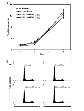
Cell Cycle Analysis
B16-BL6 cells, transiently transfected with or without PRL-3 siRNA (1 and 2μg) or Luc-siRNA (2 μg) for 24 h, were trypsinized and fixed in 75% ethanol at 4°C for 2 h. Cells were stained with 50μg/mL propidium iodide (Sigma) and assayed by flow cytometry (Becton Dickinson, San Jose, CA, USA).
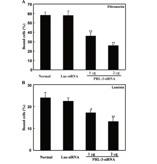
Cell Adhesion Assay
The cell adhesion assay was per-formed essentially as described (21) with some modifications. In brief, 96-well flat-bottom plates were coated with 50 μL fibronectin (10 μg/mL; Sigma) or laminin (10 μg/mL; Calbiochem) in PBS over-night at 4°C and blocked with 0.2% BSA for 2 h at room temperature followed by washing three times. Next, B16-BL6 cells, either untreated or transiently trans-fected with Luc-siRNA (2 μg) and PRL-3 siRNA (1 and 2 μg) for 24 h, were added to each well in triplicate and incubated for 30 min at 37°C. Plates were then washed three times with PBS to remove unbound cells. Cells remaining attached to the plates were fixed and stained with a solution containing 0.5% crystal violet and 2% ethanol in 100 mM borate buffer (pH 9.0). After washing, 100 μL SDS (1% wt/vol) was added, and the absorbance of the color substrate was measured with an ELISA reader (TECAN, Austria) at 592 nm. After subtraction of the back-ground cell binding to BSA-coated wells, the percentage of adherent cells was cal-culated by dividing the optical density of the adherent cells by that of the initial input cells.
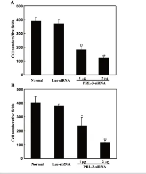
Cell Migration Assay and Invasion Assay
Cell migration and invasion assays were performed using 8.0-μm pore size Transwell inserts (Costar, Cambridge,
MA, USA) as described (10) with some modifications. For all migration essay, in brief, the undersurface of the membrane was coated with fibronectin (10 μg/mL) in PBS at 37°C for 2 h. The membrane was washed in PBS to remove excess ligand, and the lower chamber was filled with 0.6 mL DMEM with 10% FBS.
Cells were serum-starved overnight (0.5% FBS), harvested with trypsin/ EDTA, and washed twice with serum-free DMEM. Then, cells were resus-pended in migration medium (DMEM with 0.5% FBS), and 1 ×10⁵cells in 0.1 mL were added to the upper cham-ber. After 24 h at 37°C, the cells on the
upper surface of the membrane were re-moved using cotton swabs. The migrant cells attached to the lower surface were fixed in methanol at room temperature for 30 min and stained for 20 min with a solution containing 0.5% crystal violet and 2% ethanol in 100 mM borate buffer (pH 9.0). The number of migrated cells on the lower surface of the membrane was counted under a microscope in five fields at ×100. For cell invasion assay, all procedures were carried out as in the migration assay except that Matrigel was coated beforehand on the upper surface of the chambers (BD Bio-sciences) according to the manufac-turer’s protocol.
In Vivo Metastasis Assay
B16-BL6 cells in exponential growth phase were harvested by trypsinization and washed twice before injection. Cell vitality was >95% as determined by try-pan blue dye exclusion. B16-BL6 cells (5×10⁴cells in 20 μL PBS) were injected into the right hind footpads of C57BL/6 mice (100% of injected mice formed tu-mors). Ten days after injection, the mice were distributed into four groups with five mice each according to tumor size. PRL-3 siRNA (3 or 6 μg) and Luc-siRNA (6 μg) with PEI complexes (30 μL in each mouse) or PBS were injected into tumors four times every 4 days. Tumor volumes were measured every 4 days from day 10 to 22 and calculated by the following for-mula: 0.5236 ×L1 ×(L2)2, where L1 is the long axis and L2 is the short axis of the tumor. Twenty-six days later, mice were killed. The right footpads and draining popliteal lymph nodes were resected, and photos were taken (Nikon Coolpix 4500). Survival tests were made using groups of mice (n= 10) treated as above and monitored daily until all the mice died.
Statistical Analysis
Data are expressed as mean ± SEM. Student’s ttest was used to evaluate the difference between two groups. Kaplan-Meier method was used to evaluate the
survival test.P < 0.05 was considered to be significant.
RESULTS
Identification of an Efficient and Specific PRL-3 siRNA Sequence against the Expression of PRL-3
Four PRL-3 siRNA expression plas-mids (PRL-3 siRNA-1, -2, -3, and -4) tar-geting different regions of mouse PRL-3 mRNA and firefly luciferase siRNA (Luc siRNA) as an independent control were constructed using the pRNA-U6.1/Neo vector (Figure 1A). To test the efficiency
following experiments. Because PRL-1, -2, and -3 share at least 75% sequence similarity, the specific inhibitory effect on PRL-3 was tested by RT-PCR. With treat-ment of PRL-3 siRNA-1, the mRNA level of endogenous PRL-3 in B16-BL6 cells decreased in a dose-dependent manner, whereas that of endogenous PRL-1 and -2 were not influenced (Figure 1C).
No Effect of PRL-3 siRNA on the Proliferation of B16-BL6 Cells In Vitro
B16-BL6 cells were transfected with PRL-3 siRNA or Luc siRNA as control to investigate whether PRL-3 affected the tumor cell proliferation in vitro. We counted the cell number over a 4-day in-terval. The results shown in Figure 2A and B indicate that downregulation of PRL-3 does not affect the proliferation and cell cycle of B16-BL6 cells.
Inhibition of Adhesion, Migration, and Invasion of B16-BL6 Cells by PRL-3 siRNA
B16-BL6 cells were transfected with PRL-3 siRNA for 24 h, and their capabili-ties of adhesion, migration, and invasion were analyzed. As shown in Figure 3A and B, after 30-min incubation, com-pared with untreated cells, B16-BL6 cells transfected with PRL-3 siRNA (1 or 2 μg) exhibited a significant decrease in adhe-sion capability to fibronectin and laminin, by 38% and 55% for fibronectin and 29% and 45% for laminin, respec-tively. On the contrary, Luc siRNA had little effect on cell adhesion to both fibro-nectin and laminin (Figure 3). Similar to the results in the adhesion assay, cells migrating to the undersurface in the mi-gration assay were reduced by 53% and 68%, and those traversing the Matrigel in the invasion assay were reduced by 38% and 60% in B16-BL6 cells transfected with PRL-3 siRNA (1 or 2μg), respec-tively, after 24 h compared with un-treated cells (Figure 4A and B). Luc siRNA did not have such significant ef-fects.
Inhibition of Tumor Growth and Spontaneous Metastasis by
Intratumoral Injection of PRL-3 siRNA With the above findings of PRL-3 siRNA’s effects in vitro, we next investi-gated whether PRL-3 plays a critical role in tumor formation in vivo, and whether it can be used in clinical gene therapy. B16-BL6 cells were subcutaneously in-jected into footpads of C57BL/6J mice. Ten days after injection, PBS, PEI/Luc siRNA, or PEI/PRL-3 siRNA (3 or 6 μg) was injected into the tumors four times every 4 days. As shown in Figure 5B, compared with PBS, PRL-3 siRNA signif-icantly inhibited tumor growth in mice,
metastases in lungs and livers of all the groups (data not shown). Figure 5D shows the survival time of the mice. Downregulation of PRL-3 by PRL-3 siRNA (3 and 6 μg) prolonged the lifes-pan of mice bearing B16-BL6 tumor cells in a dose-dependent manner, whereas Luc-siRNA treatment showed no such ef-fect.
DISCUSSION
Recent studies have indicated that PRL-3 plays a causative role in malignant transformation and metastasis, and inhi-bition of PRL-3 expression may provide an effective means for inhibiting tumor formation and metastasis and prolonging survival. Our previous study also indi-cated that highly metastatic B16-BL6 melanoma cells express more elevated PRL-3 than less metastatic B16 cells (10).
In this study, we took B16-BL6 cells to establish a metastatic model and investi-gated its characteristics against vector-based siRNA, which was used to directly knock down the endogenous expression of PRL-3 in B16-BL6 cells (Figure 1). Al-though PRL-1 and PRL-2, which share at least 75% homologous protein sequence with PRL-3 (22,23), were also detected in B16-BL6 cells, PRL-3 siRNA treatment only specifically reduced the endogenous expression of PRL-3 in B16-BL6 cells, with no influence on the expression of PRL-1 and PRL-2 (Figure 1C). Neverthe-less, some reports have shown that over-expression of PRL-1 and PRL-2 prompts the transformation of epithelial cells and enhances the metastatic ability of cancer cells (8), and results that demonstrate the role of PRL-3 in various tumor metas-tases are accumulating. Recently, several studies using in vitro assays showed that reduced PRL-3 expression inhibited tumor growth, invasion, and metastasis in human colorectal cancer cells and ovarian cancer cells (8,9,12). In addition, Saha et al. (24) have compared the global gene expression profile of metastatic col-orectal cancers with that of primary tu-mors, benign tutu-mors, and normal col-orectal epithelium and found that PRL-3 is the only gene highly expressed in all 18 metastases examined. Overall, this ev-idence suggests that PRL-3 may be a po-tential therapeutic target for the treat-ment of malignant tumor.
Tumor metastasis is a complex process with prominent stages as follows: de-tachment of cancer cells from a primary tumor, entry into the circulatory system, and adherence and migration of the tumor cells to distant sites. The first stage of tumor cell detachment from the original site depends on adhesiveness to extracellular matrix and migration to blood capillaries and lymphatic vessels (25). Therefore, we detected these capa-bilities of PRL-3 in B16-BL6 cells. Al-though knockdown of PRL-3 by specific siRNA had little influence on the prolif-eration and cell cycle of B16-BL6 cells in vitro (Figure 2), it did inhibit B16-BL6 cells’ adhesive ability to fibronectin and Figure 4.Effect of PRL-3 siRNA on migration and invasion of B16-BL6 melanoma cells. In
laminin, and also their migration and in-vasion ability under the Transwell-based assay (Figure 3 and 4). The adhesion, mi-gration, and invasion ability of tumor cells may be regulated by multiple sig-naling cascades, including integrin-medi-ated signaling, mitogen-activintegrin-medi-ated protein kinase signaling, and cytoskeletal reor-ganization (26,27). This hypothesis was recently substantiated by the fact that PRL-3 directly interacted with integrin
α1, decreased the tyrosine phosphoryla-tion of integrin β1, and activated the Erk1/2 signal pathway (28). These find-ings further confirm that PRL-3 plays a key role in tumor cell acquisition of metastatic potential.
On the other hand, although previous studies suggest that PRL-3 regulates tumor metastasis in vivo, the function of PRL-3 in the whole process of metastasis is not totally clear. Experimental passive
attach well to fibronectin, a common ma-trix for adhesion and migration assays. The present work reconfirmed those observations and showed that PRL-3 siRNA could inhibit the adhesion, migra-tion, and invasion ability of B16-BL6 cells in vitro (Figures 3 and 4). With these results, we further examined whether PRL-3 siRNA can suppress the metasta-sis of B16-BL6 cells in vivo and prolong lifespan. B16-BL6 cells were implanted into mouse footpads to form primary tu-mors. Ten days later, intratumoral injec-tions with specific siRNA were carried out to observe that parental B16-BL6 cells with endogenous expression of PRL-3 grow more rapidly than PRL-3 knock-down cells (Figure 5B). In addition, treat-ment with PRL-3 siRNA remarkably re-duced the metastatic ability of B16-BL6 cells to popliteal lymph nodes, where no visible metastatic sites were detected in that group (Figure 5C). Furthermore, all mice treated with PBS or Luc siRNA vec-tor died within 32 days; animals with 6μg PRL-3 siRNA treatment survived through 60 days (Figure 5D). These re-sults suggest that the in vivo prolifera-tion of B16-BL6 cells in the primary tumor site, the development of macro-scopic metastases, and the survival of mice implanted with B16-BL6 cells were highly dependent on the expression level of PRL-3. Untreated B16-BL6 cells with normal endogenous expression of PRL-3 showed more apparent metastasis to draining lymph nodes than PRL-3 siRNA–treated cells (Figure 5C), whereas there were no metastatic sites visible to the lung or liver (data not shown).
Our data extend observations of PRL-3 having substantial function in sponta-neous metastasis in vivo. Although this result was consistent with the reports that PRL-3 promoted metastasis in mice models in vivo (11), we found that en-dogenous PRL-3 tended to initiate spon-taneous local lymph metastasis through lymphatic circulation but not lung or liver metastasis through blood circula-tion. Actually, the lymphatic system is optimally suited for the entry and trans-portation of tumor cells, as the smallest
lymphatic vessels are much larger than blood capillaries (29). When tumor cells released from the primary tumor mass, it is easier for them to enter lymphatic ves-sels than the blood system. Data from clinic metastatic samples also support this hypothesis. Bardelli et al. (13) have found that PRL-3 is detected in all col-orectal metastasis to the lymph nodes. These data indicate that PRL-3 plays a critical role in regional lymph node metastasis. At the mechanistic level, lymph node metastasis is closely associ-ated with lymphangiogenesis, which fa-cilitates the dissemination of tumor cells to transfer to regional lymph nodes through the lymphatic drainage network. Further studies will be necessary to un-cover whether PRL-3 regulates the ex-pression of these factors in the process of lymph node metastasis.
In this study, B16-BL6 cells treated with PRL-3 siRNA showed slow growth compared with those treated with Luc siRNA in primary tumor in vivo (Figure 5). This result contrasted with observa-tions of cell growth under in vitro cul-ture conditions, in which the level of PRL-3 did not affect proliferation rate (Figure 2). Similar effects of PRL-3 on cell growth have also been reported for human colon cancer DLD-1 cells in vitro, whose endogenous PRL-3 was abrogated by PRL-3 siRNA (9). These data indicate that the promotion of tumor growth by PRL-3 in vivo is associated with tumor microenvironment, which can provide many survival and growth factors for tumor cells. This is further supported by a recent report that PRL-3 enhanced the activation of Rho family GTPases RhoA and RhoB, following the initiation of the Rho/ROCK signal pathway necessary for invasion and motility (30). In addi-tion, this signal pathway also facilitates angiogenesis by inducing the expression of VEGF (31). Pille et al. (32) have re-ported that the angiogenesis of breast cancer, treated with RhoA-siRNA, was remarkably reduced and tumor growth was also inhibited. These data suggest that the promotion of B16-BL6 cell
prolif-eration by PRL-3 in vivo may also be as-sociated with angiogenesis.
In conclusion, our present work repro-duced the role of PRL-3 in the whole process of spontaneous metastasis from primary tumor to distant site. We also found that specific reduced expression of PRL-3 inhibited the growth of B16-BL6 cells in vivo and prevented detectable metastasis from primary tumors by de-creasing adhesive ability to extracellular matrix such as fibronectin and laminin, and by reducing the migratory ability of B16-BL6 cells. PRL-1 and PRL-2 have roles in cancer cells (9), but we found, at least in B16-BL6 cells, that PRL-3 played a more pivotal role in tumor formation and metastasis. The current results ex-tend potential therapeutic applications of PRL-3 inhibitors to the clinical treatment of malignant tumor.
ACKNOWLEDGMENTS
This study was supported by grants from the National Natural Science Foun-dation of China (no. 30300425 and 30500619) and the State Key Project Foundation of “the 10th 5-year plan of China.”
REFERENCES
1. Boyd D. (1996) Invasion and metastasis. Cancer Metastasis Rev.15:77-89.
2. Fidler IJ. (2003) The pathogenesis of cancer metastasis: the ‘seed and soil’ hypothesis revis-ited. Nat. Rev. Cancer3:453-8.
3. Miyata Y et al. (2006) Lymphangiogenesis and angiogenesis in bladder cancer: prognostic impli-cations and regulation by vascular endothelial growth factors-A, -C, and -D. Clin. Cancer Res. 12:800-6.
4. Versteeg HH, Spek CA, Peppelenbosch MP, Richel DJ. (2004) Tissue factor and cancer metas-tasis: the role of intracellular and extracellular signaling pathways. Mol. Med.10:6-11. 5. Bouzahzah B et al. (2001) Rho family GTPases
regulate mammary epithelium cell growth and metastasis through distinguishable pathways. Mol. Med.7:816-30.
6. Diamond RH, Cressman DE, Laz TM, Abrams CS, Taub R. (1994) PRL-1, a unique nuclear pro-tein tyrosine phosphatase, affects cell growth. Mol. Cell Biol.14:3752-62.
7. Cates CA et al. (1996) Prenylation of oncogenic human PTP(CAAX) protein tyrosine phos-phatases. Cancer Lett.110:49-55.
phos-phatase PRL-3 in malignant cells and endothelial cells: expression and function. Mol. Cancer Ther. 5:219-29.
9. Kato H, Semba S, Miskad UA, Seo Y, Kasuga M, Yokozaki H. (2004) High expression of PRL-3 promotes cancer cell motility and liver metastasis in human colorectal cancer: a predictive molecu-lar marker of metachronous liver and lung metastases. Clin. Cancer Res.10:7318-28. 10. Wu X et al. (2004) Phosphatase of regenerating
liver-3 promotes motility and metastasis of mouse melanoma cells. Am. J. Pathol.164:2039-54. 11. Zeng Q et al. (2003) PRL-3 and PRL-1 promote
cell migration, invasion, and metastasis. Cancer Res.63:2716-22.
12. Polato F et al. (2005) PRL-3 phosphatase is impli-cated in ovarian cancer growth. Clin. Cancer Res. 11:6835-9.
13. Bardelli A et al. (2003) PRL-3 expression in metastatic cancers. Clin. Cancer Res.9:5607-15. 14. Hammond SM, Bernstein E, Beach D, Hannon GJ. (2000) An RNA-directed nuclease mediates post-transcriptional gene silencing in Drosophila cells. Nature404:293-6.
15. Zamore PD, Tuschl T, Sharp PA, Bartel DP. (2000) RNAi: double-stranded RNA directs the ATP-de-pendent cleavage of mRNA at 21 to 23 nu-cleotide intervals. Cell101:25-33.
16. Wall NR, Shi Y. (2003) Small RNA: can RNA in-terference be exploited for therapy? Lancet 362:1401-3.
17. Ito T et al. (2006) An inducible short-hairpin RNA vector against osteopontin reduces metasta-tic potential of human esophageal squamous cell carcinoma in vitro and in vivo. Clin. Cancer Res. 12:1308-16.
18. Taulli R et al. (2005) RNAi technology and lentiviral delivery as a powerful tool to suppress Tpr-Met-mediated tumorigenesis. Cancer Gene Ther.12:456-63.
19. Saydam O et al. (2005) Herpes simplex virus 1 amplicon vector-mediated siRNA targeting epi-dermal growth factor receptor inhibits growth of human glioma cells in vivo. Mol. Ther.12:803-12. 20. Zhang X, Xu Q, Saiki I. (2000) Quercetin inhibits the invasion and mobility of murine melanoma B16-BL6 cells through inducing apoptosis via de-creasing Bcl-2 expression. Clin. Exp. Metastasis 18:415-21.
21. Qian F, Zhang ZC, Wu XF, Li YP, Xu Q. (2005) In-teraction between integrin alpha(5) and fi-bronectin is required for metastasis of B16F10 melanoma cells. Biochem. Biophys. Res. Commun. 333:1269-75.
22. Zeng Q, Hong W, Tan YH. (1998) Mouse PRL-2 and PRL-3, two potentially prenylated protein tyrosine phosphatases homologous to PRL-1. Biochem. Biophys. Res. Commun.244:421-7. 23. Stephens BJ, Han H, Gokhale V, Von Hoff DD.
(2005) PRL phosphatases as potential molecular targets in cancer. Mol. Cancer Ther.4:1653-61. 24. Saha S et al. (2001) A phosphatase associated
with metastasis of colorectal cancer. Science
294:1343-6.
25. Pepper MS, Tille JC, Nisato R, Skobe M. (2003) Lymphangiogenesis and tumor metastasis. Cell Tissue Res.314:167-77.
26. Crean JK et al. (2002) The role of p42/44 MAPK and protein kinase B in connective tissue growth factor induced extracellular matrix protein pro-duction, cell migration, and actin cytoskeletal re-arrangement in human mesangial cells. J. Biol. Chem.277:44187-94.
27. Ritz U, Seliger B. (2001) The transporter associ-ated with antigen processing (TAP): structural in-tegrity, expression, function, and its clinical rele-vance. Mol. Med.7:149-58.
28. Peng L, Jin G, Wang L, Guo J, Meng L, Shou C. (2006) Identification of integrin alpha1 as an in-teracting protein of protein tyrosine phosphatase PRL-3. Biochem. Biophys. Res. Commun.342:179-83. 29. Liotta LA, Steeg PS, Stetler-Stevenson WG. (1991) Cancer metastasis and angiogenesis: an imbal-ance of positive and negative regulation. Cell 64:327-36.
30. Fiordalisi JJ, Keller PJ, Cox AD. (2006) PRL tyro-sine phosphatases regulate rho family GTPases to promote invasion and motility. Cancer Res. 66:3153-61.
31. Mizukami Y et al. (2006) Hypoxic regulation of vascular endothelial growth factor through the induction of PI3K/Rho/ROCK and c-Myc. J. Biol. Chem.281:13957-63.