INTRODUCTION
Human β-globin gene expression is regulated tightly during development and hematopoiesis. The human β-globin locus comprises five developmentally regulated genes (5′-ε-Gγ-Aγ-δ-β-3′) whose high level and stage-specific expression depends on interactions with the locus control region (LCR), consisting of five major DNaseI hypersensitive sites (Fig-ure 1). The LCR activates β-globin gene transcription through direct interaction with promoter regions (1,2), and is a major determinant of the chromatin structure of the locus (3). Mice transgenic for the human β-globin locus express the human genes in a developmentally
regu-lated manner (4,5). Specifically, the human globin genes undergo two devel-opmental switches in their activation. Ex-pression of the embryonic ε-and fetal γ-globin genes, first activated during primitive erythropoiesis in the embry-onic yolk sac, switches to expression of the γ-genes at the start of definitive ery-thropoiesis in the fetal liver, with a small contribution by β-globin. The second switch occurs gradually around birth with the activation of the adult stage-specific δ- and β-globin genes, with δ-globin making a minor contribution, whereas γ-globin expression is gradually suppressed to very low levels (1%–2%) by the end of the first year of life.
The individual genes have been shown to be regulated by a complex interplay between cisregulatory elements, trans-acting factors, enhancer competition, and epigenetic mechanisms (6). Despite a re-markable amount of progress in this field, the exact mechanism(s) of globin gene silencing still is not understood completely.
Understanding the molecular basis of globin gene switching and discovery of strategies to efficiently express γ-globin genes in the adult is of partic-ular interest, since reactivation of the fetal γ-globin genes in the adult has been shown to ameliorate the effects of hemoglobinopathies (7).
A series of well established quantita-tive trait loci accounting for 20%–50% of the fetal hemoglobin (HbF) variabil-ity in healthy adults, such as: a) the C→T single nucleotide polymorphism at position –158 of the Gγ-gene, creating a restriction site for the enzyme XmnI (8);
following Deletion of Two Silencer Elements Located 3
′
to the
Human A
γ
-Globin Gene
Maria Gazouli,
1,2Eleni Katsantoni,
3Theodoros Kosteas,
4and Nicholas P Anagnou
1,21Laboratory of Biology, University of Athens School of Medicine, Athens, Greece; 2Laboratory of Cell and Gene Therapy, Centre of
Basic Research II, Biomedical Research Foundation of the Academy of Athens, Athens, Greece; 3Hematology Division, Biomedical Research Foundation of the Academy of Athens, Athens, Greece; and 4Institute of Molecular Biology and Biotechnology, FORTH, Heraklion, Greece
Natural deletions of the human γ-globin gene cluster lead to specific syndromes characterized by increased production of fetal hemoglobin in adult life and provide a useful model to delineate novel cis-acting elements involved in the developmental con-trol of hemoglobin switching. A hypothesis accounting for these phenotypic features assumes that silencers located within the Aγ -to δ-gene region are deleted in hereditary persistence of fetal hemoglobin (HPFH) and δβ-thalassemias, leading to failure of switching. In the present study, we sought to clarify the in vivo role of two elements, termed Enh and F, located 3′to the Aγ-globin, in silencing the fetal genes. To this end, we generated three transgenic lines using cosmid constructs containing the full length of the globin locus control region (LCR) linked to the 3.3-kb Aγ-gene lacking both the Enh and F elements. The Enh/F deletion re-sulted in high levels of Aγ-globin gene expression in adult mice in all single copy lines, whereas, the LCR-Aγsingle copy lines which retain the Enh and F elements exhibited complete normal switching of the fetal Aγ-gene. Our study documents directly for the first time the in vivo role of these two gene-proximal negative regulatory elements in silencing the fetal globin gene in the peri-natal period, and thus these data may permit their eventual exploitation in therapeutic approaches for thalassemias.
© 2009 The Feinstein Institute for Medical Research, www.feinsteininstitute.org Online address: http://www.molmed.org
doi: 10.2119/molmed.2009.00019
Address correspondence and reprint requests toNicholas P Anagnou, University of Athens School of Medicine, 75 Mikras Asias Street, 115 27 Athens, Greece. Phone: +30-210-746-2341; Fax: +30-210-746-2412; E-mail: [email protected].
b) the HBS1L-MYB intergenic region on chromosome 6q23 (9); and c) the BCL11Agene on chromosome 2, encod-ing a zinc fencod-inger transcription factor acting directly within the β-cluster (10), has been studied extensively and its contribution on the mechanisms of fetal globin expression and hemoglobin switching has been evaluated recently (11). Furthermore, the configuration (AT)9T5of the polymorphic sequence motif residing 0.5 kb 5′to the β-gene has been considered to confer high γ-globin levels in β-thalassemia patients in association with the XmnI polymor-phism (12–14), although other studies (15) have found no effect. Furthermore, analysis of mutants with rare point mu-tations either in the Gγor Aγpromoter associated with a hereditary persistence of fetal hemoglobin (HPFH) phenotype (11,16) have provided insights for the mechanisms of continued HbF synthe-sis based on the alterations in DNA-protein binding sites, resulting either in the creation of a new motif that allows
trans-activator binding, or in the abol-ishment of a repressor protein binding motif, leading to de-repression of fetal genes (11,16).
However, the most informative model for the role of cis-acting elements of the cluster involved in hemoglobin switch-ing has been provided by a number of naturally occurring deletions in the locus leading to persistent γ-globin gene expression in the adult stage, and sug-gesting that γ-gene suppression is likely a complex process but one that can be perturbed (16). The persistence of fetal hemoglobin associated with such dele-tions is classified into two related clini-cal syndromes, that is, HPFH and (δβ)°-thalassemia (16). HPFH results in a substantial (14%–30%) pancellular γ-globin gene expression, whereas (δβ)°-thalassemias give rise to lower lev-els (2%–15%) of heterocellular γ-globin expression in the adult. In a particular set of deletions where the 3′end of the locus is lost, the 5′breakpoints map within a region between the Aγ- to
δ-globin genes. Based on the above ob-servations, it has long been postulated that the Aγ- to δ-globin intergenic region harbors negative cis-acting elements in-volved in the regulation of the fetal-to-adult switch (17). It also has been sug-gested (18), without being mutually exclusive, that the juxtaposition of distal sequences located downstream of the 3′ breakpoint of these deletions with en-hancer-like function also may be in-volved in persistent γ-globin expression. The validity of the latter hypothesis for the HPFH deletions has been docu-mented conclusively (experimentally) in vivoby us (19,20) and by others (21) with the identification and functional charac-terization of a series of HPFH enhancers (19–21). A similar mechanism may be operating in the HPFH-5 and HPFH Kenya deletions via the juxtaposition of the 3′ β-globin enhancer to the proximity of the fetal genes (1,16).
Previous studies of our laboratory (22,23) have identified four elements, termed Enh, F, O, and P, located within the Aγ- to δ-globin intergenic region (see Figure 1) exhibiting silencer activity in transient transfection assays. Using elec-trophoretic mobility shift assays coupled with oligonucleotide competition and DNaseI footprinting techniques, several binding sites for the transcription factors YY1, GATA-1, and CP1 were identified in the F element. The six YY1 binding sites exhibit a variable degree of homol-ogy to the consensus motifs and bind YY1 with different affinity. Of the three strong YY1 sites, two sites residing at the 5′and 3′ends of the element, respec-tively, seem to confer its silencing activ-ity (22,23). Based on these special fea-tures, these elements were considered as candidate regulatory cis-acting se-quences for suppressing γ-globin expres-sion in the adult stage.
Subsequent transgenic mouse studies using several of these elements have been controversial as to their in vivo function. Specifically, constructs that in-cluded the LCR linked to an Aγ-globin gene fragment containing Enh and F ele-ments were silenced autonomously in
the adult stage of transgenic mice (24), indicative of their silencing activity. In contrast, deletion of a 12.5-kb region be-tween the Aγ- to δ-globin genes, in a human β-globin locus yeast artificial chromosome (YAC) which includes all four elements, has been reported to have no effect on human globin gene switch-ing in transgenic mice (25). Furthermore, deletion of Enh alone from a human β-globin locus YAC showed no observ-able effects on the regulation of the β-globin locus in transgenic mice (25,26).
Taken together, these findings, al-though not conclusive, do provide strong evidence that the identified ele-ments possess transcriptional regulatory activity at specific developmental stages and may be involved in the develop-mental regulation of globin gene switch-ing. Supporting this hypothesis, a recent study from our group (27) documented that deletion of both Enh and F elements in the context of a 185-kb human β-globin locus PAC (P1 phage artificial chromosome) results in an increase of transcription of both ε- and γ-globin genes in the embryonic yolk sac stage, whereas no effects on human globin gene expression in the fetal liver and adult blood stages were observed. The latter finding apparently reflects a known phenomenon of functional re-dundancy (28), since the rest of the so far identified silencer elements (that is, O and P elements and possibly others) remained intact and thus functional in our modified PAC, actually masking the net effect of the absence of Enh and F elements on the fetal γgenes (27).
In view of these unsettled data, in the present study, we sought to further clar-ify the in vivofunction of the Enh and F elements in the context of cosmid con-structs containing the human globin LCR directly linked to the Aγgene, either without or in the presence of the two si-lencers retained in their physical posi-tion. Our goal was to delineate the direct effect of these elements on the Aγ-gene silencing during the perinatal period, in the absence of any other silencer ele-ments of the human β-globin locus.
MATERIALS AND METHODS
DNA Constructs
The cosmid construct used was gener-ated by ligation and packaging into phage of the following fragments: a) a 27-kb PvuI-ClaI fragment from LCR-loxP-Aγ -3′HS cosmid (20) containing the cos site of the pTCFcosmid vector (1), the LCR, and the loxP site; b) a ClaI-KpnI fragment from a pBluescript-Aγplasmid contain-ing the Aγgene (3.3 kb); c) a 14-kb KpnI-NotI fragment from the LCRεcosmid (4) containing a cos site and sequences downstream of the ε-globin gene. Pack-aging was performed using Gigapack Gold extracts (Stratagene, La Jolla, CA, USA) according to the manufacturer’s instructions.
Transgenic Mice
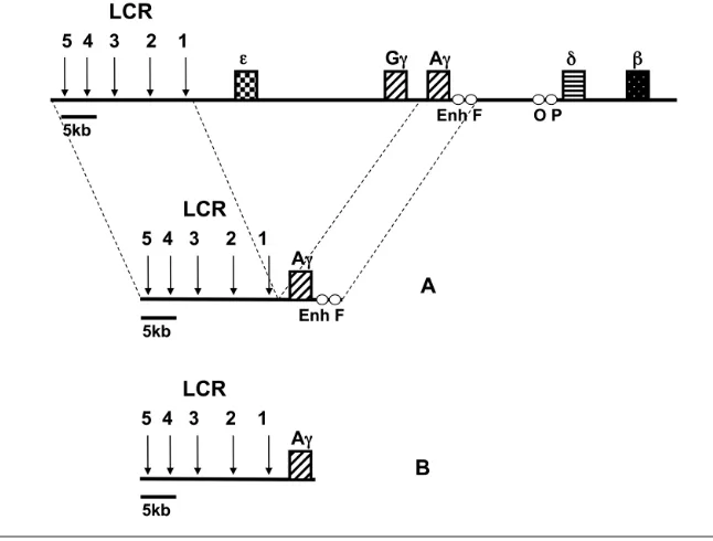
The fragment containing the 25-kb LCR-loxP-Aγinsert (see Figure 1) was re-leased by SalI–KpnI digestion, purified by Elutip D (Schleicher & Schuell, Bio-Science, Bath, UK), checked for DNA in-tegrity and concentration, and prepared for microinjection, essentially as de-scribed previously (27). Purified DNA was injected into the pronucleus of fertil-ized eggs of FVB/N mice as described previously (27). Transgenic founders were identified by polymerase chain re-action (PCR) using the primers in the HS5 site of the LCR and by Southern blotting using the LCR HS5 3.3-kb EcoRI fragment and the 5′Aγ1.7-kb EcoRI-BamHI fragment as probes. The integrity of cosmid transgenes was checked by cosmid hybridization according to Strouboulis et al. (4). Transgene copy numbers were determined by using a 5′Aγ1.7-kb EcoRI-BamHI fragment and a 0.9-kb PvuI fragment from the endoge-nous mouse carbonic anhydrase II (CA2) gene, and the ratios of the intensities of the two probes were compared with those obtained for the single copy LCR-Aγ-HPFH2C transgenic line (20). Phos-phorImager analysis was performed using ImageQuant software (Molecular Dynamics, Sunnyvale, CA, USA). Multi-copy transgenic mouse lines were bred
with the CAG-Cre transgenic lines (29) to generate single copy animals. The in-tegrity and the copy number of the single copy mice were determined simi-larly as for the multicopy lines. The con-trol transgenic mice used in this study were the LCR-loxP-Aγlines generated by our group (20). All experiments were performed according to the guidelines of the Animal Facility of IMBB, as approved by the Institutional Committee.
S1 Nuclease Protection Assays
S1 nuclease protection analysis was carried out using total RNA from blood of adult animals. RNA was isolated using the Trizol reagent according to the manufacturer’s instructions (Invitrogen, Carlsbad, CA, USA). The probes used and conditions for S1 nuclease protection assays and polyacrylamide gel elec-trophoresis were essentially as described previously (30–32). Specific activities of probes were determined as described previously (32), and are indicated in the legend of Figure 2. Quantitation of ex-pression levels was performed on a PhosphorImager using the ImageQuant software (Molecular Dynamics, Sunny-vale, CA, USA).
Real-Time Reverse Transcriptase PCR
Master Mix (Applied Biosystems, Foster City, CA, USA) according to the manu-facturer’s instructions. Real-time PCR was performed in an ABI PRISM 7000 Sequence Detection System (Applied Biosystems), as follows: initial denatura-tion for 2 min at 50°C and for 10 min at 95°C, followed by 40 cycles of PCR (95°C for 15 sec; 60°C for 1 min). Data were an-alyzed using the comparative CTmethod for the relative quantitation of results (33). Post-amplification denaturation curves showed that the primer pairs gen-erated single products.
DNA Fluorescence in situ Hybridization (FISH) Analysis
Peripheral blood cells were cultured for 72 h in RPMI 1640 medium (Invitrogen,
Carlsbad, CA, USA). Chromosome prepa-rations were made according to standard procedures. Fluorescence in situ hybridiza-tion (FISH) was carried out as described by Mulder et al. (34). The probe used to detect the transgene was the biotin-labeled 5′Aγ1.7-kb EcoRI-BamHI fragment, which was detected immunochemically with flu-orescein. Chromosomal DNA was coun-terstained with DAPI (4’,6 diamidino-2-phenylindole), which stains centromeric domains more intensely.
Linear Amplification Mediated-Polymerase Chain Reaction (LAM-PCR)
LAM-PCR was performed using the previously reported methodology (35). This newly established approach permits
the localization of the integration site of a construct in a given genome by identi-fying the sequence of an unknown ge-nomic region flanking a known gege-nomic segment. It represents a combination of linear amplification of target DNA with solid-phase second strand synthesis, fol-lowed by ligation of an oligonucleotide cassette, and then by nested exponential PCR. Briefly, genomic DNA from mouse tails was used as a template. Linear amplification was carried out with the bio -tinylated primer KN872_B (5′-TGAAG ACCTGGGGGCTGGATT-3′). The single stranded run-off DNA fragments were purified by magnetic capture. The single stranded DNA was made double stranded with random hexamers (Roche, Indianapolis, IN, USA) and the DNA polymerase Klenow fragment. The prod-ucts were digested by Tsp501Iand then ligated into a linker cassette which was generated by annealing two oligonu-cleotides: 5′-GACCCGGGAGATCTG
AATTCAGTGGCACAGCAGTTAGG-3′
and 5′-AATTCCTAACTGCTTGCCAC
TGAATTCAGATCTCCCGGGTC-3′. The
resultant ligated products were exponen-tially amplified twice using the following primers: KN87-N1: 5′-ACCTGGGGGC TGGATTGATTG-3′and 5′-GGGGCTGGAT TGATTGCAGCT-3′. The cycle sequenc-ing of the specific LAM-PCR amplicon was performed using an ABI Prism ge-netic analyzer 3100 (Applied Biosystems), according to the manufacturer’s instruc-tions. The obtained sequence data were analyzed and mapped to the appropriate mouse chromosome locations, employ-ing the Basic Local Alignment Search Tool (BLAST), developed by the National Center for Biotechnology Information (NCBI).
RESULTS
Generation of LCR-Aγ-ΔEnh/F Transgenic Lines
To investigate the effects of the dele-tion of Enh and F elements on the si-lencing of the Aγ-globin gene, we gener-ated transgenic lines by microinjecting a 25-kb construct containing the full
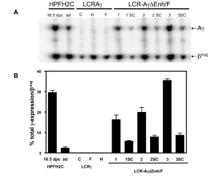
Figure 2.Expression of Aγ-globin gene in multicopy and single-copy ΔEnh/F lines. (A) Repre-sentative S1 nuclease protection analysis of expression of human Aγversus mouse βmaj glo-bin genes in adult blood RNA of single copy control lines, multicopy LCR-Aγ-ΔEnh/F-1, 2, and 3 lines, and the derived single copy (sc) lines analyzed. The type of protected fragments are indicated to the right of the panel. Controls used in this experiment included a 16.5 dpc embryo from HPFH2C line and an adult HPFH2C RNA sample (ad), expressing high lev-els of the Aγ-gene, as described by Katsantoni et al. (20). Expression levels were calculated by ImageQuant after correcting for probe-specific activities according to the ratio of 1:2.4 for Aγ:βmajprobes. (B) Quantitation of A
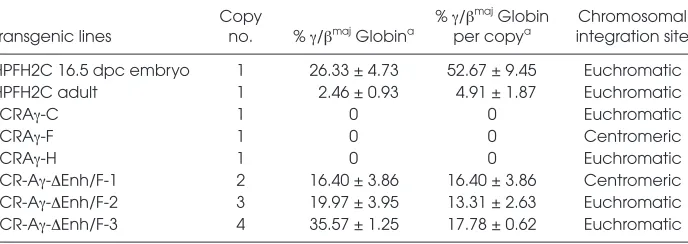
length LCR and the 3.3-kb Aγ-globin gene lacking the Enh and F elements (LCR-Aγ-ΔEnh/F) as shown in Figure 1. Four founders transgenic for the LCR-Aγ-ΔEnh/F construct were obtained. The first founder mouse never passed on the transgene. The other three LCR-Aγ-ΔEnh/F founder mice successfully transmitted the transgene to establish the corresponding transgenic lines LCR-Aγ-ΔEnh/F-1, LCR-Aγ-ΔEnh/F-2, and LCR-Aγ-ΔEnh/F-3. Further analysis showed that all three lines contained an intact transgene (data not shown) in multiple copies: LCR-Aγ-ΔEnh/F-1, two copies; LCR-Aγ-ΔEnh/F-2, three copies; and LCR-Aγ-ΔEnh/F-3, four copies (as shown in Table 1).
Aγ-Gene Expression Analysis in Adult LCR-Aγ-ΔEnh/F Multicopy Lines
We first analyzed Aγ-globin gene ex-pression levels against those of the en-dogenous βmaj-globin gene in RNAs iso-lated from adult blood of transgenic LCR-Aγ-ΔEnh/F multicopy lines by S1 nuclease protection assay (see Figure 2). As negative controls for this analysis, three LCR-Aγsingle copy lines were used (lines C, F, and H), which retain the Enh and F elements downstream of the Aγgene and exhibit normal switching, as reported previously by our group (20). Expression of Aγ-globin normally is switched off at this stage in these mice (20,24,28). This was corroborated in the three LCR-Aγcontrol lines (Table 1; see Figure 2). In contrast, all three multicopy LCR-Aγ-ΔEnh/F lines lacking the Enh and F silencers exhibited persistent Aγ -globin expression in the adult blood (see Figure 2) ranging from 13.31 ± 2.63 to 17.78 ± 0.62 per transgene copy and per copy of endogenous mouse βmaj-globin (Table 1). RNA samples from three dif-ferent adult animals from each of the three generated LCR-Aγ-ΔEnh/F lines were analyzed. The highest LCR-Aγ -ΔEnh/F Aγ-expressing line was the LCR-Aγ-ΔEnh/F-3 (Table 1; see Figure 2).
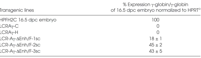
We further analyzed Aγ-gene expres-sion by quantitative real-time reverse transcriptase PCR. These results
con-firmed the S1 nuclease mapping data, with high γ-globin expression being de-tected in the adult blood of the LCR-Aγ -ΔEnh/F transgenic mice only, ranging from 24 ± 3 to 112 ± 4% of the levels of the LCR-Aγ-HPFH2C transgene at the fetal stage (20), which was used as a pos-itive control (Table 2). On the contrary, no expression was detected in the adult blood of the control LCR-Aγlines C and H containing the two silencer elements (Table 2). Since the only difference be-tween the LCR-Aγ-ΔEnh/F versusthe LCR-Aγconstructs is the selective ab-sence or preab-sence of the silencers, respec-tively, these findings directly demon-strate that the absence of Enh and F elements in the context of the containing transgene can alter the devel-opmental expression of the Aγ-gene in
adult blood and leads to its persistent and efficient expression.
Persistent Adult Stage Aγ-Globin Expression in Single Copy LCR-Aγ -ΔEnh/F Lines
To distinguish whether the persist-ence of Aγ-gene expression in the LCR-Aγ-ΔEnh/F lines was due specifically to the absence of the Enh/F silencers or to a non-specific effect of multicopy inte-grants of LCR-containing globin trans-genes, we generated single copy mice from all three multicopy animals by crossbreeding with lines expressing the Cre recombinase before the two-cell stage of embryonic development using the CAG-Cre mice (29). Three single copy (sc) LCR-Aγ-ΔEnh/F lines were generated and were designated as
LCR-Table 1.Summary of the control and multicopy lines generated, transgene copy numbers, γ-globin mRNA expression levels in adult blood, and integration sites of the transgene.
Copy % γ/βmajGlobin Chromosomal
Transgenic lines no. % γ/βmajGlobina per copya integration site
HPFH2C 16.5 dpc embryo 1 26.33 ± 4.73 52.67 ± 9.45 Euchromatic
HPFH2C adult 1 2.46 ± 0.93 4.91 ± 1.87 Euchromatic
LCRAγ-C 1 0 0 Euchromatic
LCRAγ-F 1 0 0 Centromeric
LCRAγ-H 1 0 0 Euchromatic
LCR-Aγ-ΔEnh/F-1 2 16.40 ± 3.86 16.40 ± 3.86 Centromeric
LCR-Aγ-ΔEnh/F-2 3 19.97 ± 3.95 13.31 ± 2.63 Euchromatic
LCR-Aγ-ΔEnh/F-3 4 35.57 ± 1.25 17.78 ± 0.62 Euchromatic
aExpression of the fetal A
γ-gene was calculated by S1 nuclease analysis as 1) a percentage of total βmajglobin expression and 2) per transgene copy and per copy of the endogenous βmajmouse globin gene. Mean and standard deviations are derived from three independent
RNA samples.
Table 2.Real-time PCR analysis of γ-globin expression in adult blood of the generated multicopy transgenic lines.
% Expression γ-globin/γ-globin
Transgenic lines of 16.5 dpc embryo normalized to HPRTa
HPFH2C 16.5 dpc embryo 100
LCRAγ-C 0
LCRAγ-H 0
LCR-Aγ-ΔEnh/F-1 24 ± 3
LCR-Aγ-ΔEnh/F-2 66 ± 16
LCR-Aγ-ΔEnh/F-3 112 ± 4
a
Aγ-ΔEnh/F-1sc, LCR-Aγ-ΔEnh/F-2sc, and LCR-Aγ-ΔEnh/F-3sc. We then ana-lyzed Aγ-gene expression in the adult blood by S1 nuclease protection. As controls for normal switching, we in-cluded the three single copy LCR-Aγ lines mentioned above. As expected, Aγ-gene expression was switched off in all three LCR-Aγsingle copy control lines analyzed. In contrast, Aγ-globin was expressed in the adult blood of all three LCR-Aγ-ΔEnh/F single copy lines (Table 3). The levels of expression were slightly lower and copy dependent, compared with the corre-sponding multicopy lines, as expected (Table 3; see Figure 2). These results were confirmed independently by quantitative real-time reverse transcrip-tase PCR analysis (Table 4).
Correlation of Persistent Aγ-globin Expression with Integration Sites
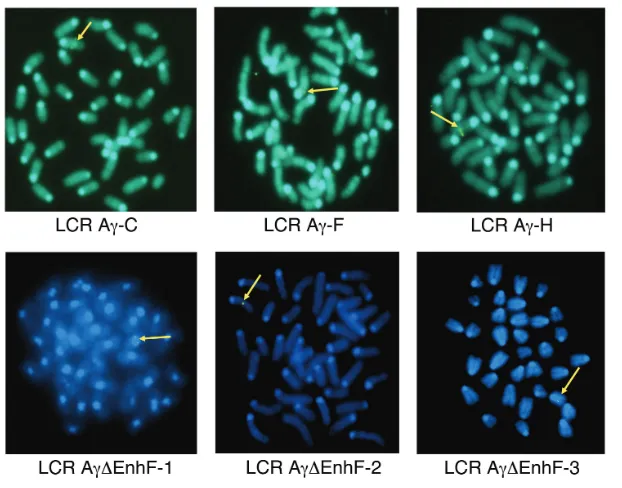
Our data demonstrate that the dele-tion of Enh and F elements can lead to persistent Aγ-expression in adult mice carrying either single or multiple copies. We wished to further investigate whether this persistent Aγ-globin gene expression in the adult stage is linked to or is affected by specific chromoso-mal integrations. Position effects depen-dent on the chromatin structure at the sites of transgene integration often are observed (30). Therefore, we initially mapped transgene integration sites in all transgenic lines using FISH analysis in metaphase spreads to exclude the possibility that Aγ-gene expression could reflect position effects and not the actual effect of the absence of Enh and F silencer elements. We classified chro-mosomal sites as centromeric and eu-chromatic, the latter including all inte-grations that did not map close to a telomere or a centromere. All integra-tion sites are shown in Figure 3 and the results are summarized in Table 1. The data documented that LCR-Aγ-ΔEnh/F-1 line is integrated in a centromeric re-gion, whereas lines 2 and 3 contain eu-chromatic integration sites (Table 1; see Figure 3).
To further determine the exact ge-nomic locations of the integration sites, we applied the recently introduced LAM-PCR method (35), which confirmed the FISH results (Table 5). The transgene of the LCR-Aγ-ΔEnh/F-1 line was inte-grated into chromosome 8 within the neuropilin-1and integrinβ1gene locus (chr8qE2), while the transgenes of the LCR-Aγ-ΔEnh/F lines 2 and 3 were inte-grated near the KIF27gene (chr13qB2) and the tyrosine phosphatase receptor type L gene (chr4qD2.3), respectively.
In the case of the integration sites near the integrinβ1, KIF27, and tyrosine phos-phatase receptor type Lgenes, their tran-scription sites were located far enough from the integrated transgene (>100 kb of surrounding region), suggesting that there should be little if any effect from their transcriptional activation. However, we cannot exclude the theoretical possi-bility that the expression levels in LCR-Aγ-ΔEnh/F-1 line could be influenced by the neuropilin-1gene, since its
transcrip-tion site was found approximately 400 bp upstream of the Aγ-gene, while it has been shown previously to be expressed in CD45+hematopoietic cells from murine fetal liver (36).
The centromeric integration site in line LCR-Aγ-ΔEnh/F-1 is correlated with slightly lower Aγ-gene expression levels compared with lines 2 and 3 associated with euchromatic integration sites. The fact that Aγ-gene is expressed even when the transgene was integrated into the centromere, excludes the possibility that the Aγ-gene expression is due to positive position effects usually associated with open chromatin euchromatic sites, and strengthens the possibility that the result-ing persistent Aγ-gene expression reflects actually altered developmental regula-tion of the gene, due solely to the ab-sence of the Enh and F silencers.
DISCUSSION
Several studies have shown that, in murine models, individual human γ- and
Table 3.Summary of the single copy (sc) lines generated, transgene copy numbers, γ-globin mRNA expression levels in adult blood, and integration sites of the transgene.
Copy % γ/βmajGlobin Chromosomal
Transgenic lines no. % γ/βmajGlobina per copya integration site
LCR-Aγ-ΔEnh/F-1sc 1 5.90 ± 0.26 11.80 ± 0.52 Centromeric LCR-Aγ-ΔEnh/F-2sc 1 7.97 ± 1.15 15.94 ± 2.30 Euchromatic LCR-Aγ-ΔEnh/F-3sc 1 8.75 ± 1.64 17.50 ± 3.28 Euchromatic
aExpression of the fetal Aγ-gene was calculated by S1 nuclease analysis as 1) a percentage
of total βmajglobin expression and 2) per transgene copy and per copy of the endogenous βmajmouse globin gene. Mean and standard deviations are derived from three independent
RNA samples.
Table 4.Real-time PCR analysis of γ-globin expression in adult blood of the generated single copy (sc) transgenic lines.
% Expression γ-globin/γ-globin
Transgenic lines of 16.5 dpc embryo normalized to HPRTa
HPFH2C 16.5 dpc embryo 100
LCRAγ-C 0
LCRAγ-H 0
LCR-Aγ-ΔEnh/F-1sc 18 ± 1
LCR-Aγ-ΔEnh/F-2sc 45 ± 2
LCR-Aγ-ΔEnh/F-3sc 43 ± 5
a
β-globin transgenes are expressed at high levels in the presence of the LCR, and are regulated appropriately at the fetal and adult stages of development (37,38). The δβand Aγδβ-thalassemias and the deletion forms of hereditary persistence of fetal he-moglobin (HPFH) are naturally occurring mutations associated with persistent ex-pression of fetal hemoglobin in adult life, albeit at variable levels. It has been
pro-posed that the Aγ-δglobin intergenic se-quences harbor negative regulatory ele-ments that are involved in suppressing γ-globin expression in the adult stage (17). In accordance to this notion, we previ-ously described two elements Enh and F, located 3′to the Aγgene as silencers in transient transfection assays (22,23).
To further elucidate the direct in vivorole of the Enh and F elements on Aγ-globin
gene silencing under the control of LCR, we generated transgenic mice using cosmid constructs containing the full length LCR linked to the 3.3-kb Aγ-gene, but lacking selectively both the Enh and F silencers. The deletion of Enh and F ele-ments resulted in persistent high levels of Aγ-expression in the adult stage in all three multicopy and three single copy lines tested. Furthermore, according to our results of FISH analysis, the deletion of Enh and F elements can lead to persis-tent Aγ-gene expression in the adult stage in all types of chromatin environments, not only in “permissive” ones.
Our observations are in agreement with earlier results using μLCR 3.3-kb Aγ (39) and LCR HS2 3.3-kb Aγlines (40) ex-hibiting adult stage Aγ-gene expression in constructs lacking the region harbor-ing the Enh and F silencers, and in con-trast to earlier reports describing Enh ini-tially as an enhancer element in transient reporter assays using a different setting (41). Additional studies by the same group have documented the mixed func-tional features of the Enh element (42).
Our previous experiments (27) have documented that the selective deletion of Enh and F elements from a 185-kb human β-globin locus PAC results in an increase of εand γ-globin mRNA levels in the embryonic stage (that is, yolk sac stage) of erythropoiesis. This phenome-non was shown to be due to an increase
Table 5.The precise transgene integration sites identified by LAM-PCR in LCR-Aγ-ΔEnh/F lines used in this study.
Position to
Transgenic line Sequence (5′ →3′)a Locus TSS (bp)b Genec E-valued
LCR-Aγ-ΔEnh/F-1 GACCTGTAGCCCAGTGCCCAGAGCATATTATCATAACCAC chr8qE2 >100 5′side: neuropilin-1
9e–23
ATTTCAGGGGACGCCAACGT >100,000 3′side: integrinβ1
LCR-Aγ-ΔEnh/F-2 GAATTCCAGCACACTGGCGGCCGTTACTAGTGGATCCGAG chr13qB2 >100,000 Kinesin-related
1e–10 protein 27 (KIF27 )
LCR-Aγ-ΔEnh/F-3 GACTCAGTTTCTTCATCTGTAGAATGG chr4qD2.3 >100,000 5′side: hypothetical protein
2e–04 >100,000 3′side: tyrosine
phosphatase, receptor type L
a
The sequences shown are part of the LAM-PCR amplicons located 5′of the linker cassette.
bTSS, transcription start site.
c
Precise chromosomal locations were defined by BLAST search for the mouse genome.
dE-value, expectation value (by BLAST search).
in the rate of transcription rather than to an increase in the number of cells tran-scribing the human globin locus (27). However, the human developmental switching from fetal γ-globin to adult β-globin gene expression was not af-fected by this particular deletion, thus identifying Enh and F as locus-wide reg-ulatory elements capable of downregu-lating transcription of the human β-globin locus in an embryonic-specific manner. These results however, also im-plied the operation of a well known mechanism of functional redundancy (28), and, in the context of the full human β-globin locus, additional regula-tory elements in the region between Aγ and δgenes (that is, O and P silencer ele-ments we have described previously [22,23] located upstream of the δgene) or in other regions of the cluster, might be needed to act synergistically with Enh and F elements to silence γ-globin genes in the adult stage of transgenic mice. This notion is corroborated by the phe-notype of Corfu (δβ)° thalassemia, a nat-ural deletion of a similar size and loca-tion, removing O and P elements, leading to significant levels of γ-globin in heterozygotes (43).
Nevertheless, in our previous study (20), we showed that in the presence of LCR and the two Enh and F silencers, and in the absence of additional regula-tory elements or competition with the β-globin gene, Aγ-globin gene is silenced autonomously in the adult.
Furthermore, elements of the proximal and the distal Aγ-gene promoter (44), combined with a competition by the other globin genes, are important to downregu-late γ-globin gene expression in the adult stage (45,46). Additionally, it is well docu-mented that the distance or gene order relative to the LCR is an essential determi-nant for the type of globin gene to be acti-vated selectively during the primitive stage of erythropoiesis (47–51). In our study, the LCR was linked directly to the Aγ-globin gene and the lines tested versus the control lines differed solely on either the absence or presence of the Enh and F elements, respectively.
The function of these silencer elements in the context of the entire β-globin clus-ter, the proper spacing of the LCR, and the high order chromatin structure can be addressed by considering the features of the active chromatin hub (ACH) model (52,53). Specifically, the model refers to the erythroid-specific spatial clustering of cisregulatory elements, consisting of the 5′hypersensitive sites HS1-6 of the LCR, the downstream hypersensitive site 3′HS1 and of the active globin genes. According to the ACH model, stable enhancer-promoter interactions determine the es-tablishment of a functional expression module. Depending on the competitive nature of the surrounding chromatin, ad-ditional cisregulatory elements, such as the Enh and F silencers, may have evolved to stabilize the promoter interactions in the ACH and maintain the required expression level at a particular developmental stage. The transcriptional outcome of an ACH criti-cally depends on the productive interac-tion between cisregulatory elements and specific developmental transcription fac-tors, such as Ikaros and its family mem-ber Eos, which seem to participate in the regulation of hemoglobin switching by facilitating the DNA looping between the LCR and the upstream region of the δ-globin gene (54). Interestingly, the tran-scriptional pattern of globin gene switch-ing seems to correlate with the switchswitch-ing of interactions of the individual globin genes with this spatial cluster (55). Fur-thermore, the ACH model can explain why the γ-globin expression pattern might differ in the case of the Enh and F deletion in the context of the full β-globin locus construct (27), possessing all the regulatory elements of the locus versus the deletion of the same elements in the context of a cosmid construct containing just the LCR and the Aγgene; it is con-ceivable that the entry of new regulatory elements may further stabilize or destabi-lize existing interactions and alter the ex-pression levels of genes present in the ACH. Further investigation on the role of Enh and F elements on γ-globin gene transcription can be accomplished by
applying the chromosome conformation capture carbon copy (5C) technology, which permits the analysis of millions of chromatin interactions in parallel (56), to understand how such elements interact with γ-globin promoters, the LCR, and other genes within the ACH.
All the above studies strongly indicate that apart from the elements located within the Aγ- to δ-globin intergenic re-gion, hemoglobin switching is a conse-quence of a complex interplay between developmental stage-specific transcription factors which interact both within the pro-moters of the various genes and the 5′LCR, as well as of specific modifications of the physical location of a gene within the locus (47,57–59). Thus, sequence-specific transcription factors (activators and/or repressors), such as the recently identified developmental stage-specific re-pressor BCL11A (10), can regulate ε- and γ-globin activation and repression through nearby ciselements in a gene autono mous manner, independently of the amplifying effects of the LCR.
In addition, our studies employing the specific and selective Enh/F deletion, permitted for the first time the direct in vivoevaluation of the effect of the Enh and F elements on the expression of the fetal γ-globin gene in adult life and pro-vide the first experimental proof that these two elements represent bona fide re-pressing elements capable of altering the developmental pattern of expression of the fetal γ-genes. Furthermore, previous studies described by our group (27) and by others (59,60) identified a new class of gene-proximal regulatory elements within the human β-globin locus that are involved in regulating the levels of glo-bin gene transcription either positively (59,60) or negatively (27), in a develop-mental-stage specific manner.
Further delineation of the molecular mechanisms of fetal-to-adult switch are expected to contribute to designing fu-ture therapeutic strategies for tha-lassemias and sickle cell anemia. To this end, eventual manipulation of Enh, F, and other regulatory elements of the locus or of stage-specific repressors (10) aiming to the derepression and reactiva-tion of the fetal gene expression in the adult erythropoietic stem cells is ex-pected to lead to an effect which has been shown to represent a potent thera-peutic approach (7).
ACKNOWLEDGMENTS
We thank Kostas Kourouniotis and Athanasios Stavropoulos for expert tech-nical assistance. This work was sup-ported by a 70-3-9209/03 Grant from the Central Council of Health of the Greek Ministry of Health and Social Welfare (to NP Anagnou).
DISCLOSURES
The authors declare that they have no competing interests as defined by Molecu-lar Medicine, or other interests that might be perceived to influence the results and discussion reported in this paper.
REFERENCES
1. Grosveld F, van Assendelft GB, Greaves DR, Kol-lias G. (1987) Position-independent, high-level
expression of the human β-globin gene in trans-genic mice. Cell. 51:975–85.
2. Ragoczy T, Bender MA, Telling A, Byron R, Groudine M. (2006) The locus control region is required for association of the murine β-globin locus with engaged transcription factories during erythroid maturation. Genes. Dev. 20:1447–57. 3. Felsenfeld G, Groudine M. (2003) Controlling the
double helix. Nature. 421:448–53.
4. Strouboulis J, Dillon N, Grosveld F. (1992) Devel-opmental regulation of a complete 70-kb human
β-globin locus in transgenic mice. Genes. Dev. 6:1857–64.
5. Townes TM, Behringer RR. (1990) Human globin locus activation region (LAR): role in temporal control. Trends Genet. 6:219–23.
6. Brand M, et al. (2004) Dynamic changes in tran-scription factor complexes during erythroid dif-ferentiation revealed by quantitative proteomics.
Nat. Struct. Mol. Biol. 11:73–80.
7. Ley TJ, et al. (1982) 5-azacytidine selectively in-creases γ-globin synthesis in a patient with β+ thalassemia. N. Engl. J. Med. 307:1469–75. 8. Gilman JG, Huisman THJ. (1985) DNA sequence
variation associated with elevated fetal Gγglobin production. Blood. 66:783–7.
9. Creary LE, et al. (2009) Genetic variation on chro-mosome 6 influences F cell levels in healthy indi-viduals of African descent and HbF levels in sickle cell patients. PLoS ONE. 4:e4218. 10. Sankaran VG, et al. (2008) Human fetal
hemoglo-bin expression is regulated by the developmen-tal stage-specific repressor BCL11A. Science. 322:1839–42.
11. Thein SL, Menzel S. (2009) Discovering the ge-netics underlying foetal haemoglobin production in adults. Brit. J. Haematol. 145:455–67.
12. Ragusa A, et al. (1992) Genetic epidemiology of
β-thalassemia in Sicily: do sequences 5′to the Gγ
gene and 5′to the βgene interact to enhance HbF expression in β-thalassemia? Am. J. Hematol. 40:199–206.
13. Gonçalves I, et al. (1998) Combined effect of two different polymorphic sequences within the βglobin gene cluster on the level of HbF.
Am. J. Hematol. 57:269–76.
14. Gonçalves I, et al. (2002) Fetal hemoglobin eleva-tion in Hb Lepore heterozygotes and its correla-tion with β-globin cluster linked determinants.
Am. J. Hematol. 69:95–102.
15. Bandyopadhyay S, et al. (2005) Two β-globin cluster-linked polymorphic loci in thalassemia patients of variable levels of fetal hemoglobin.
Eur. J. Haematol. 75:47–53.
16. Wood WG. (1993) Increased HbF in adult life.
Baillieres Clin. Haematol. 6:177–213.
17. Huisman TH, et al. (1974) The present status of the heterogeneity of fetal hemoglobin in β-thalassemia: an attempt to unify some observations in tha-lassemia and related conditions. Ann. N. Y. Acad.
Sci. 232:107–24.
18. Flavell RA, et al. (1983) Structure and expression of the human globin genes and murine
histocom-patibility antigen genes. Cold Spring Harb. Symp.
Quant. Biol. 47:1067–78.
19. Anagnou NP, et al. (1995) Sequences located 3′to the breakpoint of the hereditary persistence of fetal hemoglobin-3 deletion exhibit enhancer ac-tivity and can modify the developmental expres-sion of the human fetal Aγ-globin gene in trans-genic mice. J. Biol. Chem. 270:10256–63. 20. Katsantoni EZ, et al. (2003) Persistent γ-globin
ex-pression in adult transgenic mice is mediated by HPFH-2, HPFH-3, and HPFH-6 breakpoint se-quences. Blood. 102:3412–9.
21. Arcasoy MO, et al. (1997) High levels of human
γ-globin gene expression in adult mice carrying a transgene of deletion-type hereditary persistence of fetal hemoglobin. Mol. Cell Biol. 17:2076–89. 22. Kosteas T, Manifava M, Moschonas N, Anagnou
NP. (1993) Functional analysis of the Aγto ψβ
globin gene region of the human β-locus: evi-dence for negative regulatory elements. Clin. Res. 41:38A.
23. Kosteas T, Manifava M, Moschonas N, Anagnou NP. (1994) Functional analysis of the Aγto δ glo-bin gene region of the β-cluster: evidence for negative regulatory elements. Blood. 84:506A. 24. Dillon N, Grosveld F. (1991) Human γ-globin genes silenced independently of other genes in the β-globin locus. Nature. 350:252–4. 25. Gaensler KM, et al. (2003) Sequences in the Aγ-δ
intergenic region are not required for specific regulation of the human β-globin gene locus. Proc. Natl. Acad. Sci. U. S. A. 100:3374–9. 26. Liu Q, Tanimoto K, Bungert J, Engel JD. (1998) The Aγ-globin 3′element provides no unique function(s) for human β-globin locus gene regu-lation. Proc. Natl. Acad. Sci. U. S. A. 95:9944–9. 27. Katsantoni EZ, et al. (2004) An embryonic-specific
repressor element located 3′to the Aγ-globin gene influences transcription of the human β-globin locus in transgenic mice. Exp. Hematol. 32:224–33. 28. Dillon N, Grosveld F. (1993) Transcriptional
regu-lation of multigene loci: multilevel control. Trends Genet. 9:134–7.
29. Sakai K, Miyazaki J. (1997) A transgenic mouse line that retains Cre recombinase activity in ma-ture oocytes irrespective of the cre transgene transmission. Biochem. Biophys. Res. Commun. 237:318–24.
30. Milot E, et al. (1996) Heterochromatin effects on the frequency and duration of LCR-mediated gene transcription. Cell. 87:105–14.
31. Lindenbaum MH, Grosveld F. (1990) An in vitro
globin gene switching model based on differenti-ated embryonic stem cells. Genes. Dev. 4:2075–85. 32. Fraser P, Hurst J, Collis P, Grosveld F. (1990)
DNaseI hypersensitive sites 1, 2 and 3 of the human β-globin dominant control region direct position-independent expression. Nucleic Acids Res. 18:3503–8.
33. Pfaffl MW. (2001) A new mathematical model for relative quantification in real-time RT-PCR. Nu-cleic Acids Res. 29:e45.
loci in the DiGeorge critical region at chromo-some 22q11 using a new marker (D22S183). Hum.
Genet. 96:133–41.
35. Schmidt M, et al. (2002) Polyclonal long-term re-populating stem cell clones in a primate model.
Blood. 100:2737–43.
36. Yamada Y, et al. (2003) Neuropilin-1 on hematopoi-etic cells as a source of vascular development.
Blood. 101:1801–9.
37. Kollias G, Wrighton N, Hurst J, Grosveld F. (1986) Regulated expression of human Aγ-, β-, and hybrid γβ-globin genes in transgenic mice: manipulation of the developmental expression patterns. Cell. 46:89–94.
38. Collis P, Antoniou M, Grosveld F. (1990) Defini-tion of the minimal requirements within the human β-globin gene and the dominant control region for high level expression. EMBO J. 9:233–40.
39. Enver T, Ebens AJ, Forrester WC, Stamatoy-annopoulos G. (1989) The human β-globin locus activation region alters the developmental fate of a human fetal globin gene in transgenic mice.
Proc. Natl. Acad. Sci. U. S. A. 86:7033–7. 40. Lloyd JA, Krakowsky JM, Crable SC, Lingrel JB.
(1992) Human γ- to β-globin gene switching using a mini construct in transgenic mice. Mol.
Cell Biol. 12:1561–7.
41. Bodine DM, Ley TJ. (1987) An enhancer element lies 3′to the human Aγglobin gene. EMBO J. 6:2997–3004.
42. Purucker M, Bodine D, Lin H, McDonagh K, Nienhuis AW. (1990) Structure and function of the enhancer 3′to the human Aγglobin gene.
Nucleic Acids Res. 18:7407–15.
43. Chakalova L, et al. (2005) The Corfu δβ tha-lassemia deletion disrupts γ-globin gene silenc-ing and reveals post-transcriptional regulation of HbF expression. Blood. 105:2154–60.
44. Anagnou NP, Karlsson S, Moulton AD, Keller G, Nienhuis AW. (1986) Promoter sequences re-quired for function of the human γ-globin gene in erythroid cells. EMBO J. 5:121–6.
45. Ragoczy T, Telling A, Sawado T, Groudine M, Kosak ST. (2002) A genetic analysis of chromo-some territory looping: diverse roles for distal regulatory elements. Chromosome Res. 11:513–25. 46. Omori A, Tanabe O, Engel JD, Fukamizu A,
Tani-moto K. (2005) Adult stage γ-globin silencing is mediated by a promoter direct repeat element.
Mol. Cell Biol. 25:3443–51.
47. Harju S, Navas PA, Stamatoyannopoulos G, Peter-son KR. (2005) Genome architecture of the human
β-globin locus affects developmental regulation of gene expression. Mol. Cell Biol. 25:8765–78. 48. Dillon N, Trimborn T, Strouboulis J, Fraser P,
Grosveld F. (1997) The effect of distance on long-range chromatin interactions. Mol. Cell. 1:131–9. 49. Hanscombe O, et al. (1991) Importance of globin gene order for correct developmental expression.
Genes. Dev. 5:1387–94.
50. Peterson KR, et al. (1993) Transgenic mice con-taining a 248-kb yeast artificial chromosome
car-rying the human β-globin locus display proper developmental control of human globin genes.
Proc. Natl. Acad. Sci. U. S. A. 90:7593–7. 51. Tanimoto K, Liu Q, Bungert J, Engel JD. (1999)
Effects of altered gene order or orientation of the locus control region on human β-globin gene ex-pression in mice. Nature. 398:344–8.
52. Noordermeer D, de Laat W. (2008) Joining the loops: β-globin gene regulation. IUBMB Life. 60:824–33.
53. Palstra R-J, et al. (2003) The β-globin nuclear compartment in development and erythroid dif-ferentiation. Nat. Genet. 35:190–4.
54. Keys JR, et al. (2008) A mechanism for Ikaros reg-ulation of human globin gene switching. Brit. J.
Haematol. 141:398–406.
55. Stamatoyannopoulos G, Grosveld F. (2001) He-moglobin switching. In: The Molecular Basis of Blood Diseases. Stamatoyannopoulos G, Majerus P, Perlmutter R, Varmus H (eds.) W.B. Saunders, Philadelphia, pp. 135–82.
56. van Bekkum NL, Dekker J. (2009) Determining spatial chromatin organization of large genomic regions using 5C technology. Methods Mol. Biol. 567:189–213.
57. Raich N, Clegg CH, Grofti J, Roméo PH, Stama-toyannopoulos G. (1995) GATA1 and YY1 are de-velopmental repressors of the human ε-globin gene. EMBO J. 14:801–9.
58. Rupon JW, Wang SZ, Gaensler K, Lloyd J, Ginder GD. (2006) Methyl binding domain protein 2 medi-ates γ-globin gene silencing in adult human βYAC transgenic mice. Proc. Natl. Acad. Sci. U. S. A. 103:6617–22.
59. Liu Q, Bungert J, Engel JD. (1997) Mutation of gene-proximal regulatory elements disrupts human ε-, γ-, and β-globin expression in yeast ar-tificial chromosome transgenic mice. Proc. Natl.
Acad. Sci. U. S. A. 94:169–74.
60. Calzolari R, McMorrow T, Yannoutsos N, Langeveld A, Grosveld F. (1999) Deletion of a re-gion that is a candidate for the difference be-tween the deletion forms of hereditary persist-ence of fetal hemoglobin and δβ-thalassemia affects β- but not γ-globin gene expression.