INTRODUCTION
Antibodies to a wide variety of au-toantigens are a hallmark of systemic lupus erythematosus (SLE) (1). In partic-ular, anti-double-stranded DNA (anti-dsDNA) antibodies are associated with lupus nephritis and disease activity (2–5). Two distinct models have been proposed to explain the origin of patho-genic antibodies in SLE. One model suggests that pathogenic anti-dsDNA autoantibodies arise from naïve autore-active B cells through polyclonal B cell activation, which is antigen-independent; the alternative model proposes that anti-dsDNA antibodies acquire autoreac-tivity by somatic mutation and the
anti-dsDNA response in SLE is antigen driven.
Analysis of various murine anti-dsDNA antibodies has demonstrated that somatic mutation, often with the in-troduction of basic amino acids, can re-sult in dsDNA binding (6). Although this observation is consistent with antigen-dependent affinity maturation, the na-ture of the triggering and selecting anti-gens remains to be elucidated. Whether nucleosomes, DNA, or phospholipid antigens released by apoptotic cells, or alternatively, foreign antigens trigger the response is unclear. Back mutation of four human IgG anti-DNA antibodies de-rived from lupus patients demonstrates
that in their germline configuration these antibodies may fail to bind DNA (7–9). This observation would suggest that polyclonal activation is not the mecha-nism for the generation of pathogenic au-toantibodies and that self-DNA is a criti-cal eliciting antigen. These antibodies are the only anti-dsDNA antibodies from lupus patients that have been analyzed for antigenic specificity in their germline configuration.
It has been reported recently that pa-tients with SLE have a defect in early B cell tolerance checkpoints, leading to the accumulation of many autoreactive B cells in the mature naïve B cell com-partment (10). This observation again raises the question whether pathogenic anti-dsDNA antibodies might originate from these naïve autoreactive B cells. Naïve B cells can become IgM memory B cells through antigen-dependent mech-anisms and IgG-expressing cells through antigen-independent pathways (11). IgM memory B cells are diminished in lupus patients (12), probably due to increased
Are Derived from Both Self-Reactive and Non-Self–Reactive
B Cells
Jie Zhang, Annett M Jacobi, Tao Wang, and Betty Diamond
The Center for Autoimmune and Musculoskeletal Disease, Feinstein Institute for Medical Research, North Shore-LIJ Health System, Manhasset, New York, United States of America
Previous studies have shown that both murine and human anti-double-stranded DNA (anti-dsDNA) antibodies can develop from non-DNA–reactive B cells and suggest a crucial role for somatic mutation in dsDNA binding. However, since only a limited number of human anti-dsDNA antibodies have been analyzed previously, we could not exclude other mechanisms for the generation of anti-dsDNA antibodies in patients with systemic lupus erythematosus (SLE). Therefore, we isolated IgM anti-dsDNA antibodies from peripheral blood B cells of a patient with SLE. Three somatically mutated IgM anti-DNA antibodies with pathogenic potential (glomerular binding) were reverted to their germline configuration. Although all three IgM anti-dsDNA antibodies came from the same lupus patient, they displayed different profiles. Reversion to the germline sequence of autoantibodies A9 and B5 resulted in decreased dsDNA binding. In contrast, the germline form of G3-recognized dsDNA as well as the mutated counterpart. These re-sults suggest that mutated IgM anti-dsDNA antibodies may develop from both DNA- and non-DNA–reactive B cells. The implica-tions are that B cell activation occurs in response to self and non-self antigens, while selection after activation may be mediated by self antigen in SLE. Moreover, ineffective tolerance checkpoints may exist before and after antigen activation in SLE.
Online address: http://www.molmed.org doi: 10.2119/2008-00066.Zhang
Address correspondence and reprint requests toBetty Diamond, The Center for Auto-immune and Musculoskeletal Disease, Feinstein Institute for Medical Research, North Shore-LIJ Health System, 350 Community Drive, Manhasset, NY, USA, 11030. Phone: 516-562-3830; Fax: 516-562-2921; Email: [email protected].
Ig-class switching of IgM B cells which may be caused by elevated BAFF-levels, overexpression of costimulatory mole-cules, and certain cytokines, such as IL-10 and IL-21 (13–15). This subset, therefore, may be a major precursor population for pathogenic antibodies in SLE. We, there-fore, chose to examine this subset to de-termine the origins of IgM anti-dsDNA antibodies and their antigenic cross-reactivity, because of the possibility that they might undergo class switch recom-bination even in the absence of cognate T-cell interaction.
So far, only a few mutated IgM anti-dsDNA antibodies have been isolated from lupus patients (16), and none have been back-mutated to assess germline-encoded antigenic specificity. Therefore, we isolated IgM anti-dsDNA antibodies from peripheral blood B cells of a patient with SLE. Three somatically mutated IgM anti-DNA antibodies with patho-genic potential, as reflected in their abil-ity to bind to isolated glomeruli, were re-verted to their germline configuration. Reactivity against dsDNA and other anti-gens was determined in both somatically mutated and reverted antibodies in order to understand the impact of somatic mutation on the generation of dsDNA-reactive IgM memory B cells in SLE, and to determine whether pathogenic human anti-DNA autoantibodies are derived from DNA- or non-DNA–reactive B cells.
MATERIALS AND METHODS
Production of Human Anti-dsDNA Monoclonal Antibodies from Peripheral Blood of Lupus Patients
G3, A9, and B5 are human monoclonal antibodies derived from peripheral blood B cells of a lupus patient, M55, who met the revised ACR criteria for SLE (17). Patient M55 was 37 years old and presented no signs of active disease at the time of the blood draw. The pa-tient exhibited elevated serum titers of anti-dsDNA antibodies and was being treated with hydroxychloroquine and low dose prednisone. In brief, individual B cells, identified by reactivity with a
fluorochrome-tagged peptide mimetope of dsDNA (18), were sorted into 96-well PCR plates and IgH (μonly) and IgL chain gene rearrangements were ampli-fied in two rounds of PCR (50 cycles each) before being cloned into human Ig
γ1 and κexpression vectors (gift of MC Nussenzweig, Rockefeller University, New York, NY, USA). Human embry-onic kidney fibroblast 293T cells were cotransfected with IgH- and IgL-encod-ing plasmid DNA by calcium phosphate precipitation as described previously (10,19). Supernatants were collected after 5 d of culture.
Reversion of Somatic Hypermutations into Germline Sequences
The plasmids with G3, A9, and B5 Ig gene segments were used as templates for the QuikChange Mutagenesis Kit (Stratagene, La Jolla, CA, USA). Muta-tion and juncMuta-tion analyses were con-ducted using the JOINSOLVER and IMGT/V-QUEST programs. Mutated IgH and IgL chain genes were reverted to their germline sequences and were se-quenced to confirm the reversions before being expressed in vitroas described above.
Immunofluorescence Assay (IFA) and Glomerular Binding Assay
IFAs were performed following the manufacturer’s instructions using anti-bodies at 7-45 μg/mL for 1 h, followed by FITC anti-human IgG (Bion Enter-prises Ltd., Des Plaines, IL, USA).
Murine glomeruli were isolated and attached to glass slides as described pre-viously (20). Monoclonal antibodies were applied at 7 to 30 μg/mL for 1 h at room temperature and visualized with FITC anti-human IgG (Inova Diagnostics Inc, San Diego, CA, USA).
Crithidia assays were performed ac-cording to the manufacturer’s instruc-tions. (IMMCO Diagnostics Inc, Buffalo, NY, USA)
Monoclonal antibody B1, which does not recognize dsDNA by Enzyme-Linked ImmunoSorbent Assay (ELISA), was cho-sen as a negative control (45 μg/mL for
IFA and 30 μg/mL for glomeruli and crithidia assays).
ELISA
Antibody concentrations in super-natants were determined by using human IgG1 kappa as a standard. The capture antibody and detection antibody were unlabeled goat anti-human IgG and alkaline phosphatase conjugated goat anti-human kappa, respectively (Southern Biotechnology, Birmingham, AL, USA).
Ninety six-well plates (Corning Life Science, Pittsburgh, PA, USA) were coated with 100 μg/mL of calf thymus dsDNA (Sigma-Aldrich, St. Louis, MO, USA), single-stranded DNA (ssDNA), 10 μg /mL of lipopolysaccharide (LPS), or 5 μg/mL of recombinant human in-sulin (Sigma-Aldrich). Antibody bind-ing to phosphatidylserine (10 μg/mL) or cardiolipin (50 μg/mL) with β2-GPI was measured as described previously (21). In addition, a commercial anti-nu-cleosome assay was performed (IBL-America, Minneapolis, MN, USA). All ELISAs were developed with alkaline phosphatase-conjugated goat anti-human IgG (Southern Biotechnology) and OD405 was measured using a Vic-tor microplate reader (Perkin Elmer, Waltham, MA, USA). Clone 53 (gift of MC Nussenzweig) was used as a nega-tive control (10,19).
RESULTS
Characterization of Mutated Antibodies
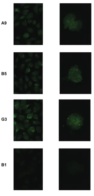
com-plementarity determining region 3 (CDR3) of the heavy chains, all se-quences were analyzed by IMGT junc-tion analysis tool to distinguish somatic mutations from junctional diversity in CDR3. We chose three antibodies (A9, B5, and G3) for reversion study, as the IMGT analysis showed they had unam-biguous germline sequences in CDR3 and different mutation profiles. Further-more, all of these had more than three mutations making it unlikely that the mutations were introduced by PCR error. In their mutated configuration, these an-tibodies displayed anti-nuclear reactivity on HEp-2 cells. In addition, they bound to isolated mouse glomeruli (Figure 1). This tissue binding suggested a
patho-genic potential of these antibodies once they undergo heavy chain class switch-ing to IgG. Furthermore, DNase treat-ment did not diminish glomerular bind-ing (data not shown), demonstratbind-ing cross-reactivity to glomerular antigens. It is important to note that glomerular binding is a surrogate for pathogenicity, but it is not clear that all glomerular-binding antibodies, in fact, trigger an in-flammatory process.
The amino acid sequences of these an-tibodies are shown in Figure 2. The heavy chain of antibody A9 was encoded by the IGHV1-69, D6-13, and J4; the light chain by IGKV3-20 and J2. A high ratio of replacement to silent mutations (R/S) (5:1 in the heavy chain, and 4:0 in the light chain) and a clustered distribution of replacement mutations in the CDRs of the heavy chain gene were additional features of A9, suggestive of antigen se-lection. Three basic amino acids (Arg, Lys, and His) were acquired through so-matic mutation at amino acid residues 50, 58, and 64, respectively.
Antibody G3 was encoded by IGHV3-30, D3-3, and J6, and IGKV1-5 and J1. In contrast to A9, the heavy chain of anti-body G3 had only one replacement mu-tation, A40G, located in framework re-gion 2 (FR2), and four silent mutations. Thus, this antibody displays no strong evidence of antigen selection.
The heavy and light chain variable re-gions of antibody B5 were encoded by IGHV3-07, D3-10, and J4, and IGKV1-NL1 and J1, respectively. Two replace-ment mutations and two silent mutations were present in the heavy chain variable region; one introduced the basic amino acid Lys at position 76.
The Effects of Somatic Mutation on Self- and Non-Self Reactivity
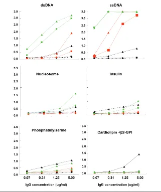
To evaluate the role of somatic muta-tion in the generamuta-tion of dsDNA binding, we reverted the mutations of the above three anti-dsDNA antibodies to their germline sequence and tested the vari-ants for self-reactivity (Figure 3). The three germline counterparts of the mu-tated antibodies differed with respect to
antigenic specificity. While the germline form of G3 still recognized dsDNA, the germline forms of the other two antibod-ies displayed minimal dsDNA binding (Figure 3, top panel); thus, somatic muta-tion was essential for dsDNA binding of A9 and B5, but not G3.
We also tested all antibodies for bind-ing to dsDNA in the crithidia assay which has been shown to be specific for high affinity antibodies. None displayed reactivity in this assay (data not shown) Figure 1.Potential pathogenicity of IgM
DNA antibodies. Three mutated anti-bodies were tested for binding to HEp-2 cells (left panel) and to isolated mouse glomeruli (right panel). Monoclonal anti-body B1 was used as a negative control.
suggesting that these antibodies bind dsDNA with relatively low affinity.
Previous studies have reported the presence of a large number of auto- and polyreactive antibodies in the transitional B cell repertoire of non-autoimmune in-dividuals. A defect in B cell tolerance during B cell maturation in lupus pa-tients results in an increased frequency of auto and polyreactive B cells in the naïve B cell repertoire. To ascertain the polyreactivity of the antibodies we iso-lated, we assayed the germline and mu-tated antibodies for binding to a set of
antigens previously used to characterize polyreactivity (10,19).
Both mutated and germline-encoded G3 and B5 exhibited strong ssDNA bind-ing. While the mutated A9 antibody bound to ssDNA, the unmutated coun-terpart did not. Thus, G3 bound both ssDNA and dsDNA prior to the acquisi-tion of mutaacquisi-tion; B5 bound ssDNA in the germline configuration but bound dsDNA only after mutation; A9 failed to bind ssDNA or dsDNA in the germline configuration but displayed both ssDNA and dsDNA binding after mutation. It
has been reported in models of SLE that anti-chromatin antibodies arise before antibodies to isolated DNA. G3 bound nucleosomes, exhibiting higher binding in the mutated than the germline config-uration. Mutated A9 displayed a low level of nucleosome binding. Thus, these antibodies did not appear to begin as anti-nucleosome antibodies, and acquire binding to naked dsDNA only as a con-sequence of somatic mutation. Rather, it appeared that even nucleosome binding was acquired or enhanced by somatic mutation. In addition, G3 displayed weak binding to human recombinant in-sulin in both the germline and mutated configuration; mutated A9 bound insulin weakly and B5 did not bind insulin ei-ther in the germline or the mutated con-figuration (see Figure 3). No binding to LPS was observed by any of the three an-tibodies, either in the germline or mu-tated configuration (data not shown). Thus, only G3 can be considered to have arisen from a polyreactive precursor.
Some anti-dsDNA antibodies cross-react with phospholipids, which have been suggested to be triggering antigens in SLE. We, therefore, tested the binding of the mutated antibodies and their germline variants to phospholipid anti-gens (see Figure 3). Mutated A9 exhib-ited detectable binding to both phos-phatidylserine and cardiolipin, while mutated G3 bound to phosphatidylserine only. All germline variants lacked detect-able binding to phospholipids, suggest-ing that phospholipids are not elicitsuggest-ing antigens, although they might be select-ing antigens after somatic mutation.
It is important to note that we tested all antibodies as IgG1, but they were present in the patient as IgM antibodies. In solution, IgM might be pentameric and display increased avidity for anti-gen. On the membrane of the B cell, however, the avidity for antigen would depend on the density of the BCR on the cell membrane.
Interestingly, while A9 required mu-tation for dsDNA binding and the R/S ratio suggests antigen selection, this an-tibody does not display the greatest Figure 3.Antigenic reactivity of mutated antibodies in comparison to their unmutated
dsDNA binding. Perhaps DNA was not the selecting antigen or there was a high density of antibody of the B cell membrane providing a high avidity for antigen.
Basic Amino Acids Contribute to dsDNA Binding
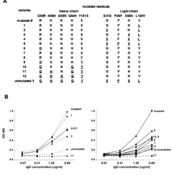
To understand which mutations were crucial for the acquisition of dsDNA binding, we produced a set of variants of A9 and B5, each with different muta-tions. As shown in Figure 4 (lower left panel), the A9 mutated heavy chain with an unmutated light chain (variant 5) ex-hibited diminished, but still detectable, binding to dsDNA. No difference was observed in dsDNA binding between variants 4 and 5, suggesting the mutation P46F does not affect dsDNA reactivity. However, when we introduced muta-tions S31G and S66N to generate variant 1, the dsDNA reactivity was similar to the wild-type A9. Thus, light chain muta-tions contribute to an increase in dsDNA binding.
We then analyzed the effects of muta-tions in the heavy chain of A9. As shown in Figure 4 (lower right panel), variant 12, which possessed the unmutated heavy and mutated light chain, com-pletely lost binding to dsDNA. The mu-tations G55N and Q64H had no effect on dsDNA binding. In contrast, the muta-tions N58K, G50R, and Y101S signifi-cantly enhanced dsDNA binding (vari-ants 8, 9, and 10). The back mutation of any of these three mutations to their germline sequences (variants 6, 7, and 8) resulted in significantly decreased dsDNA binding.
Lys at position 76 is essential for dsDNA reactivity of B5, as the variant containing a K76N reversion signifi-cantly lost dsDNA binding (data not shown).
All variants derived from G3 showed identical dsDNA binding (data not shown).
DISCUSSION
Sequence analysis of IgG anti-dsDNA antibodies derived from autoimmune
mouse models has revealed extensive so-matic mutation in the CDRs and a role for basic amino acids, generally acquired by mutation, in DNA-binding. The germline variants of these antibodies often fail to bind dsDNA, although they may bind ssDNA, suggesting a crucial role for somatic mutation in dsDNA binding.
Only four human IgG anti-dsDNA an-tibodies from patients with SLE have been analyzed previously and all of these also fail to bind DNA in the germline configuration (7–9). Since the number of sequenced dsDNA-binding antibodies derived from lupus patients is still very
limited compared with those isolated from different autoimmune mouse mod-els, it remains a question whether con-clusions drawn from murine models can be applied fully to the human dsDNA-antibody repertoire.
lated and characterized, and the effect of somatic mutations on human IgM anti-dsDNA antibodies has not been studied.
IgM producing cells displaying evi-dence of somatic mutation are considered to be IgM memory cells. These cells are diminished in lupus patients (12), most likely reflecting increased Ig-class switch-ing of these B cells in a pro-inflammatory environment. We were interested in the characterization of these IgM antibodies as potential precursors of class-switched anti-dsDNA antibodies in lupus patients. We were particularly interested in deter-mining whether these antibodies are de-rived from autoreactive naïve B cells that mature to immunocompetence due to an ineffective tolerance checkpoint, or whether they acquired autoreactivity through somatic mutation and reflect in-effectiveness of a later checkpoint, acting after antigen activation.
Although the three IgM anti-dsDNA antibodies in this report came from the same lupus patient, they displayed dif-ferent profiles. They all differed with re-spect to R/S ratio and, therefore, differed with respect to evidence for antigen se-lection. Reversion of mutations in au-toantibodies A9 and B5 resulted in de-creased dsDNA binding. In contrast, the germline form of G3 recognized dsDNA. These results suggest that mutated IgM anti-dsDNA antibodies with pathogenic glomerulotropic potential may develop from both DNA- and non-DNA–reactive naïve B cells.
It is of interest to speculate that clinical disease may become apparent as these antibodies actually switch to IgG, and, then, can activate Fc-receptor-bearing cells and transport DNA to toll-like re-ceptor 9 to lead to dendritic cell activa-tion, especially in the pro-inflammation milieu in lupus patients which promotes class-switch recombination even outside of germinal center.
It has been reported that there is an ac-cumulation of arginine, lysine, and as-paragine (Arg, Lys, and Asn) residues at contact sites of anti-dsDNA antibodies. Arg and Lys are basic amino acids with positive charges, and might, therefore,
in-crease the affinity of an antibody to nega-tively charged DNA by electrostatic inter-actions and hydrogen bonds. Asn is un-charged, but may interact with DNA either by donating or by accepting hydro-gen bonds (16). Consistent with this para-digm, antibody A9 and B5 acquired sev-eral basic amino acids through somatic mutations. The Arg and Lys at position 50 and 58 of the heavy chain was crucial for the binding of A9 to dsDNA, and Lys at position 76 was essential for anti-dsDNA binding of B5. As Lys 76 was lo-cated in FR3, it is clear that not only CDRs but also FRs can contribute to the dsDNA binding. In addition, Asn at posi-tion 66 of the light chain was important for dsDNA binding of A9, whereas the acquisition of other Asn residues, such as G55N and S55N in the heavy chains of A9 and B5, and S93N and D17N in the light chains of B5 and G3, had no signifi-cant effect on dsDNA binding. Therefore, it is not possible to predict the effects of particular amino acid residues on reactiv-ity to DNA without crystal structure. The data, however, are consistent with self antigen functioning in positive selection after B cell activation.
While some studies of murine anti-DNA antibodies have suggested a major contribution of the heavy chain to DNA reactivity, others have reported that the light chain also can play a critical role in DNA binding in some antibodies (6,25,26). Moreover, a recent analysis of two human IgG anti-dsDNA antibodies shows that reverting somatic mutations of either heavy or light chains indepen-dently results in a loss of dsDNA bind-ing (9). This also was seen with antibody A9, as mutations in both heavy and light chains play an important role for dsDNA binding. Decoding the relative contribu-tion of each chain will require the analy-sis of many more antibodies. Such an analysis also may reveal if there are par-ticular heavy or light chains that are highly likely to confer DNA binding in-dependent of their partner.
It has been reported that self-reactive antibodies rarely are found in the IgM memory B cell compartment in healthy
humans, and that the rare self-reactive IgM memory B cells that were present are most likely not derived from naïve autoreactive B cells (22). DNA-binding IgM-producing B cells can exist in SLE because of a failure at an early tolerance checkpoint, as seen in antibody G3. Al-ternatively, their presence may reflect a failure in tolerance of B cells that acquire autoreactivity by somatic mutation, such as antibodies B5 and A9. The analysis of these three antibodies suggests that both these checkpoints might be defective in a single patient with SLE.
It will be necessary to study a large number of patients in detail in a similar fashion to determine if multiple toler-ance checkpoints, some preceding anti-gen-activation and some after antigen-activation, must fail for a lupus-like phenotype to develop, or if it is possible to display defects only in early or late tolerance checkpoints and still develop disease.
This study also suggests that the trig-gering antigen(s) in SLE may be more promiscuous than some studies suggest. In the context of multiple defects in gen-eration and maintenance of B cell toler-ance, both self and non-self antigens may activate or sustain autoreactivity.
ACKNOWLEDGMENTS
The study was supported by a grant from the National Institutes of Health (BD). AMJ is supported by the Irvington Institute Fellowship Program of the Can-cer Research Institute.
REFERENCES
1. Tan EM. (1991) Autoantibodies in pathology and cell biology. Cell67:841–2.
2. Renaudineau Y, et al. (2006) Association of alpha-actinin-binding double-stranded DNA anti-bodies with lupus nephritis. Arthritis Rheum. 54:2523–32.
3. Vlahakos D, et al. (1992) Murine monoclonal anti-DNA antibodies penetrate cells, bind to nuclei, and induce glomerular proliferation and protein-uria in vivo. J. Am. Soc. Nephrol. 2:1345–54. 4. Vlahakos DV, et al. (1992) Anti-DNA antibodies
form immune deposits at distinct glomerular and vascular sites. Kidney Int. 41:1690–700.
glomerular eluates from patients with systemic lupus erythematosus. Association of high avidity antinative DNA antibody with glomerulonephri-tis. J. Clin. Invest. 59:90–6.
6. Radic MZ, et al. (1993) Residues that mediate DNA binding of autoimmune antibodies. J. Im-munol. 150:4966–77.
7. Lambrianides A, et al. (2007) Arginine mutation alters binding of a human monoclonal antibody to antigens linked to systemic lupus erythemato-sus and the antiphospholipid syndrome. Arthritis Rheum. 56:2392–401.
8. Rahman A, et al. (2001) The importance of so-matic mutations in the V(lambda) gene 2a2 in human monoclonal anti-DNA antibodies. J. Mol. Biol. 307:149–60.
9. Wellmann U, et al. (2005) The evolution of human anti-double-stranded DNA autoantibod-ies. Proc. Natl. Acad. Sci. U. S. A. 102:9258–63. 10. Yurasov S, et al. (2005) Defective B cell tolerance
checkpoints in systemic lupus erythematosus. J.Exp. Med. 201:703–11.
11. Bernasconi NL, Traggiai E, Lanzavecchia A. (2002) Maintenance of serological memory by polyclonal activation of human memory B cells. Science298:2199–202.
12. Wehr C, et al. (2004) A new CD21low B cell popu-lation in the peripheral blood of patients with SLE. Clin. Immunol. 113:161–71.
13. Briere F, Servet-Delprat C, Bridon JM, Saint-Remy JM, Banchereau J. (1994) Human interleukin 10 in-duces naive surface immunoglobulin D+ (sIgD+) B cells to secrete IgG1 and IgG3. J. Exp. Med. 179:757–62.
14. Litinskiy MB, et al. (2002) DCs induce CD40-independent immunoglobulin class switching through BLyS and APRIL. Nat. Immunol. 3:822–9. 15. Pene J, et al. (2004) Cutting edge: IL-21 is a switch
factor for the production of IgG1 and IgG3 by human B cells. J. Immunol. 172:5154–7. 16. Rahman A, Giles I, Haley J, Isenberg D. (2002)
Systematic analysis of sequences of anti-DNA antibodies—relevance to theories of origin and pathogenicity. Lupus11:807–23.
17. Hochberg MC. (1997) Updating the American College of Rheumatology revised criteria for the classification of systemic lupus erythematosus. Arthritis Rheum. 40:1725.
18. Zhang J, et al. (2008) Identification of DNA-reactive B cells in patients with systemic lupus erythematosus [technical note]. J. Immunol. Methods. 338:79–84.
19. Wardemann H, et al. (2003) Predominant autoan-tibody production by early human B cell precur-sors. Science301:1374–7.
20. Zhao Z, et al. (2005) Cross-reactivity of human lupus anti-DNA antibodies with alpha-actinin and nephritogenic potential. Arthritis Rheum. 52:522–30.
21. Seal SN, Monestier M, Radic MZ. (2000) Di-verse roles for the third complementarity
deter-mining region of the heavy chain (H3) in the binding of immunoglobulin Fv fragments to DNA, nucleosomes and cardiolipin. Eur. J. Im-munol. 30:3432–40.
22. Tsuiji M, et al. (2006) A checkpoint for autoreac-tivity in human IgM+ memory B cell develop-ment. J. Exp. Med. 203:393–400.
23. Pankewycz OG, Migliorini P, Madaio MP. (1987) Polyreactive autoantibodies are nephritogenic in murine lupus nephritis. J. Immunol. 139:3287–94. 24. Xie C, Liang Z, Chang S, Mohan C. (2003) Use of
a novel elution regimen reveals the dominance of polyreactive antinuclear autoantibodies in lupus kidneys. Arthritis Rheum. 48:2343–52.
25. Ibrahim SM, Weigert M, Basu C, Erikson J, Radic MZ. (1995) Light chain contribution to specificity in anti-DNA antibodies. J. Immunol. 155:3223–33. 26. Radic MZ, Mascelli MA, Erikson J, Shan H,