R E S E A R C H
Open Access
Modelling effects of internalized antibody:
a simple comparative study
Vladas Skakauskas
1, Pranas Katauskis
1, Alex Skvortsov
2*and Peter Gray
2*Correspondence:
[email protected] 2Defence Science and Technology Organisation, 506 Lorimer st., VIC 3207 Melbourne, Australia Full list of author information is available at the end of the article
Abstract
Background: The modelling framework is proposed to study protection properties of antibodies to neutralize the effects of the plant toxin (ricin). The present study extends our previous work by including (i) the model of intracellular transport of toxin to the Endoplasmic Reticulum and (ii) the model of the internalised antibodies (when antibody is delivered directly into the cytosol).
Method: Simulation of the receptor-toxin-antibody interaction is implemented by solving the systems of PDEs (advection-diffusion models) or ODEs (rate models) for the underlying transport coupled with mass-action kinetics.
Results: As the main application of the enhanced framework we present a comparative study of two kinds (external and internalised) of antibodies. This
comparison is based on calculation of the non-dimensional protection factor using the same set of parameters (geometry, binding constants, initial concentrations of species, and total initial amount of the antibody).
Conclusion: This research will provide a framework for consistent evaluation and comparison of different types of antibodies for toxicological applications.
Keywords: Toxin, Antibody, Cell receptor, Intracellular transport
Background
The plant toxin ricin made from the seeds of the castor oil plant is highly toxic to mam-malian cells. It is one of the deadliest toxins known and is classified as a potential bioterror agent for which no treatment is available. Some promising results have been shown recently in the immunotherapeutic approach, i.e. application of antibodies to neutralise the effects of ricin [1-4]. With the recent progress in bio-engineering, antibodies with high affinity have been generated. The development and production of new antibodies is still an expensive process that usually includes extensive experimental studies with continu-ous experimental refinement of antibodies properties. Evidently, that such a retrospective evaluation of different antibodies aiming at selection of the best candidate may become very time and resource consuming.
In order to reduce this experimental burden a simple (but scientifically consistent) mod-elling framework has been recently proposed [5-9]. This framework enables extensive theoretical optimization studies to increase the protective potential of antibodies before proceeding with targeted experimental studies.
The mechanism of ricin intracellular transport involves a number of steps each with its complex phenomenology which are well-documented (see [1-4] and Refs. therein). Ricin consists of anA(RTA) andB(RTB) chain linked by a disulphide bond. RTB binds to a cell surface receptor triggering uptake and retrograde transport to the Endoplasmic Reticulum (ER). In the ER, the RTA and RTB chains are separated and the RTA is translo-cated across the ER membrane into the cytosol. Subsequently RTA reaches ribosome and damages the protein production machinery of the cell resulting in the cell death. In this context, the toxin concentration in the cytoplasm near ER becomes the critical quantity to estimate the toxicological impact of ricin on the cell and evaluate the protective potential of the antibody. This was a motivation to introduce a consistent quantitative characteristic for antibody comparison (see below).
For the sake of parametrisation simplicity the coarse-grained modeling framework pro-posed in [5-9] seemingly ignores these fine details of toxin binding and internalisation. In fact, it is aimed at capturing the complexity of these processes by means of a small number of ‘aggregated’ rate constants that can be (or have been) evaluated experimen-tally or numerically. Such kind of models becomes a conventional tool in pharmacological modeling (for example, see [10] and Refs. therein). From the chemical point of view the framework is similar to one well-established in electrochemistry where it is used for esti-mation of uptake rates of the heavy metal ions from the environment, see [11-13]. A practical application of the proposed models involves a numerical (or sometimes ana-lytical) solution of a nonlinear system of PDEs (diffusion kinetics) for a given set (or range) of antibody parameters (i.e. binding rates, concentration) to infer the effect of these parameters on the protective potential of the antibody.
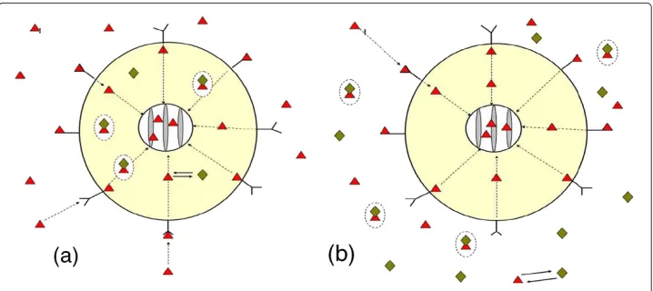
In the present paper we extend our previous work [5-9] by refining models for intra-cellular transport and chemical interaction of species. Motivated by experimental studies available in the literature [1-4] and possible toxicological applications we consider two scenarios of antibody delivery. In the first scenario (below refer to asScenario I) the anti-body is placed inside the cell between the ER and cell membrane. The toxin initially is delivered outside the cell (in the extracellular domain). Then it moves toward the cell and interacts with receptors on the cell membrane. Some of toxin penetrates into the cell, where it further moves toward the ER (see Figure 1a) and eventually interacts with antibody.
In the second scenario (Scenario II) the antibody is delivered outside the cell where it initially interacts with the toxin partially neutralising it. The free toxin moves toward the cell membrane where it interacts with membrane receptors. Some of toxin penetrates into the cell where it is subsequently transported toward ER (see Figure 1b).
To model biologically complex phenomenology of species transport in cell we employ the following physical mechanisms: (i) diffusion (for species in extracellular space), (ii) diffusion and advection (direction transport via microtubule network) for toxin and only diffusion for antibody and toxin-antibody complex inside the cell, (iii) mass-action kinetics (to form complexes and describe binding processes).
Figure 1 The schematic diagram of receptor-toxin-antibody system:(a)- Scenario I and
(b)- Scenario II.– toxin,♦– antibody, conglutinatedand♦in circle – toxin-antibody complex, external sphere – cell membrane, internal sphere – ER envelope.
for each scenario. The simplified (sometimes called compartment) models are based on the ‘well-mixed’ assumption which states that all species have uniform concentration across the modeling domain. This assumption eliminates the a necessity to calculate the gradient driven fluxes in the models (i.e. diffusion) leading to a significant simplification (translation from PDEs to ODEs). By comparing outputs of complete and reduced mod-els we can infer and defensively comment on the trade-off between simplicity and fidelity in modeling approach to an evaluation of each scenario.
As the main application of this framework we present a comparative study of external (conventional) and internalised antibodies. More specifically, for the same set of parame-ters (geometry, binding constants, initial concentration or initial amount of antibody) we calculate the non-dimensional parameter
δ(t)= uT(t,ρn)|A∈i
uT(t,ρn)|A∈e
, (1)
which is a ratio of reductions of toxin concentration in the cytoplasm due to introduc-tion of two kinds of antibodies. HereuT(t,ρn)|A∈i,uT(t,ρn)|A∈e is the concentration
of internalized toxin near ER for the case of internalized (i) and external antibody (e). The time evolution of this aggregated parameterδ(t)enables a consistent comparison of two scenarios of antibody treatment (i.e. either delivered extracellularly or into the cytosol). For instance, the conditionδ < 1 indicates that internalised antibody performs better than conventional antibody, while atδ >1 the conventional antibody outperforms. This comparison is the main outcome of our study.
The paper is organised as follows. In Section ‘The models’ we introduce models for toxin-antibody interaction outside and inside the cell. Results are discussed in Section ‘Numerical results’. Conclusions and summarising remarks are presented in Section ‘Conclusions’.
Notation
We use the notation of papers [5-7]:
T and A – the toxin and antibody;
ρ– the distance to the origin;
Sm= {ρ:ρ=ρm}– the surface of the membrane of a spherical cell;
Se= {ρ:ρ=ρe},ρe> ρm– the surface of the external sphere (external surface of e);
e= {ρ:ρ∈(ρm,ρe)}– the extracellular domain;
Sn= {ρ:ρ=ρn},ρn< ρm– the surface of the spherical envelope of the domain
occupied by ER;
i= {ρ:ρ∈(ρn,ρm)}– the intracellular domain;
r0– the concentration of receptors confined to the cell membrane; θ– the fraction of the toxin-bound receptors;
r0θ – the concentration of the toxin-bound receptors;
r0(1−θ)– the concentration of the free receptors;
uAanduC– the concentrations of the antibody and toxin-antibody complex, respectively, in the domainsiore;
uT– the toxin concentration ine∪i;
u0
T,u0Ae,uA0i– the initial concentrations of the toxin and antibody ineandi,
respectively;
κT,κA,κC– the diffusivity of the toxin, antibody, and toxin-antibody complex, respectively;
k1,k−1– the forward and reverse constants of the toxin-antibody reaction rate;
k2andk−2– the forward and reverse binding rate constants of the toxin and receptor confined on the membrane;
k – the toxin internalization rate constant fromeacross the membrane into the cell; γ – the toxin absorption rate constant describing toxin influx into ER;
v(ρ),v>0– the toxin advective velocity; ∂t=∂/∂t, =ρ−2∂
∂ρ(ρ2∂ρ∂ )– the Laplace operator.
The models
As mentioned above we study two scenarios of antibody delivery. In Scenario I the
antibody is delivered inside the cell, i.e. in domaini(space between the ER and cell mem-brane) while inScenario IIit is delivered outside the cell (space between cell membrane and external boundary of the extracellular domain,e). In theScenario Itoxin being ini-tially ine(extracellular domain) moves toward the cell and interacts with receptors on the cell membrane. Subsequently some of toxin penetrates into the intracellular domain,
i, where it moves toward ER and interacts with antibody. Toxin competitively reacts with antibody in the extracellular domain (Scenario II) or inside the cell (Scenario I). This ‘blocking’ reaction results in a reduced toxin concentration on the ER envelope and is the main bio-chemical mechanism of toxin neutralization by antibody.
In order to extend our results from a single cell model to many-cell systems we impose the no-toxin flux boundary condition on the external surface Se of the extracellular domaine(to mimic periodicity of the infinite system). This also accounts for conserva-tion of species in the system and enables consistent simulaconserva-tion of depleconserva-tion effects (see [13] for details).
have been modified to include toxin directional transport and diffusion of all species in the intracellular domain (by adding appropriate advection and diffusion terms, e.g. see [15,16]). To describe extracellular transport and toxin binding by cell receptors we model the cell as a partially absorbing sphere with distributed binding sites that follow the Langmuir adsorption model. In the intracellular domain of theScenario Ithe toxin transport is modelled by the advection-diffusion equation in which an advective velocity (drift) is introduced to account for directional transport via microtubule network. For the transport of the antibody and antibody-toxin complexes we still use the pure diffusion mechanism. We also assume that antibody and toxin-antibody complexes do not inter-nalize and never cross the cell membrane or the ER envelope (internal concentric sphere). The schematic diagram of receptor-toxin-antibody system is presented in Figure 1.
Since the process of receptor binding is very rapid (seconds) and toxin internalization is relatively slow (hours) we can effectively decoupled the model for extracellular and intracellular domains and solve them sequentially. The relative values of time scales for receptor binding and toxin internalization have also been validated retrospectively and found to be consistent with the initial assumption of domains decoupling (see below).
The Advection-Diffusion Model used to evaluateScenario Iis an extension of the mod-els previously published [4-9]. The following new processes have been included in our simulations: (i) the intracellular traffic that captures the diffusion of all species and the main features of the toxin microtubule transport, (ii) chemical interaction of all species inside the cell (see [15] and Refs. therein), and (iii) process of the toxin transport to ER.
In order to model toxin transport towards the ER a special boundary condition was introduced on the ER envelope. The radius of that spherical envelope was about the size of the domain occupied by the ER. It is assumed that the mixed (or radiation) bound-ary condition for species concentrations can capture (at least phenomenologically) the complexity of toxin kinetics inside the ER [17].
Now we can formulate the equations for the Advection-Diffusion Model (refer to as the ADM1 model). In extracellular spaceewe have the following set of equations:
⎧ ⎪ ⎪ ⎪ ⎪ ⎪ ⎪ ⎨ ⎪ ⎪ ⎪ ⎪ ⎪ ⎪ ⎩
∂tuT=κT uT, ρ∈(ρm,ρe), t>0,
∂ρuT =0, ρ=ρe, t>0,
∂ρuT = r0
κT(k2(1−θ)uT−k−2θ), ρ=ρm, t>0, uT|t=0=u0T, ρ∈(ρm,ρe),
(2)
∂tθ =k2(1−θ)uT−k−2θ −kθ, ρ=ρm, t>0,
θ|t=0=0, ρ=ρm.
(3)
Function θ determined from solution by these equations is used as the boundary
condition in the equations in intracellular domaini: ⎧
⎪ ⎪ ⎪ ⎪ ⎪ ⎪ ⎪ ⎪ ⎨ ⎪ ⎪ ⎪ ⎪ ⎪ ⎪ ⎪ ⎪ ⎩
∂tuT= −k1uTuA+k−1uC+κT uT+∂ρ(vuT)+2vuT/ρ, ρ∈(ρn,ρm), t>0,
κT∂ρuT =r0kθ−vuT, ρ=ρm,t>0, κT∂ρuT =(γ−v)uT, ρ=ρn, t>0, uT|t=0=0, ρ∈(ρn,ρm),
⎧ ⎪ ⎪ ⎪ ⎪ ⎪ ⎨ ⎪ ⎪ ⎪ ⎪ ⎪ ⎩
∂tuA= −k1uTuA+k−1uC+κA uA, ρ∈(ρn,ρm), t>0,
∂ρuA=0, ρ=ρm,t>0,
∂ρuA=0, ρ=ρn, t>0, uA|t=0=u0Ai, ρ∈(ρn,ρm),
(5) ⎧ ⎪ ⎪ ⎪ ⎪ ⎪ ⎨ ⎪ ⎪ ⎪ ⎪ ⎪ ⎩
∂tuC=k1uTuA−k−1uC+κC uC, ρ∈(ρn,ρm), t>0,
∂ρuC =0, ρ=ρm,t>0,
∂ρuC =0, ρ=ρn, t>0, uC|t=0=0, ρ∈(ρn,ρm).
(6)
The set of Eqs. (2)–(6) composes the ADM1 model. The main parameter of interest for theScenario Iis the antibody protection factor (a relative reduction of the internalized toxin due to application of antibody) defined by the expression
μ1(t)= uT(t
,ρn;u0
A)|A∈i
uT(t,ρn; 0) . (7)
By definition, 0≤μ1(t)≤1 with the lower values ofμ1(t)corresponding to the more
profound therapeutic effect of antibody treatment.
To simplify the ADM1 model we employ the ‘well-mixed’ assumption [5]. This assump-tion implies that all species (toxin, antibody, and toxin-antibody complex) are uniformly distributed in the calculation domain for all time, so there is no spatial gradients of con-centrations. Under this condition all flux terms disappear from the equations of ADM1. The process of toxin internalization (i.e. flux of toxin through the cell surfaceSmand ER envelopeSn) can be modelled as an appropriate rate of toxin removal fromeandi, respectively.
It is worth emphasizing that a reduction to the ‘well-mixed’ (or compartment) mod-els requires fulfilment of some condition that can be formulated in terms of a smallness of the ratio = τκ/τR, where τκ andτR is a diffusion and reaction time scale in the system, respectively (diffusion can quickly restore any spatial inhomogeneity of species distribution maintaining uniform concentration). In the simplest case τκ ∼ a2/κ and
τR∼1/k, whereais a characteristic length scale,κis a scale of diffusivity andkis the scale of reaction rate. This is condition for what is called diffusion- or reaction–dominated regimes in diffusion kinetics [18]. Indeed, this condition does not always hold and we do not assume that it satisfies automatically in the context of our study. Since our models are characterized by a number of spatial scales (extracellular and intracellular domains, ER envelope), hierarchy of reaction rates and diffusion coefficients, it is very difficult (or even impossible) to formulate and validate any general criteria for the feasibility of the well-mixed assumption. For this reason we take a heuristic approach in which this assumption is validated retrospectively by comparing the output of the models, viz. the full (advection-diffusion) and reduced (compartment) models.
Under the well-mixed assumption the ADM1 model reduces to two ODEs. ⎧ ⎪ ⎪ ⎪ ⎪ ⎪ ⎨ ⎪ ⎪ ⎪ ⎪ ⎪ ⎩ ˙
uT = −k3r0(k2(1−θ)uT−k−2θ), t>0,
uT|t=0=u0T, ˙
θ =k2(1−θ)uT−k−2θ−kθ, t>0, θ|t=0=0,
for extracellular domaineand three ODEs for inracelluar domain: ⎧ ⎪ ⎪ ⎪ ⎪ ⎪ ⎪ ⎪ ⎪ ⎪ ⎪ ⎪ ⎨ ⎪ ⎪ ⎪ ⎪ ⎪ ⎪ ⎪ ⎪ ⎪ ⎪ ⎪ ⎩ ˙
uT = −k1uTuA+k−1uC+kk4r0θ−k5γuT, t>0,
uT|t=0=0, ˙
uA= −k1uTuA+k−1uC, t>0,
uA|t=0=u0Ai, ˙
uC =k1uTuA−k−1uC, t>0,
uC|t=0=0,
(9)
withk3=3ρm2/(ρe3−ρm3),k4=3ρm2/(ρm3 −ρn3),k5=3ρn2/(ρm3 −ρn3).
We call systems (8), (9) as the Well-Mixed Model 1 (WM1).
Analogously we formulate two models used to evaluate antibody protection properties inScenario II. For the extracellular domainewe employ the Diffusion Model proposed in [5-7]: ⎧ ⎪ ⎪ ⎪ ⎪ ⎪ ⎨ ⎪ ⎪ ⎪ ⎪ ⎪ ⎩
∂tuT= −k1uTuA+k−1uC+κT uT, ρ∈(ρm,ρe),t>0, ∂ρuT =0, ρ=ρe,t>0,
κT∂ρuT =r0{k2(1−θ)uT−k−2θ}, ρ=ρm, t>0,
uT|t=0=u0T, ρ∈(ρm,ρe),
(10) ⎧ ⎪ ⎪ ⎪ ⎪ ⎪ ⎨ ⎪ ⎪ ⎪ ⎪ ⎪ ⎩
∂tuA= −k1uTuA+k−1uC+κA uA, ρ∈(ρm,ρe), t>0, ∂ρuA=0, ρ=ρe,t>0,
∂ρuA=0, ρ=ρm, t>0, uA|t=0=u0Ae, ρ∈(ρm,ρe),
(11) ⎧ ⎪ ⎪ ⎪ ⎪ ⎪ ⎨ ⎪ ⎪ ⎪ ⎪ ⎪ ⎩
∂tuC=k1uTuA−k−1uC+κC uC, ρ∈(ρm,ρe), t>0,
∂ρuC =0, ρ=ρe,t>0,
∂ρuC =0, ρ=ρm, t>0, uC|t=0=0, ρ∈(ρm,ρe),
(12)
∂tθ =k2(1−θ)uT−k−2θ −kθ, ρ=ρm, t>0,
θ|t=0=0, ρ=ρm, (13)
with toxin concentration inibeing described by the Advection-Diffusion equation ⎧ ⎪ ⎪ ⎪ ⎪ ⎪ ⎨ ⎪ ⎪ ⎪ ⎪ ⎪ ⎩
∂tuT=κT uT+∂ρ(vuT)+2vuT/ρ,ρ∈(ρn,ρm), t>0,
κT∂ρuT =r0kθ−vuT, ρ=ρm,t>0,
κT∂ρuT =(γ−v)uT, ρ=ρn, t>0, uT|t=0=0, ρ∈(ρn,ρm).
(14)
We refer to systems (10), (13) as the Advection-Diffusion Model 2 (ADM2). The main parameter of interest evaluated with this model is the antibody protection factor
μ2(t)= uT(t
,ρn;u0A)|A∈e
Similarly to theScenario I we can apply the well-mixed assumption to simplify this model and reduce it to a set of ODEs
⎧ ⎪ ⎪ ⎪ ⎪ ⎪ ⎪ ⎪ ⎪ ⎪ ⎪ ⎪ ⎪ ⎪ ⎪ ⎪ ⎪ ⎨ ⎪ ⎪ ⎪ ⎪ ⎪ ⎪ ⎪ ⎪ ⎪ ⎪ ⎪ ⎪ ⎪ ⎪ ⎪ ⎪ ⎩ ˙
uT = −k1uTuA+k−1uC−k3r0(k2(1−θ)uT−k−2θ), t>0,
uT|t=0=u0T, ˙
uA= −k1uTuA+k−1uC, t>0,
uA|t=0=u0Ae, ˙
uC =k1uTuA−k−1uC, t>0,
uC|t=0=0, ˙
θ =k2(1−θ)uT−k−2θ−kθ, t>0, θ|t=0=0
(16)
ineand
˙
uT =kk4r0θ−γk5uT, t>0,
uT|t=0=0
(17)
ini. We call system (16) and (17) as the Well-Mixed Model 2 (WM2).
To compare the antibody protection capability in both scenarios (i.e. to infer which scenario of antibody delivery is more beneficial for a particular type of antibody) we use functionδ(t)defined in Eq. (1) in whichuT(t,ρn)|A∈ianduT(t,ρn)|A∈eare the toxin
concentrations near ER determined for theScenario I andII, respectively. It is evident that the following identity holds,δ(t)=μ1(t)/μ2(t).
It is convenient to translate Eqs. (1)–(17) to the non-dimensional form by using scales ofτ∗ (time),l(length), and u∗(concentration). By substituting new variables, ρ = lρ¯, t = τ∗¯t,r0 = lu∗¯r0,uT = u∗uT¯ , uA = u∗uA¯ ,uC = u∗uC¯ , u0T = u∗u¯0T,u0A = u∗u¯0A, ¯
k1=τ∗u∗k1,k¯2=τ∗u∗k2,k¯−1=τ∗k−1,k¯−2=τ∗k−2,k¯=τ∗k,κ¯T =τ∗κTl−2,γ =lτ∗−1γ¯, v=lτ−1
∗ v¯,κ¯A=τ∗κAl−2,κ¯C =τ∗κCl−2,k¯3=lk3,k¯4=lk4,k¯5=lk5, into (1)–(17) we can
deduce the same systems, but now in non-dimensional form. Therefore, for simplicity in what follows, we treat system (1)–(17) as non-dimensional.
Numerical results
We treated Eqs. (2)–(6), (8), (9), (10)–(14), and (16), (17) numerically fort >0 in spher-ically symmetric domainsρ ∈ (ρm,ρe)andρ ∈ (ρn,ρm), respectively. We solve PDEs, Eqs. (2)–(6) and Eqs. (10)–(14), by an implicit finite-difference scheme [14]. To solve ODEs, Eqs. (8), (9) and Eqs. (16), (17), we apply the Runge–Kutta scheme.
Our selection of the values of parameters for the models (2)–(6), (8), (9), (10)–(14), and (16), (17) was motivated by the values available in the literature [2,5,19-21] with an extended range to allow exploration and illustration of the various transport regimes that are possible inside the cell. The following values were used in simulations [18]: u∗ = 6.02·1013cm−3, τ∗ = 1 s,r0 = 1.6·104/Sm, where 1.6·104is the total number of
receptors of the cell,l=10−2cm,Sm=4πρm2 =4π·10−6cm2,¯r0=2.115·10−3. Values
of the dimensionless parameters were the following:ρn = 0.02,k = 3.3·10−5,k1 =
1.3·10−2, k−
1 = 1.4·10−4,k2 = 1.25·10−2, k−2 = 5.2·10−2,κA = 10−2,κC =
We expect that the chosen values of the parameters were representative enough to illus-trate a rich variety of possible scenarios of the evolution of the Receptor-Toxin-Antibody system and provide a reasonable estimate of time scales of the associated dynamics.
The Advection-Diffusion Models enables the fine spatial-temporal resolution of species concentration and provide valuable insights into the phenomenology of species interac-tion in our system. We found them to be useful tools for understanding the evoluinterac-tion of aggregated parametersμ1,μ2, δ predicted by our simulations. More specifically, by
means of the Advection-Diffusion Models we were able to identify a number of important regimes of toxin penetration that may have significant implications for the assessment for the antibody protection properties.
These regimes can be best described by using the ‘transport phenomena’ terminol-ogy which is well-established in chemical engineering, for instance see [22,23]. Initially all species are concentrated in spherical layers between the external sphere, cell mem-brane, and ER envelope (i.e.(ρm,ρe)and(ρn,ρm)). This layer structure results in strong radial gradient of toxin concentration which facilitates the development of diffusion fluxes across the system (since cell membrane and ER envelope are ‘penetrable’). Without anti-body toxin would eventually be flushed out of the system when it reaches the ER envelope driven by the pure diffusion mechanism. The advection velocity will change toxin trans-port at one part of the system (from diffusion dominated to advection dominated). How much toxin can be blocked by antibody during this transition to ER depends on a number of factors. They include the initial reactants concentration (toxin and antibody), relative value of toxin advection flux, and reaction time of toxin-antibody binding. It also depends on the availability of antibody in the areas of high toxin concentration to maximize the effective toxin-antibody binding. The last condition (local availability of antibody for binding toxin) is determined by the antibody diffusivity and can be validated by inspection of the solutions of Advection-Diffusion Models (spatial-temporal outputs). This valida-tion enables justifiable comments on the relative reducvalida-tion of toxin in the output of the system (on ER envelope) and the specific values of antibody protection parametersμ1, μ2. This approach was employed in our study to identify and investigate the most
‘vulner-able’ regimes of toxin penetration and comment on the shapes of functionsμ1(t),μ2(t).
Some of these regimes are presented in examples below.
Furthermore such a ‘chemical engendering’ approach provides a clear path for the sys-tem optimization. Indeed, from the formal point of view our syssys-tem (cell) can be cast as a chemical reactor with the only output (product), being the toxin concentration at the ER envelope. In this context, the aim of antibody treatment is to reduce this output by dis-tributing the minimal amount of a given antibody across the system. We anticipate that in such settings the optimization problem can be tacked in reduced parameter space by using the established framework for chemical reactor design [23,24].
u0i
A =ηu0Ae, η= (ρe/ρm)
3−1
1−(ρn/ρm)3 (18)
which follows from the simple geometrical arguments. All calculations were performed forρe =0.18,ρm = 0.1,ρn = 0.02 for whichη= 4.871. This condition becomes espe-cially important for the comparison of full and reduced models. Unless stated otherwise the value of all parameters (except depicted in plots) are assumed to be fixed and equal to those given above.
As mentioned, our simulations reveal a number of interesting regimes of toxin prop-agation. In particular, in Scenario I we observed that there was a range of parameters (γ,κT,v) for which the diffusive flux of toxin,κT∂ρuT|ρ=ρm = (r0kθ − vuT)|ρ=ρm,
becomes negative near the cell membrane, i.e., it is opposite to the advective flux directed toward ER. This may reduce the total toxin flux inside the cell and provide more favorable condition for toxin-antibody binding.
0 200 400 600 800 1000
0,4 0,6 0,8 1,0
0 1000 2000 3000 4000
0,7 0,8 0,9 1,0
t [s]
(b)
1 2
3 2
1
t [s]
1 2
3
(a)
Figure 2 Influence of toxin diffusivity and absorption constant on antibody protection factor.
(a)Scenario I. Solid and dashed lines correspond to value ofuTdetermined forγ=0.1 andγ=0.05. Toxin
0 1000 2000 3000 4000 1
2 3
δ
t [s]
1
3
3
2
1
2
Figure 3 Effect of variation of toxin diffusivity on parameterδin caseu0,Ae=1,u0,Ai=4.87.Solid and dashed lines correspond to value ofδdetermined forγ =0.1 andγ=0.01. Toxin diffusivityκT: 10−2(1),
10−3(2), 5·10−4(3).
In other words this phenomenon is a direct result of toxin transport being dominated by directional advection (due to velocityv). In our simulations this regime occurred for slow toxin transport characterized by parametersγ,κT,vand the slow forward reaction rate for toxin-antibody binding. It is worth mentioning that the similar phenomenon has been reported in toxicological studies [17]. We found that the development of this ‘advection
10
-110
010
110
210
310
410
510
60 1 2 3 4 5
t [s]
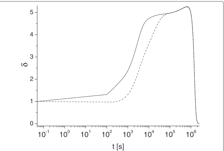
Figure 4 Long-time asymptotic behavior of metric parameterδ(t).Solid and dashed lines correspond to
0 200 400 600 800 1000 0,0
0,5 1,0 1,5 2,0 2,5
0 200 400 600 800 1000
0,5 1,0 1,5 2,0 2,5
t [s]
(a)
1
1
2
1
2
2
1
2
2
t [s]
1
2
2 1
1
1
2
(b)
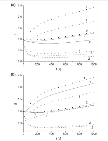
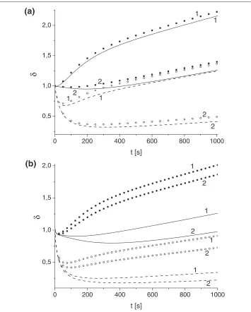
Figure 5 Effect of variation of toxin diffusivity and absorption constant on parameterδ.Toxin diffusivity:(a)κT=10−4,(b)κT=10−3. Solid (u0,e
A =u
0,i
A =1) and dashed (u
0,e A =1,u
0,i
A =4.87) lines
correspond toδdetermined by advection-diffusion models, and bullets (u0,Ae=uA0,i=1) and circles (u0,Ae=1, u0,Ai=4.87) toδdetermined by well-mixed models forγ: 0.1 (1), 0.01 (2).
dominated’ region of toxin transport with reverse diffusive flux has a positive effect on antibody protection properties since it effectively increases the efficiency toxin-antibody binding (by increasing time available for toxin-antibody reaction). We also solved problem (2)–(9) using position-dependent advective velocityv(ρ)which is zero at the membrane and then rapidly tends to a constant value asρ→ρn. In this case because of the boundary condition(4)2, the diffusive toxin flux near the cell membrane is directed to ER.
0 200 400 600 800 1000 0,5
1,0 1,5 2,0
0 200 400 600 800 1000
0,5 1,0 1,5 2,0
t [s]
(b)
12
1
2 1
2
1
2
(a)
t [s]
2 1
1 2
1
2 2
1
Figure 6 Effect of variation of toxin advection velocityvon parameterδ.Advection-diffusion models (solid lines):v=0.001,γ=0.01 (1),v=0.001,γ=0.1 (2),v=0.05,γ=0.01 (3),v=0.05,γ=0.1 (4); toxin diffusivity:(a)κT=10−3,(b)κT=10−4. Well-mixed models (dashed lines):γ=0.01 (1),γ=0.1 (2).
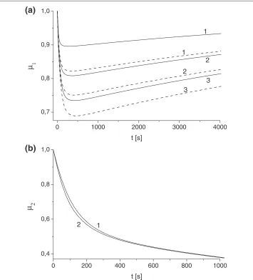
results demonstrate the general trend ofδ(t)increase with the increase ofκT andγ. For functionμ1(t)(Scenario I, Eq. (7)) we found noticeable non-monotonic behavior for all κT andγ. This implies that there is a time interval for which the protection potential of antibody inScenario Ireaches its maximum and then decays. According to Figure 2a this time interval is of order of 500 s.
Figure 2b demonstrates the evolution of antibody protection factorμ2(t)(Scenario II,
Eq. (15)) for a range of parametersκT andγ. Although the shape of functionμ2is very
different (it is always monotonically decreasing) we can conclude that the value of μ2
conclusion of insignificance of parametersκT andγ for the prediction of antibody pro-tection factors inScenario IIwhich is a direct consequence of equation decoupling for extracellular and intracellular domain discussed above.
The plots in Figure 3 illustrate the effect of variation of parametersκT andγ on the comparative protective skills of the same antibody. This effect is described in terms of metric factorδ(t), Eq. (1). Our simulations generally demonstrate the decrease of param-eterδ(t)asγ orκT decrease. The simulations have been performed for the same initial amount of antibody (Eq. (18)) as well as for the same initial concentration (results not pre-sented here). We observe that depending on value ofκTandγ there exists a time interval in whichδ(t) <1, i.e. the application of the antibody inScenario Iis more efficient than inScenario II. We found that this interval grows asκTorγ decreases. Overall we found that the conditionδ(t) <1 is rather sensitive to the ER absorption rateγ. This conclusion is intuitively clear since by reducing value ofγ we also reduce the toxin flux through the system facilitating more effective antibody-toxin binding which, in turn, leads to more favorable output forScenario I.
Figure 4 demonstrates some universal behaviors of functionδ(t)that we identified for a broad range parameters of our study. Our simulations reveal that for some intermediate time (t ∈(2·104; 106)seconds) functionδ(t)may approximately saturates to a constant plateau (depending on values ofγ andκ), before it rapidly decays to zero. This is illus-trated by two plots presented in Figure 4 (note different time scales). In particular we found that foru0Ai = u0Ae,γ = 10−1,κT = 10−2functionδ(t)varies very insignificantly (less that 10%, between 4.9 and 5.2) for the time interval (t∈[2·104; 5·105]) seconds, then it decreases and vanishes ast→ ∞. Note, the limitδ(t)→0 occurs at the unpractically long times, so it has more methodologically interest. We do not have a clear explanation of the specific numerical value of this saturation limit (δ(t)≈5); this will require further investigation.
Figure 5 shows the comparative results of the performance of the full Advection-Diffusion models (ADM1, ADM2) and the reduced models (WM1, WM2). We compare estimations of the metric parameterδ(t) provided by these models for different toxin diffusivityκT, ER absorbtion rateγ, and initial antibody concentration. We found that for the high toxin diffusivity (κT ≥ 10−2) the outputs of full model and reduced mod-els are almost identical. The differences between the estimations ofδ(t)increases asκT decreases and forκT <10−4the reduced models fails to provide any meaningful
predic-tions (Figure 5b). We found that even forκT >10−4the reduced models usually poorly handle the non-monotonic behavior of functionδ(t)at short times. At the long times we observed that for the broad range of parameters the full and reduced models are reason-ably aligned and can yield reasonable estimation ofδast → ∞(within 20% accuracy), Figure 5a.
vthe toxin neutralization time decreases and thereforeScenario I becomes less favor-able. We also noticed the improved accuracy of reduced models asvincreased. So, for givenκT andγ, the difference between δ(t)determined by Advection-Diffusion mod-els and Well-Mixed ones is small only if advective velocity,v, is small. But, for smallγ, it dramatically increases as vgrows, while for largeγ it increases but not so dramati-cally. This is due to the switching to the advection dominated transport of toxin in the system.
Conclusions
We present a new modeling framework to evaluate the protective potential of various antibodies. This framework is based on the equation for the diffusion- and advection-diffusion transport of all species in the extracellular and intracellular domains and include mass-action kinetics for toxin, receptor and antibody. The advection term and radiation boundary condition on the Endoplasmic Reticulum envelope were used to model the toxin transport via the microtubule network. We estimated the protection factor of antibody (relative reduction of toxin concentration near the ER) for two sce-narios of operational relevance, viz., when the antibody is delivered externally to the cell and when it is delivered directly into the cytosol. To provide a consistent com-parison of antibody performance we estimated the evolution of the metric factorδ(t), which is defined as the ratio of the protections factors of an antibody for each sce-narios. Based on this definition the condition δ(t) < 1 over a time span implies that the internally introduced antibody has better protective capability during this time while in the opposite case (δ(t) > 1) the internalized antibody is more effective. In this study simulated both cases. Our models reveal that depending on values of some parameters (primary toxin diffusivity and ER absorbtion rate) the plot of function δ(t) can significantly change its shape while keeping the same short- and long-time limits, by undergoing a saturation stage at the intermediate times with a universal value of
δ≈5.
To reduce the computational burden of the Advection-Diffusion Model (a system of nonlinear PDEs) we investigated its possible simplification. By employing the well-mixed assumption we reduced the Advection-Diffusion Model to the compartment or rate model (set of ODEs) which can be easily solved with any numerical solver. For an extended range of parameters we found that predictions of the compartment model can provide about±20% accuracy in estimation of antibody protection potential (depending on values of some other parameters), but with decreasing toxin diffusivity limit the compartment model become a poor predictor and should not be used in this context. This finding can provide some appreciation of a possible trade-off between simplification and fidelity in toxicological modelling and can be an important criterion for selection of operational models.
We would like to emphasise that the comparative study reported in the present paper can be treated only as the first step for the application of the proposed modelling framework to the practical pharmacological studies. More extensive simulations (to pro-vide statistically viable outcomes), data fitting and an established strategy for parameter optimization using toxicological observations will be necessary.
Competing interests
Authors’ contributions
The work presented here was carried out in collaboration between ors. VS extended previous work by including the model of intracellular transport via diffusion and advection. PK developed the computation algorithms and carried out simulations. AS co-designed the model and validated its outcome against available analytical results. PG conducted a literature review, defined the research theme and interpreted the results. All authors contributed to the writing of the article and read and approve the final manuscript.
Acknowledgements
The first two authors were supported by the Research Council of Lithuania (project No. MIP-052/2012).
Author details
1Faculty of Mathematics and Informatics, Vilnius University, 24 Naugarduko st., LT-03225 Vilnius, Lithuania.2Defence
Science and Technology Organisation, 506 Lorimer st., VIC 3207 Melbourne, Australia.
Received: 9 September 2013 Accepted: 31 January 2014 Published: 13 February 2014
References
1. Reisler BB, Smith LA:The need for continued development of ricin countermeasures.Adv Prev Med2012, 2012:149737.
2. Prigent J, Panigai L, Lamourette P, Sauvaire D, Devilliers K, Plaisance M, Volland H, Créminon C, Simon S: Neutralising antibodies against ricin toxin.PLoS ONE2011,6:e20166.
3. Pelat T, Avril A, Chahboun S, Mathieu J, Thullier P:Development of anti-toxins antibodies for biodefense.
J Bioterr Biodef2011,S7:001.
4. Song K, Mize RR, Marrero L, Corti M, Kirk JM, Pincus SH:Antibody to ricin A chain hinders intracellular routing of toxin and protects cells even after toxin has been internalized.PLoS ONE2013,8:e62417.
5. Skakauskas V, Katauskis P, Skvortsov A:A reaction-diffusion model of the receptor-toxin-antibody interaction.
Theor Biol Med Model2011,8:32.
6. Skakauskas V, Katauskis P, Skvortsov A:Numerical simulation of a receptor-toxin-antibody interaction.InProc. 12th Int. Conf. on Computational and Mathematical Methods in Science and Engineering (CMMSE): 2–5 July 2012. Edited by Vigo-Aguiar J. Murcia, Spain; 2012:1111–1117. http://gsii.usal.es/~CMMSE/images/stories/congreso/1-cmmse-2012.pdf.
7. Katauskis P, Skakauskas V, Skvortsov A:On the reaction-diffusion model of the receptor-toxin-antibody interaction.InProc. Int. Conf. on Mech. (VI Polyakhov Readings): 31 January–3 February 2012; St. Petersburg. Edited by Morozov NF, et al. Moscow: Balabanov IV; 2012:127–132.
8. Skvortsov A, Gray P:A simple model for assessment of anti-toxin antibodies.Biomed Res Int2013,2013:230906. 9. Skvortsov A, Gray P:Modeling and simulation of receptor-toxin-antibody interaction.InProc. 18th World
IMACS/MODSIM Congress: 13–17 July 2009; Cairns, Australia. Edited by Anderssen RS, Braddock RD, Newham LTH. Modelling and Simulation Society of Australia and New Zealand and International Association for Mathematics and Computers in Simulation; 2009:185–191. http://www.mssanz.org.au/modsim09/F12/kragt.pdf.
10. Ruiz-Herrero T, Estrada J, Guantes R, Miguez DJ:Coarse-grained model for ligand-receptor systems.PLOS Comput Biol2013,9:e1003274.
11. van Leeuwen HP:Revisited: The conception of lability of metal complexes.Electroanalysis2001,13:826–830. 12. van Leeuwen HP, Town RM, Buffle J, Cleven RF, Davison W, Puy J, van Riemsdijk WH, Sigg L:Dynamic speciation
analysis and bioavailability of metals in aquatic systems.Environ Sci Technol2005,39:8545–8556. 13. Pinheiro JP, Minor M, van Leeuwen HP:Metal speciation dynamics in colloidal ligand dispersions. Part 2:
electrochemical lability.J Elec Chem2006,587:284–292.
14. Samarskii AA:The Theory of Difference Schemes. New York: Marcel Dekker; 2001.
15. Lagache T, Dauty E, Holcman D:Physical principles and models describing intracellular virus particle dynamics.Curr Opin Microbiol2009,12:439–445.
16. Lord JM, Deeks E, Marsden CJ, Moore K, Pateman C, Smith DC, Spooner RA, Watson P, Roberts LM:Retrograde transport of toxins across the endoplasmic reticulum membrane.Bioch Soc Trans2003,31:1260–1262. 17. Spooner RA, Smith DC, Easton AJ, Roberts LM, Lord JM:Retrograde transport pathways utilised by viruses and
protein toxins.Virology J2006,3:26.
18. Truskey GA, Yuan F, Katz DF:Transport Phenomena in Biological Systems. Upper Saddle River: Pearson Prentice Hall; 2009.
19. Wu F, Fan S, Martiniuk F, Pincus S, Muller S, Kohler H, Tchou-Wong KM:Protective effects of anti-ricin A-chain antibodies delivered intracellularly against ricin-induced cytotoxicity.World J Biol Chem2010,1:188–195. 20. Sandvig K, Olsnes S, Pihl A:Kinetics of binding of the toxic lectins abrin and ricin to surface receptors of
human cells.J Biol Chem1976,251:3977–3984.
21. McGuinness CR, Mantis NJ:Characterization of a novel high-affinity monoclonal immunoglobulin G antibody against the ricin B subunit.Infect Immun2006,74:3463–3470.
22. Brenner H, Edwards DA:Macrotransport Processes. Stoneham: Butterworth-Heinemann; 1993. 23. Bird RB, Stewart WE, Lightfoot EN:Transport Phenomena. New York: John Wiley and Sons; 2006. 24. Thomas WJ, Crittenden BD:Adsorption Technology and Design. Oxford: Butterworth-Heinemann; 1998.
doi:10.1186/1742-4682-11-11
Cite this article as:Skakauskaset al.:Modelling effects of internalized antibody: a simple comparative study.Theoretical