R E V I E W
Open Access
Drug-induced uveitis
Nikolas JS London
1, Sunir J Garg
2*, Ramana S Moorthy
3,4and Emmett T Cunningham Jr
5,6Abstract
A number of medications have been associated with uveitis. This review highlights both well-established and recently reported systemic, topical, intraocular, and vaccine-associated causes of drug-induced uveitis, and assigns a quantitative score to each medication based upon criteria originally described by Naranjo and associates.
Review Introduction
Although most cases of uveitis are due to autoimmune disorders or infections, medications are recognized as an increasingly important cause of uveitis. A number of medications encompassing various forms of administra-tion, including topical formulations, periocular and in-traocular injections, and systemic medications, have been associated with uveitis. The recent availability of treatments for neovascular diseases of the retina and choroid with anti-vascular endothelial growth factor (anti-VEGF) agents has increased the prevalence and recognition of drug-induced uveitis. The mechanism(s) underlying drug-induced uveitis are generally unclear, al-though both inflammatory and toxic reactions have been suggested to play a role [1-3]. As such, for each agent summarized below, we only discuss the mechanism of drug-induced uveitis when specific studies have provided additional insight. This review highlights both well-established and recently reported systemic, topical, intra-ocular, and vaccine-associated causes of drug-induced uveitis. Although many drugs have been reported as causing uveitis, only those drugs with multiple inde-pendent publications to help confirm causation were fur-ther ranked. Using an algorithm originally proposed by Naranjo and associates, we quantitatively describe the association of various drugs to uveitis as‘definite,’ ‘ prob-able,’ ‘possible,’ and ‘doubtful’ (Table 1) [4]. Naranjo scores of 9 or higher imply a definite association, scores of 5 to 8 a probable association, scores of 1 to 4 a pos-sible association, and scores of 0 make an association
doubtful. Table 2 lists the drugs most strongly associated with uveitis. Table 3 provides a list of those reviewed in the current paper and their likelihood of causing uveitis based on the Naranjo scoring system. In addition, the likelihood of causation per the Naranjo criteria is listed in parentheses next to the name of the medication in each subsection. Current updates regarding specific agents may be found at www.eyedrugregistry.com.
Systemic medications
Cidofovir (Naranjo score 11, definite)
Cidofovir is an infrequently administered antiviral medi-cation initially utilized for the treatment of cytomegalo-virus (CMV) retinitis in patients with profound immune suppression due to human immunodeficiency virus (HIV)/ acquired immune deficiency syndrome (AIDS). It select-ively inhibits viral DNA polymerase, preventing virus repli-cation. Cidofovir can be administered intravenously or directly into the vitreous cavity through an intravitreal in-jection. The efficacy of intravenous cidofovir for the treat-ment of previously untreated CMV retinitis in patients with HIV/AIDS has been demonstrated in a multicenter randomized trial and in a dose-ranging study of cidofovir in patients with HIV/AIDS and previously treated relapsing CMV retinitis [5,6].
Compared to other treatments for CMV retinitis, cido-fovir has several advantages, including a long intracellular half-life [7]. However, it is rarely used today due to several important side effects, the most serious of which is poten-tially irreversible nephrotoxicity. In addition, cidofovir has been shown to induce a non-granulomatous anterior uveitis in 17% to 89% of cases and hypotony in approximately 10% of cases [8-14]. These side effects occurred more frequently if patients received concomitant protease inhibitors, were previously treated for CMV retinitis, had chronic or recur-rent retinitis, or had immune recovery [13]. Cidofovir-* Correspondence:sunirgarg@yahoo.com
2MidAtlantic Retina, The Retina Service of Wills Eye Institute, Thomas Jefferson University, 840 Walnut Street, Suite 1020, Philadelphia, PA 19107, USA
Full list of author information is available at the end of the article
associated uveitis is more common following intravitreal administration and typically occurs after multiple injections, with a mean of 4.2 injections in one study [11]. Although nearly all cases of cidofovir-associated uveitis occur in the eyes with a history of active or treated CMV retinitis, there is one report in a patient with non-ocular CMV [15] and a second report of 5 of 14 patients treated with low-dose intravenous cidofovir for polyomavirus-associated nephrop-athy [16]. The incidence of ocular side effects decreases significantly with concomitant administration of oral pro-benecid, with one study reporting a decrease from 71% to 18% [8]. Probenecid is believed to inhibit intraocular
secretion of cidofovir from the ciliary body, thereby minim-izing its intraocular concentration [13]. Previously, the pathogenesis of cidofovir-associated uveitis was hypothe-sized to be due to a direct reaction with the CMV; however, uveitis has been associated with the use of cidofovir in non-CMV-related viral infections as well [16].
Treatment involves prompt initiation of a topical cor-ticosteroid and a cycloplegic/mydriatic agent. Severe in-flammation may require discontinuation of cidofovir [12], but this is not required in most cases. Hypotony is typically only seen in eyes with uveitis and may be asso-ciated with choroidal detachment, retinal detachment,
Table 1 The Naranjo scoresheet for assessing the association between a medication and an adverse reaction
Question Yes No Not known Score
1. Are there previous conclusive reports on this reaction? +1 0 0
2. Did the adverse event appear after the suspected drug was administered? +2 −1 0
3. Did the adverse reaction improve when the drug was discontinued or a specific antagonist was administered?
+1 0 0
4. Did the adverse reaction reappear when the drug was readministered? +2 −1 0
5. Are there alternative causes (other than the drug) that could on their own have caused the reaction?
−1 +2 0
6. Did the reaction reappear when a placebo was given? −1 +1 0
7. Was the drug detected in the blood (or other fluids) in concentrations known to be toxic?
+1 0 0
8. Was the reaction more severe when the dose was increased, or less severe when the dose was decreased?
+1 0 0
9. Did the patient have a similar reaction to the same or similar drugs in any previous exposure?
+1 0 0
10. Was the adverse event confirmed by any objective evidence? +1 0 0
TOTAL
The maximal possible score is 13. Naranjo scores of 9 or higher imply a definite association, scores of 5 to 8 a probable association, scores of 1 to 4 a possible association, and scores of 0 make an association doubtful.
Table 2 Drugs associated with uveitis (modified from Cunningham et al. [2])
Systemic Topical Intraocular Vaccines
TNF-αantagonistsa Prostaglandin analogsa Antibioticsa Bacille Calmette-Guérin (BCG)a,c
Rifabutina Metipranolola Cidofovira Influenzaa
Bisphosphonatesa Corticosteroidsa Urokinasea Hepatitis Ba
Sulfonamidesa Cholinomimeticsa Plasmin/microplasmina Measles, mumps, and rubella (MMR)b
Diethylcarbamazinea Brimonidinea Antibodies (ranibizumab, bevacizumab, etc.)a Diphtheria, tetanus, and pertussis (DPT)b
Fluoroquinolonesa Antibioticsa Triamcinolone acetonidea Varicellab
Oral contraceptivesb Betaxololb Smallpoxb
Topiramateb Cholinesterase inhibitorsb
Trifluoperazineb
Quinidineb
Ibuprofenb
Reserpineb
Sildenafilb
Clomiphene citrateb
A comprehensive list of drugs reported to be associated with uveitis may be found atwww.eyedrugregistry.com.a
Strong evidence;b
anecdotal evidence;c
macular folds, or visual acuity reduction. In most of the reported cases, hypotony was treated with topical and/or periocular corticosteroids as well as avoidance of add-itional cidofovir injections when possible [9,11,17]. Visu-ally significant hypotony occasionVisu-ally can persist for an extended period of time [11,17].
Rifabutin (Naranjo score 10, definite)
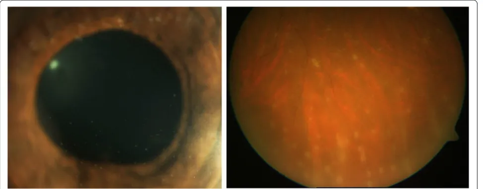
Rifabutin is an oral bacteriocidal antibiotic used as prophylaxis against Mycobacterium avium complex (MAC), typically for immunocompromised patients and particularly those infected by the HIV. It is most com-monly associated with anterior uveitis with hypopyon (Figure 1), although intermediate uveitis, panuveitis, and retinal vasculitis have been reported [18,19].
Most of our understanding of rifabutin-induced uveitis comes from cases series reported in the early- to mid-1990s [20-23]. Saran and associates described the clinical features of seven patients with HIV/AIDS who received between 300 to 600 mg of rifabutin daily along with clarithromycin and fluoconazole, and the majority of patients also received concomitant ethambutol. In this report, five patients presented with acute hypopyon uve-itis 51 to 393 days (median 79 days) after starting the medications. All patients eventually developed bilateral anterior uveitis. Vision recovered to 20/30 in all patients
within 3 weeks of starting topical corticosteroid treat-ment alone, although three of the five patients required rifabutin dose reduction and/or discontinuation. In a lar-ger group of 24 patients on 600 mg of rifabutin per day along with clarithromycin and ethambutol, Shafran and colleagues described the development of ocular irritation and redness in 75% and 54% of patients, respectively, after a median of 42 days of rifabutin use. Photophobia occurred in 33% of patients, and a hypopyon developed in 29% of patients [24]. In the same study, patients who were on a lower dose of rifabutin (300 mg/day) seldom developed uveitis, and when it occurred, it took at least 7 months of medication use for the uveitis to develop. Skinner and Blaschke subsequently confirmed that drug-related uveitis was unusual at the recommended dose of 300 mg/day [25].
Risk factors for the development of rifabutin-associated uveitis include dosage and duration of rifabutin therapy, low body weight, and use of concomitant medications, in-cluding clarithromycin and ritonavir [21]. In a multivariate analysis of patients taking 600 mg of rifabutin daily, Shafran and colleagues found that uveitis occurred in 64% of patients weighing less than 55 kg, in 45% of patients 55 to 65 kg, and in only 14% of patients weighing over 65 kg [24]. Several medications, such as clarithromycin and rito-navir, may exacerbate rifabutin-related side effects such as
Table 3 Quantitative assessment of causation of drug-induced uveitis using Naranjo algorithm
Route Drug Score per Naranjo criterion Total
score
Likelihood of causation
1 2 3 4 5 6 7 8 9 10
Systemic Cidofovir 1 2 1 2 2 0 1 1 0 1 11 Definite
Rifabutin 1 2 1 2 2 0 0 1 0 1 10 Definite
Bisphosphonates 1 2 1 1 2 0 0 0 1 1 10 Definite
Sulfonamides 1 2 1 2 2 0 0 0 1 1 10 Definite
TNF-αinhibitors 1 2 1 2 0 0 0 0 0 1 7 Probable
Fluoroquinolones 1 2 1 0 0 0 0 0 1 1 6 Probable
DEC 1 2 1 0 −1 1 0 0 0 1 5 Probable
Topical Metipranolol 1 2 1 2 2 0 0 1 0 1 10 Definite
Glucocorticosteroids 1 2 1 2 0 1 0 0 1 1 9 Definitea
Brimonidine 1 2 1 2 2 0 0 0 0 1 9 Definite
Prostaglandin analogs 1 2 1 2 2 0 0 0 0 1 9 Definite
Intraocular Cidofovir 1 2 1 2 2 0 1 1 0 1 11 Definite
Anti-VEGF agents 1 2 1 2 2 1 0 0 1 1 11 Definite
Triamcinolone acetonide 1 2 1 2 −1 0 0 0 1 1 7 Probable
Vaccines BCG 1 2 1 2 2 0 0 0 0 1 9 Definite
Influenza 1 2 1 0 2 0 0 0 0 1 7 Probable
MMR 1 2 1 0 2 0 0 0 0 1 7 Probable
Hepatitis B 1 2 0 0 2 0 0 0 0 1 6 Probable
Varicella 1 2 1 0 −1 0 0 0 0 1 4 Possible
BCG, bacille Calmette-Guérin; DEC, diethylcarbamazine; MMR, measles, mumps, and rubella; TNF, tumor necrosis factor.a
uveitis through inhibition of hepatic cytochrome P-450 [26-28]. Although systemic azoles, such as fluoconazole, also inhibit cytochrome P-450, Shafran and associates found no evidence that concurrent use of systemic azoles increased the risk of uveitis [21].
Rifabutin-induced uveitis likely results from direct rifabutin toxicity. The association between rifabutin and uveitis is supported by an association with dosage and with the duration of use, as well as bilateral involvement, limited rechallenge data [29], and reversibility with drug discontinuation.
Bisphosphonates (Naranjo score 10, definite)
Bisphosphonates are primarily used to treat osteoporosis and to prevent fractures due to malignant bone disease. While they are generally well tolerated [30], several med-ications in this class have been associated with uveitis. Moreover, bisphosphonates have been noted to cause scleritis/episcleritis in some patients [31].
Intravenous pamidronate sodium is the bisphosphonate most frequently associated with ocular side effects, and symptoms typically develop 1 to 6 days after starting treat-ment [32]. Conjunctivitis appears to be the most common ocular manifestation, followed by anterior uveitis and scler-itis/episcleritis [31-37]. Among 23 cases reported by the Ciba-Geigy Central Epidemiology and Drug Safety Center in 1993, 13 (57%) had non-specific conjunctivitis, 7 (30%) had anterior uveitis (most commonly bilateral), and 3 (13%) had unilateral episcleritis or scleritis [32]. The daily dose of pamidronate in this study ranged from 30 to 120 mg. A positive rechallenge reaction was noted in 11/15 pa-tients (73%) in whom it was tested. Most papa-tients with
non-specific conjunctivitis recovered spontaneously within several days. In patients with anterior uveitis, the severity of inflammation ranged from minimal, resolving spontan-eously within 24 h, to severe and required topical corti-costeroids with or without a cycloplegic agent and, occasionally, hospitalization. Episcleritis or scleritis was noted in three patients between 1 and 6 days following intravenous pamidronate disodium treatment. All patients recovered fully with treatment. The patient with episcleritis was treated with oral indomethacin, and the patient with posterior scleritis was treated with oral corticosteroids. The treatment for the patient with anterior scleritis was not elaborated upon beyond‘hospitalization.’
More recently, Fraunfelder and Fraunfelder reported 17 cases of unilateral scleritis associated with intraven-ous pamidronate with doses ranging from 30 to 60 mg daily [38]. All of these cases required the discontinuation of pamidronate therapy for full resolution of the inflam-mation. Positive rechallenge data were provided for six of these cases.
Although less common, similar side effects have been observed with the oral agents alendronate [39-41], risedronate [42,43], etidronate [44], and intravenous zoledronic acid [45-47]. Although rechallenge data are not available, alendronate has been associated with scleritis and acute non-granulomatous anterior uveitis [40-42]. Only one case of risedronate-associated anter-ior uveitis has been reported [42].
a systemic acute phase reaction, including the develop-ment of transient influenza-like symptoms, in approxi-mately 30% of patients following the first intravenous infusion [49]. It should be noted, however, that inflamma-tory reactions, including anterior uveitis, can occur follow-ing administration of non-nitrogen, halogen-containfollow-ing bisphosphonates, such as clodronate [50]. Patients receiv-ing bisphosphonates may also develop leukopenia as well as elevation of C-reactive protein, interleukin-1, and interleukin-6, all of which may contribute to the develop-ment of uveitis.
Treatment depends on the type and severity of the ocular inflammation. The conjunctivitis is typically self-limiting, although artificial tears and/or topical non-steroidal anti-inflammatory drugs may provide symp-tomatic relief. More substantial inflammation may require topical or systemic corticosteroid therapy. Either a change of drug to a non-nitrogen-containing bisphosphonate or cessation of bisphosphonates altogether should be consid-ered in some cases of anterior uveitis and in nearly all cases of scleritis [31,47,50].
Sulfonamides (Naranjo score 10, definite)
Sulfonamides are antibiotics used to treat a number of common infections, including urinary tract infections and toxoplasmosis. Newer sulfonamides are used as anti-convulsants, diuretics, and as dermatologic agents. Sev-eral reports have linked sulfonamide use with a mild non-granulomatous anterior uveitis [51,52].
The largest case series of sulfonamide-associated uve-itis was reported by Tilden and associates in 1991 [51]. Using the data on 12 patients obtained from the Na-tional Registry of Drug-Induced Ocular Side Effects, the US Food and Drug Administration, and two additional case reports, they described 14 patients who developed non-granulomatous anterior uveitis. Six (43%) of the pa-tients had bilateral involvement, and most developed symp-toms within a week of drug initiation. Patients rechallenged with trimethoprim-sulfamethoxazole, a common sulfona-mide, developed recurrent inflammation within 24 h. Five patients also had evidence of other sulfonamide-induced side effects such as Stevens-Johnson syndrome (SJS), ery-thema multiforme, stomatitis, glossitis, vesicular rash, or abnormal liver function.
Often associated with sulfanomide use, SJS and toxic epidermal necrolysis are acute, severe, self-limited but potentially life-threatening drug reactions which may present with dermatologic findings such as widespread mucosal erosions with numerous erythematous macules and, eventually, epithelial detachment. Common oph-thalmic manifestations of SJS include lid, conjunctival, and corneal inflammation and scarring, which may re-sult in severe visual loss [53]. Uveitis is uncommon in SJS, but has been reported [54].
Non-sulfonamide medications, particularly trimetho-prim, may play an under-appreciated role in some cases of presumed sulfonamide-induced uveitis. Notably, most of the reported cases involve trimethoprim-sulfamethoxazole. This includes at least 12 of the 14 patients in the ori-ginal case series by Tilden and colleagues [51]. Several case reports have also implicated trimethoprim alone, including two with positive rechallenge data as de-scribed below [55-57].
Pathak and Power described a 41-year-old woman who developed bilateral non-granulomatous anterior uveitis 2 days after receiving 200 mg of trimethoprim twice daily. Her work-up was unremarkable, and her inflammation promptly resolved with topical therapy and drug discon-tinuation, only to recur 45 min following rechallenge with 200 mg of trimethoprim [55]. On two separate occasions, a 38-year-old woman developed flu-like symptoms as well as bilateral non-granulomatous anterior uveitis within days of starting trimethoprim-sulfamethoxazole [56]. She subsequently developed bilateral non-granulomatous an-terior uveitis within a day of starting trimethoprim alone. All episodes resolved promptly with topical corticosteroid therapy [56].
Tumor necrosis factor inhibitors (Naranjo score 7, probable)
Tumor necrosis factor (TNF)-α is a proinflammatory cytokine strongly implicated in the pathogenesis of auto-immune disease. Inhibition of TNF-αis an effective therapy for treating a variety of inflammatory conditions, including rheumatoid arthritis, juvenile idiopathic arthritis, Crohn's disease, psoriatic arthritis, and ankylosing spondylitis. These agents have been used to treat ocular inflammation associated with these diseases as well as for the off label to treat both uveitis and scleritis [2,3,58-65]. The TNF-α in-hibitors most commonly used for uveitis are infliximab, adalimumab, and less commonly etanercept. Infliximab is a chimeric mouse-human monoclonal antibody administered as an intravenous infusion. Adalimumab is a fully human-ized monoclonal antibody administered subcutaneously. In contrast, etanercept is a genetically engineered soluble TNF receptor conjugated to the Fc region of human IgG im-munoglobulin administered as a subcutaneous injection.
therapy, and the large majority of cases completely resolved after discontinuing the medication. Of the cases reviewed, 87 involved paradoxical new intraocular inflammation after starting the medication. Even more striking, another subset of patients who developed intraocular inflammation was being treated for diseases not commonly associated with uveitis. The authors specifically described onset of intraoc-ular inflammation after starting anti-TNF agents as one with a relatively poor outcome.
More recently, additional anecdotal reports and meta-analyses have implicated all three agents, particularly etanercept, as a cause of uveitis [66-75]. In 2007, Lim and colleagues reviewed all cases of uveitis associated with etanercept, infliximab, and adalimumab reported to national databases between 1998 and 2006, and excluded patients with underlying disease likely to be associated with uveitis [76]. In total, they found 59 cases thought to have drug-induced uveitis: 43 with etanercept, 14 with infliximab, and 2 with adalimumab. Etanercept was sig-nificantly more likely to be associated with uveitis than either infliximab (odds ratio 5.375) or adalimumab (odds ratio 8.6). There were also dechallenge/rechallenge data for a limited number of patients within the etanercept group, which strengthened the association of the drug with uveitis causation [76].
The anti-TNF agents, particularly etanercept, have also been noted to induce paradoxical, biopsy-proven sarcoidosis (or sarcoid-like granulomatosis), including ocular sarcoidosis (Figure 2) [2,77-81]. Affected patients had varied presentations. The onset of uveitis ranged from 3 weeks to 6 years and included anterior and/or posterior ocular inflammation as well as periphlebitis or chorioretinitis. Most patients recovered spontaneously once the offending agent was discontinued, but some required systemic corticosteroids.
The cause of paradoxical inflammation induced by anti-TNF agents is not clear. The inverse relationship between TNF-α and the pro-inflammatory cytokine interferon may promote immune cell activation, auto-antibody formation, and immune complex deposition, eventually leading to the development of autoimmune disease [2,82]. It has also been suggested that TNF-α in-hibition may promote infection by organisms associated with non-caseating granuloma formation [2]. The reason for the difference between the various TNF inhibitors and risk of developing uveitis is unknown, but it may re-flect a differential alteration of the cytokine milieu and/ or differential effectiveness in treating intraocular in-flammation [2].
Oral fluoroquinolones (Naranjo score 6, probable)
Uveitis associated with systemic fluoroquinolone therapy was first described in 2004 [83]. Since that time, at least 40 cases have been described, ranging from isolated case
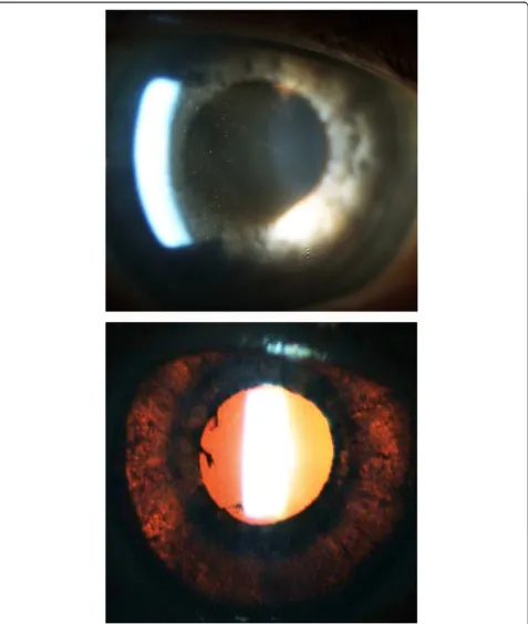
reports to a spontaneous large case series identified from reporting databases [70,83-87]. Of note, identical cases have been reported with no identified exposure to sys-temic fluoroquinolone therapy, suggesting that other factors might contribute [88]. Most of the commonly pre-scribed oral fluoroquinolones have been implicated, in-cluding ciprofloxacin, ofloxacin, gatifloxacin, levofloxacin, and norfloxacin. However, the most commonly implicated agent is moxifloxacin. In all cases, the medication was given at the standard oral dose. Uveitis developed a me-dian of 13 days (range 0 to 20 days) after drug initiation. Patients presented with bilateral ocular pain and visual impairment. Examination often revealed fine, pigmented keratic precipitates with prominent pigment in the anter-ior chamber with only minimal non-pigmented cell (Figure 3). Another common feature included diffuse iris transillumination defects with atonic pupils (Figures 3 and 4). Up to 50% of patients developed ocular hypertension or glaucoma requiring intraocular pressure-lowering ther-apy - presumably related to drug-induced pigment disper-sion [86,87].
The pathophysiology of fluoroquinolone-associated uveitis is largely unknown but may involve phototoxicity, autoimmune predisposition, and/or concurrent viral in-fection. Phototoxicity has been proposed as a mechan-ism but is unlikely, given the restriction of the findings to the eyes as opposed to other sun-exposed areas [85]. There also may be a predisposition to autoimmune disorders. Of the 40 patients described by Hinkle and as-sociates, 27 (69%) were women, the median age was 54 years, and HLA-B27 and HLA-B51 haplotypes were identified in 20% and 40% of tested individuals, respect-ively [84]. Concurrent viral infection is another possibil-ity but is unlikely to adequately account for the findings [84-86]. A single case report described a concave iris configuration leading to a potential mechanical explan-ation; however, this was not described in other cases [86]. Interestingly, this constellation of signs and symp-toms has not been described with topical or intracameral injection of fluoroquinolones.
Diethylacarbamazine (Naranjo score 5, probable)
side effects [94]. Thus, the Mazzotti reaction alone is not enough to explain the uveitis associated with DEC. With limited data on rechallenge or dosing, DEC may be a pos-sible cause of drug-induced uveitis.
Topical medications
Metipranolol (Naranjo score 10, definite)
Metipranolol is a non-selective topical beta-blocker used for the treatment of ocular hypertension and glau-coma. Shortly after its introduction, reports of associ-ated granulomatous anterior uveitis began to appear [95-102]. Akingbehin and Villada presented the findings of 56 episodes of granulomatous anterior uveitis in 26 eyes of 15 patients [96]. Nearly all cases of uveitis oc-curred with the 0.6% dose compared to the 0.3% dose, leading to the withdrawal of the 0.6% dose from the market in 1990. Most cases developed after 7 to 31 months of therapy and patients typically presented with cell, flare, and granulomatous keratic precipitates, and 2% developed posterior synechiae. Over 50% of eyes also developed ocular hypertension. With discontinuation of metipranolol and initiation of topical corticosteroids, patients generally did quite well, with resolution of the inflammation after 3 to 5 weeks. In addition to a large number of case reports, positive rechallenge data confirm the association between metipranolol and the inflammation [103]. Initial hypotheses regarding the pathogenesis of metipranolol-induced uveitis centered around sterilization methods (i.e., gamma irradiation) used for multidose bottles; however, adverse reactions have been seen with single-dose bottles as well as with other sterilization methods [98,100].
Glucocorticosteroids (Naranjo score 9, definite)
There are several reports of uveitis developing or wors-ening in otherwise non-inflamed eyes during or imme-diately following withdrawal of topical corticosteroids [104-106]. Although earlier reports mainly indicated dexamethasone sodium phosphate 0.1%, corticosteroid withdrawal-associated uveitis has been described with different topical corticosteroid formulations, including prednisolone acetate 1% and dexamethasone 0.1% [104,105,107].
cases, the inflammation resolved quickly with a topical cycloplegic/mydriatic agent alone. One patient was rechallenged with prednisolone acetate 1% for 10 days and developed recurrent uveitis 2 days after discontinuing the medication. Importantly, the medication vehicles alone failed to induce recurrent inflammation.
While the mechanism for corticosteroid-induced uveitis is unknown, its occurrence in patients with a positive fluor-escent treponemal antibody absorption (FTA-ABS) serology has been noted [106]. Of note, there have been no new re-ports of corticosteroid-induced uveitis since 1979.
Brimonidine (Naranjo score 9, definite)
Brimonidine tartrate is a selective alpha2-adrenergic re-ceptor agonist that is used for the treatment of glaucoma
all four patients developed recurrent inflammation within 3 weeks of restarting the medication.
Stopping the drug is the most effective treatment and usually results in rapid resolution of the inflamma-tion. The use of topical corticosteroid therapy and a cycloplegic/mydriatic agent may be needed in some cases.
Prostaglandin analogs (Naranjo score 9, definite)
The topical prostaglandin analogs latanoprost, travoprost, and bimatoprost lower intraocular pressure primarily by increasing uveoscleral outflow [115-117]. These medica-tions have been associated with several side effects, includ-ing conjunctival hyperemia, eyelash growth, iris and periocular skin pigmentation, iris cyst formation, cystoid macular edema, reactivation of herpes simplex keratitis, and anterior uveitis [118,119].
While high levels of endogenous prostaglandins are known to cause intraocular inflammation and induce breakdown of the blood-aqueous barrier, the topical prostaglandin analogs were specifically designed to minimize potential prostaglandin-related side effects [120,121]. Despite this, several studies have described the development of anterior uveitis in a small percent-age of patients. One study found the development of low-grade acute anterior uveitis in 8 of 163 eyes treated with latanoprost [122]. None of the patients had a previous history of uveitis or a medical condition po-tentially associated with uveitis. The inflammation de-veloped after several months of latanoprost use in six of the eight eyes and after 1 day in both eyes of one pa-tient [122]. The uveitis resolved promptly following discontinuation of the medication, although some eyes were also treated with a topical corticosteroid. In two of the three eyes tested, the inflammation recurred with rechallenge. Fechtner and associates presented positive dechallenge and rechallenge data of four patients with latanoprost-associated uveitis [123].
The mechanism by which prostaglandin analogs might cause anterior uveitis may involve the downstream stimulation of proinflammatory eicosanoids [123]. More-over, prostaglandin analogs can increase IL-1 and IL-6 levels in the tears, and potentially in the aqueous humor, of treated patients [124].
Interestingly, when used in eyes with active endogenous anterior uveitis and ocular hypertension/glaucoma, topical prostaglandins do not appear to cause increased inflamma-tion or cystoid macular edema when compared to other intraocular pressure (IOP)-lowering agents [125]. This may be in part due to the common use of concomitant topical, regional, or systemic corticosteroids in these pa-tients that may blunt or mask the pro-inflammatory effect of the prostaglandin analogs.
Intraocular medications
Cidofovir (Naranjo score 11, definite)
In addition to systemic administration, intraocular injec-tion of cidofovir has been associated with secondary in-flammation. In phase I/II clinical trials, intravitreal cidofovir without concurrent systemic administration was associated with hypotony and a mild uveitis that was easily controlled with topical medications [9,126]. Other studies found a 26% incidence of mild to moder-ate non-granulomatous uveitis after intravitreal injection of cidofovir [8,17]. Similar to uveitis seen with systemic administration of cidofovir, the incidence of uveitis was substantially lower in patients who used concurrent sys-temic probenecid plus a topical corticosteroid [8,17]. In most cases, the hypotony was mild, with an IOP reduc-tion from a mean baseline IOP of 9.3 to 7.4 mmHg 2 weeks after a 20-μg intravitreal injection [126]. In one study, only 1 out of 24 eyes developed clinically signifi-cant hypotony (with an IOP of 1 mmHg) that was asso-ciated with choroidal detachment and decreased visual acuity [126]. This patient improved spontaneously.
Anti-VEGF agents (Naranjo score 11, definite)
Anti-VEGF agents are monoclonal antibodies (ranibizumab, bevacizumab), RNA aptamers (pegaptanib), or VEGF decoy receptors (afilbercept) that bind to and effectively inhibit the activity of VEGF. These agents are used for the treat-ment of a number of retinal disorders, including exudative age-related macular degeneration (AMD), diabetic macular edema, neovascular glaucoma, and macular edema associ-ated with retinal vein occlusion (RVO) [127-143]. Despite an otherwise relatively strong safety profile in several large, multicenter studies, there have been several reports of uve-itis associated with intraocular injection of these medica-tions [130,144-153].
In 2004, pegaptanib sodium was the first anti-VEGF agent approved for the treatment of exudative age-related macular degeneration. It is an RNA-based aptamer that binds to VEGF-165 [154]. In a multicenter trial of 890 eyes treated with varying doses of pegaptanib, 14% of eyes developed some degree of anterior chamber inflammation compared to only 6% of eyes treated with a sham injection (p = 0.001) [154]. The authors attributed this relatively high incidence in both groups to the injection procedure and/or preparation. A follow-up study of 147 patients treated with doses up to 3 mg (ten times higher than the approved dose) reported no cases of ‘clinically relevant’ ocular inflammation [155].
of treated eyes in the ANCHOR study developed some post-injection inflammation [128,133]. More recently, the CRUISE, BRAVO, PIER, and CATT studies reported ocular inflammation in 1% to 1.5% of treated eyes at some point during the study period [129,131,133,134,140,156]. How-ever, in the BRAVO and CRUISE trials, inflammation oc-curred less frequently in ranibizumab-treated eyes than in sham-treated eyes [131,134]. The phase I/II FOCUS study, a study of ranibizumab plus PDT vs. PDT alone, reported a relatively high incidence of severe uveitis in 12.4% of eyes treated with ranibizumab plus PDT compared to 0% of pa-tients who received PDT alone [157,158]. The authors at-tributed this difference to the formulation of ranibizumab used (lyophilized in the FOCUS study versus the Food and Drug Administration (FDA)-approved and commercially available liquid formulation) as well as the temporal rela-tionship between ranibizumab injection and PDT treatment. Bevacizumab appears to have a more variable rate of drug-associated inflammation. In a retrospective study of 2,000 injections (1,275 bevacizumab and 725 ranibizumab), Ladas and associates reported 38 instances of intraocular inflammation. This constituted 6% of treated eyes and 1.9% of injections [147]. Most of the cases (30) were mild cases of anterior uveitis that presented within a few days after the injection and were easily controlled with topical corti-costeroids. Eight cases (constituting 1.3% of treated eyes and 0.4% of injections) presented within 1 to 3 weeks with moderate to severe inflammation. The inflammation in these cases was also easily controlled with a short course of topical and systemic corticosteroids. Only one eye expe-rienced recurrent inflammation with each of three subse-quent injections. More recently, the recent CATT study was a head-to-head comparison between ranibizumab and bevacizumab. The reported rates of inflammation in treated eyes were low at 0.3% for ranibizumab and 0.5% for bevacizumab [129,130]. Similarly, in a large, retrospective trial of 1,173 eyes treated with bevacizumab over 12 months, only 4 eyes (0.3% of treated eyes, 0.09% of injec-tions) developed severe uveitis [159]. All of these cases were managed with topical corticosteroids.
While most cases of uveitis following anti-VEGF injec-tions are mild and transient, sporadic case series have described a severe inflammatory response, more com-monly with bevacizumab compared to ranibizumab [146,149,160-162]. Sato and associates reported a series of five eyes that developed severe uveitis presenting as a toxic anterior segment syndrome following injection with bevacizumab all from the same lot [149]. Four of the five eyes had previously received intraocular bevacizumab without developing uveitis. The authors proposed that these cases were likely due to a toxic byproduct present in that lot of injections. In another series, Kay and colleagues described seven eyes of six patients that developed non-infectious uveitis from a
cohort of 978 consecutive bevacizumab injections [161]. Of note, 338 ranibizumab injections were performed over the same time frame with no associated inflamma-tion. Within 24 h, all affected eyes developed signs and symptoms typical of anterior uveitis that rapidly improved with topical corticosteroid treatment. All eyes received bevacizumab from separate lots. Three eyes received sub-sequent injections and did not develop recurrent uveitis. Anterior chamber aspirate from one affected eye revealed evidence for foreign particulate matter, which the authors speculated might have been causally related.
Larger series appear to reflect the data from large tri-als, where special formulations (i.e., distinct from those commercially available) of the medications may be used. An internet-based survey of doctor-reported adverse events noted mild uveitis in 0.14% of eyes receiving bevacizumab injections [153]. In a retrospective review of 1,278 injections of bevacizumab, Wickremasinghe and associates reported that 14 cases (1.1%) developed acute inflammation, but that study excluded those suspicious for endophthalmitis [163]. Most eyes had received prior injections (mean 2.7) and presented within 1 to 3 days with reduced vision and mild discomfort. Inflammation was mild (1 to 2+) in most cases, with equal inflamma-tion present in both the anterior chamber and vitreous. All eyes responded well to intensive topical corticoster-oid therapy. Shah and associates reported 16 cases of culture-negative endophthalmitis among 27,736 ranibizumab or bevacizumab injections, with no differ-ence in inciddiffer-ence between the two medications [164]. Day and colleagues identified 6,154 patients receiving 40,903 anti-VEGF injections in the Medicare claims data-base and evaluated them for post-injection adverse out-comes compared to disease-matched controls [165]. At 2 years, uveitis was significantly more common in the anti-VEGF group (0.73% vs. 0.37%,p< 0.01). The incidence of uveitis per injection was 0.11% in their series.
Aflibercept is a soluble decoy receptor fusion protein approved for the treatment of exudative AMD and macu-lar edema associated with RVO. Early experience sug-gested that aflibercept may be less frequently associated with medication-related uveitis compared to ranibizumab or bevacizumab, affecting 0.6% of patients or fewer in the CLEAR-IT 2 and DA VINCI studies [137,139], with the COPERNICUS and EXCITE studies reporting zero cases [138,166].
Triamcinolone acetonide (Naranjo score 7, probable)
following intravitreal injection of TA [152,167,168,170-180], which is often called non-infectious or sterile endoph-thalmitis. Patients may present with reduced vision with lit-tle or no conjunctival injection or pain, but there may be substantial anterior chamber cell involvement, often with a hypopyon. Patients may also have moderate to significant vitritis. The incidence of this presumed inflammatory re-sponse is variable in the literature, occurring in 0.5% to 9.7% of injections [170,172-175,178]. In one study of 922 intravitreal (IV) TA injections [171], the authors reported presumed non-infectious endophthalmitis in eight eyes (0.87%). Patients presented within 1 to 7 days of the injec-tion with poor visual acuity and hypopyon. Treatment var-ied from systemic antibiotics to vitreous tap and injection of antibiotics to pars plana vitrectomy. None of the vitreous samples were culture positive, and the patients tended to do well, with an average final acuity of 20/75. Infectious endophthalmitis should be suspected in all patients with intraocular inflamma-tion following IVTA, particularly those with worse conjunctival injection or pain, poorer vision, and/or involvement of the vitreous at presentation.
Some investigators have suggested that intraocular in-flammation following IVTA may represent a toxic reac-tion to the drug, the vehicle, or a contaminant [172]. Jonas and colleagues administered 1,135 intravitreal in-jections of filtered 20 mg TA and observed no cases of non-infectious endophthalmitis [181]. Given the high dose of TA, this argues against a direct toxic effect of drug. Supporting this hypothesis, one retrospective review of 646 injections of 4 mg of TA noted significantly more cases of non-infectious endophthalmitis among patients who received TA with preservative than without [180]. Further support comes from two large studies that used a preservative-free, single-use formulation of IVTA in 457 patients with branch or central retinal vein occlusion. The
authors reported no cases of post-injection inflammation [167,168]. Patients with a previous history of uveitis ap-pear more likely to develop ocular inflammation after in-jection [182]. However, some authors found that a presumed non-infectious inflammatory reaction can occur even with preservative-free TA [179].
Another distinct entity that can be confused with infectious or non-infectious endophthalmitis is pseudo-endophthalmitis due to the migration of the TA crystals into the anterior chamber, where they settle into a pseudohypopyon (Figure 5) [183]. Pseudo-endophthalmitis occurs less commonly in phakic patients and pseudophakic patients with an intact posterior capsule [170].
Vaccines
Bacille Calmette-Guérin (Naranjo score 9, definite)
Bacille Calmette-Guerin (BCG) is a vaccine used for prophylaxis againstMycobacterium tuberculosisand is fre-quently administered in countries with a high prevalence of infection. Bacille Calmette-Guerin vaccination is pri-marily used to prevent childhood tuberculous meningitis and miliary disease, and also has utility in the treatment of bladder cancer. Multiple case reports have implicated both BCG vaccination and intravesical BCG in the develop-ment of acute bilateral non-granulomatous or granuloma-tous anterior uveitis [184-192]. The inflammation may be associated with loss of iris pigmentation and typically re-sponds within days to weeks to treatment with topical cor-ticosteroids and a cycloplegic/mydriatic agent. Patients may also present with systemic symptoms resembling re-active arthritis [191-193]. Less commonly, patients may present with bilateral panuveitis [194,195], chorioretinitis [196], vitiligo [189], and/or optic neuritis [195].
Some authors have argued for an induced abnormal immune response due to molecular mimicry [184,185]. The authors of a case report of a 13-year-old girl who
developed chronic bilateral anterior uveitis following BCG vaccination implicated her HLA-DR4 positivity, noting that HLA-DR4 is highly responsive to M.
tuber-culosis antigens [184]. A separate report described a 72-year-old woman who developed bilateral granuloma-tous anterior uveitis following intravesical BCG for blad-der carcinoma [185]. The authors noted evidence for a polyclonal autoimmune reaction elicited by BCG as well as amino acid sequence homology between proteins from M. tuberculosis, BCG, and retinal antigens [185]. Of note, the patient was negative for HLA-B27 but posi-tive for HLA-B8 and HLA-B38.
Several cases of uveitis following tuberculin skin testing (purified protein derivative or PPD) have been reported [197]. Reactions have ranged from panuveitis [197] to presentation resembling multifocal choroiditis and Vogt-Koyanagi-Harada disease with serous retinal detach-ment [198]. The delay in onset of uveitis following skin test placement supports an immune-mediated, delayed-type hypersensitivity reaction, perhaps to resident immune cells previously sensitized toM. tuberculosis[197].
Measles, mumps, and rubella (Naranjo score 7, probable)
Both anterior uveitis and panuveitis have been reported in association with measles, mumps, and rubella (MMR) vaccination in isolated case reports [199,200]. These reports have been sporadic, with variable severity. In two cases reported by Islam and associates, patients presented with bilateral panuveitis 4 and 6 weeks following MMR vaccination [199]. Both patients were HLA-B27 negative. Sedaghat and colleagues reported a 17-year-old girl who developed bilateral panuveitis associated with dermal vas-culitis and arthritis shortly after MMR vaccination [200]. The inflammation may take 12 months or longer to re-solve [199] and may require both topical and systemic cor-ticosteroid treatment [199,200]. The mechanism may involve antigenic mimicry or processes involved in the preparation of the vaccine such as contamination or faulty preservation [199].
Influenza (Naranjo score 7, probable)
The influenza vaccination has been implicated in the de-velopment of secondary uveitis in several case reports, including bilateral panuveitis [201-204], recurrent panuveitis [205], and acute posterior multifocal placoid pigment epitheliopathy (APMPPE) [206]. Anterior uve-itis can be controlled with topical corticosteroids and a cycloplegic/mydriatic agent, whereas posterior involve-ment may require systemic corticosteroids. Rothova and associates reported reactivation of acute retinal necrosis (ARN) following vaccination for the H1N1 strain of in-fluenza [207].
Hepatitis B (Naranjo score 6, probable)
The hepatitis B virus (HBV) vaccine is approved by the FDA for the prevention of hepatitis B infection in children and for at-risk populations. A few case reports have impli-cated HBV vaccination in the development of uveitis, in-cluding a single case report of post-vaccination APMPPE [208-211]. The bulk of our information regarding hepatitis B vaccine-related uveitis comes from a review of 32 cases reported to a national database [211]. In this series, the mean age of patients was 29 years (1 to 57 years), with a predominance of female patients (24 females vs. 8 males). The majority of cases developed within a few days follow-ing vaccination and more often after the initial vaccination than subsequent vaccinations, although several patients developed recurrent inflammation with each vaccination. A majority of patients (75%) also reported a flu-like illness. It was hypothesized that this reaction represented either an immune-mediated delayed-type hypersensitivity reac-tion or a reacreac-tion to the vaccine carrier meant to potenti-ate the vaccine's immunogenic activity [211].
Varicella (Naranjo score 4, possible)
Several case reports describe the development of uveitis following varicella vaccination. Various presentations have been described, including isolated anterior uveitis [210], anterior uveitis associated with a vesicular rash [212], keratouveitis with inflammatory glaucoma and Hutchinson sign [213], sclerokeratitis with anterior uve-itis (herpes zoster ophthalmicus) [214], and ARN [215]. In general, patients responded to standard treatment regi-mens, including topical corticosteroids and oral acyclovir. However, the two patients with ARN both required more aggressive treatment, including oral valacyclovir, intraocu-lar ganciclovir or foscarnet, intravenous acyclovir, and eventually vitrectomy with silicone oil tamponade due to progressive disease [215]. Uveitis may occur due to reacti-vation of varicella virus or, alternatively, due to primary in-fection with the attenuated virus used in the vaccine.
Conclusions
Competing interests
The authors declare that they have no competing interests.
Authors’contributions
NL drafted the manuscript. SG, RM, and EC provided guidance, and reviewed and edited the manuscript. All authors read and approved the final manuscript.
Acknowledgments
This work was supported in part by The Pacific Vision Foundation (ETC) and The San Francisco Retina Foundation (ETC).
Author details
1
Retina Consultants San Diego, 9850 Genesee Avenue, Suite 700, La Jolla, CA 92037, USA.2MidAtlantic Retina, The Retina Service of Wills Eye Institute, Thomas Jefferson University, 840 Walnut Street, Suite 1020, Philadelphia, PA 19107, USA.3Associated Vitreoretinal and Uveitis Consultants, St. Vincent Hospital and Health Services, Indianapolis, IN 46260, USA.4Associate Clinical Professor of Ophthalmology, Indiana University School of Medicine, Indianapolis, IN 46202, USA.5Department of Ophthalmology, California Pacific Medical Center, San Francisco, CA 94115, USA.6Department of Ophthalmology, Stanford University School of Medicine, Stanford, CA 94305-5101, USA.
Received: 4 February 2013 Accepted: 13 March 2013 Published: 25 March 2013
References
1. Moorthy RS, Valluri S, Jampol LM (1998) Drug-induced uveitis. Surv Ophthalmol 42:557–570
2. Cunningham ET Jr, Pasadhika S, Suhler EB, Zierhut M (2012) Drug-induced inflammation in patients on TNFαinhibitors. Ocul Immunol Inflamm 20:2–5 3. Cunningham ET Jr, Zierhut M (2010) TNF inhibitors for uveitis: balancing
efficacy and safety. Ocul Immunol Inflamm 18:421–423
4. Naranjo CA, Busto U, Sellers EM, Sandor P, Ruiz I, Roberts EA, Janecek E, Domecq C, Greenblatt DJ (1981) A method for estimating the probability of adverse drug reactions. Clin Pharmacol Ther 30:239–245
5. Lalezari JP, Drew WL, Glutzer E, James C, Miner D, Flaherty J, Fisher PE, Cundy K, Hannigan J, Martin JC, Jaffe HS (1995) (S)-1-[3-hydroxy-2-(phosphonylmethoxy)propyl]cytosine (cidofovir): results of a phase I/II study of a novel antiviral nucleotide analogue. J Infect Dis 171:788–796 6. Lalezari JP (1997) Cidofovir: a new therapy for cytomegalovirus retinitis.
J Acquir Immune Defic Syndr Hum Retrovirol 14(Suppl 1):S22–S26 7. Ho HT, Woods KL, Bronson JJ, De Boeck H, Martin JC, Hitchcock MJ (1992)
Intracellular metabolism of the antiherpes agent (S)-1-[3-hydroxy-2-(phosphonylmethoxy)propyl]cytosine. Mol Pharmacol 41:197–202 8. Chavez-de la Paz E, Arevalo JF, Kirsch LS, Munguia D, Rahhal FM, De Clercq
E, Freeman WR (1997) Anterior nongranulomatous uveitis after intravitreal HPMPC (cidofovir) for the treatment of cytomegalovirus retinitis. Analysis and prevention. Ophthalmology 104:539–544
9. Kirsch LS, Arevalo JF, De Clercq E, Chavez de la Paz E, Munguia D, Garcia R, Freeman WR (1995) Phase I/II study of intravitreal cidofovir for the treatment of cytomegalovirus retinitis in patients with the acquired immunodeficiency syndrome. Am J Ophthalmol 119:466–476 10. Tseng AL, Mortimer CB, Salit IE (1999) Iritis associated with intravenous
cidofovir. Ann Pharmacother 33:167–171
11. Davis JL, Taskintuna I, Freeman WR, Weinberg DV, Feuer WJ, Leonard RE (1997) Iritis and hypotony after treatment with intravenous cidofovir for cytomegalovirus retinitis. Arch Ophthalmol 115:733–737
12. Akler ME, Johnson DW, Burman WJ, Johnson SC (1998) Anterior uveitis and hypotony after intravenous cidofovir for the treatment of cytomegalovirus retinitis. Ophthalmology 105:651–657
13. Ambati J, Wynne KB, Angerame MC, Robinson MR (1999) Anterior uveitis associated with intravenous cidofovir use in patients with cytomegalovirus retinitis. Br J Ophthalmol 83:1153–1158
14. The Studies of Ocular Complications of AIDS Research Group in
collaboration with the AIDS Clinical Trials Group (2000) Long-term follow-up of patients with AIDS treated with parenteral cidofovir for cytomegalovirus retinitis: the HPMPC peripheral cytomegalovirus retinitis trial. The studies of ocular complications of AIDS research group in collaboration with the AIDS clinical trials group. AIDS 14:1571–1581
15. Scott RA, Pavesio C (2000) Ocular side-effects from systemic HPMPC (Cidofovir) for a non-ocular cytomegalovirus infection. Am J Ophthalmol 130:126–127
16. Lopez V, Sola E, Gutierrez C, Burgos D, Cabello M, Garcia I, Florez P, Lopez J, Gonzalez-Molina M (2006) Anterior uveitis associated with treatment with intravenous cidofovir in kidney transplant patients with BK virus nephropathy. Transplant Proc 38:2412–2413
17. Rahhal FM, Arevalo JF, Munguia D, Taskintuna I, Chavez de la Paz E, Azen SP, Freeman WR (1996) Intravitreal cidofovir for the maintenance treatment of cytomegalovirus retinitis. Ophthalmology 103:1078–1083
18. Smith WM, Reddy MG, Hutcheson KA, Bishop RJ, Sen HN (2012) Rifabutin-associated hypopyon uveitis and retinal vasculitis with a history of acute myeloid leukemia. J Ophthalmic Inflamm Infect 2:149–152
19. Skolik S, Willermain F, Caspers LE (2005) Rifabutin-associated panuveitis with retinal vasculitis in pulmonary tuberculosis. Ocul Immunol Inflamm 13:483–485 20. Saran BR, Maguire AM, Nichols C, Frank I, Hertle RW, Brucker AJ, Goldman S,
Brown M, Van Uitert B (1994) Hypopyon uveitis in patients with acquired immunodeficiency syndrome treated for systemic Mycobacterium avium complex infection with rifabutin. Arch Ophthalmol 112:1159–1165 21. Shafran SD, Deschenes J, Miller M, Phillips P, Toma E (1994) Uveitis and
pseudojaundice during a regimen of clarithromycin, rifabutin, and ethambutol. MAC study group of the Canadian HIV trials network. N Engl J Med 330:438–439
22. Siegal FP, Eilbott D, Burger H, Gehan K, Davidson B, Kaell AT, Weiser B (1990) Dose-limiting toxicity of rifabutin in AIDS-related complex: syndrome of arthralgia/arthritis. AIDS 4:433–441
23. Frank MO, Graham MB, Wispelway B (1994) Rifabutin and uveitis. N Engl J Med 330:868
24. Shafran SD, Singer J, Zarowny DP, Deschenes J, Phillips P, Turgeon F, Aoki FY, Toma E, Miller M, Duperval R, Lemieux C, Schlech WF 3rd (1998) Determinants of rifabutin-associated uveitis in patients treated with rifabutin, clarithromycin, and ethambutol for Mycobacterium avium complex bacteremia: a multivariate analysis. Canadian HIV Trials Network Protocol 010 Study Group. J Infect Dis 177:252–255
25. Skinner MH, Blaschke TF (1995) Clinical pharmacokinetics of rifabutin. Clin Pharmacokinet 28:115–125
26. Kuper JI, D’Aprile M (2000) Drug-drug interactions of clinical significance in the treatment of patients with Mycobacterium avium complex disease. Clin Pharmacokinet 39:203–214
27. Cato A 3rd, Cavanaugh J, Shi H, Hsu A, Leonard J, Granneman R (1998) The effect of multiple doses of ritonavir on the pharmacokinetics of rifabutin. Clin Pharmacol Ther 63:414–421
28. Jacobs DS, Piliero PJ, Kuperwaser MG, Smith JA, Harris SD, Flanigan TP, Goldberg JH, Ives DV (1994) Acute uveitis associated with rifabutin use in patients with human immunodeficiency virus infection. Am J Ophthalmol 118:716–722
29. Schimkat M, Althaus C, Becker K, Sundmacher R (1996) Rifabutin-associated anterior uveitis in patients infected with human immunodeficiency virus. Ger J Ophthalmol 5:195–201
30. Watts NB (2003) Bisphosphonate treatment of osteoporosis. Clin Geriatr Med 19:395–414
31. Fraunfelder FW, Fraunfelder FT, Jensvold B (2003) Scleritis and other ocular side effects associated with pamidronate disodium. Am J Ophthalmol 135:219–222
32. Macarol V, Fraunfelder FT (1994) Pamidronate disodium and possible ocular adverse drug reactions. Am J Ophthalmol 118:220–224
33. Fraunfelder FW, Solomon J, Druker BJ, Esmaeli B, Kuyl J (2003) Ocular side-effects associated with imatinib mesylate (Gleevec). J Ocul Pharmacol Ther 19:371–375
34. De S, Meyer P, Crisp AJ (1995) Pamidronate and uveitis. Br J Rheumatol 34:479
35. Stewart GO, Stuckey BG, Ward LC, Prince RL, Gutteridge DH, Constable IJ (1996) Iritis following intravenous pamidronate. Aust N Z J Med 26:414–415 36. Ghose K, Waterworth R, Trolove P, Highton J (1994) Uveitis associated with
pamidronate. Aust N Z J Med 24:320
37. O’Donnell NP, Rao GP, Aguis-Fernandez A (1995) Paget’s disease: ocular complications of disodium pamidronate treatment. Br J Clin Pract 49:272–273 38. Fraunfelder FW, Fraunfelder FT (2003) Bisphosphonates and ocular
inflammation. N Engl J Med 348:1187–1188
40. Mbekeani JN, Slamovits TL, Schwartz BH, Sauer HL (1999) Ocular inflammation associated with alendronate therapy. Arch Ophthalmol 117:837–838
41. Salmen S, Berrueta L, Sanchez N, Montes H, Borges L (2002)
Nongranulomatous anterior uveitis associated with alendronate therapy. Invest Clin 43:49–52
42. Siris ES (1993) Bisphosphonates and iritis. Lancet 341:436–437
43. Tan YL, Sims J, Chee SP (2009) Bilateral uveitis secondary to bisphosphonate therapy. Ophthalmologica 223:215–216
44. Fraunfelder FW, Fraunfelder FT (2004) Adverse ocular drug reactions recently identified by the National Registry of Drug-Induced Ocular Side Effects. Ophthalmology 111:1275–1279
45. Moore MM, Beith JM (2008) Acute unilateral anterior uveitis and scleritis following a single infusion of zoledronate for metastatic breast cancer. Med J Aust 188:370–371
46. Banal F, Briot K, Ayoub G, Dougados M, Roux C (2008) Unilateral anterior uveitis complicating zoledronic acid therapy in prostate cancer. J Rheumatol 35:2458–2459
47. Benderson D, Karakunnel J, Kathuria S, Badros A (2006) Scleritis complicating zoledronic acid infusion. Clin Lymphoma Myeloma 7:145–147
48. Sharma NS, Ooi JL, Masselos K, Hooper MJ, Francis IC (2008) Zoledronic acid infusion and orbital inflammatory disease. N Engl J Med 359:1410–1411 49. Black DM, Delmas PD, Eastell R, Reid IR, Boonen S, Cauley JA, Cosman F, Lakatos P, Leung PC, Man Z, Mautalen C, Mesenbrink P, Hu H, Caminis J, Tong K, Rosario-Jansen T, Krasnow J, Hue TF, Sellmeyer D, Eriksen EF, Cummings SR (2007) Once-yearly zoledronic acid for treatment of postmenopausal osteoporosis. N Engl J Med 356:1809–1822
50. Fietta P, Manganelli P, Lodigiani L (2003) Clodronate induced uveitis. Ann Rheum Dis 62:378
51. Tilden ME, Rosenbaum JT, Fraunfelder FT (1991) Systemic sulfonamides as a cause of bilateral, anterior uveitis. Arch Ophthalmol 109:67–69
52. Northrop CV, Shepherd SM, Abbuhl S (1996) Sulfonamide-induced iritis. Am J Emerg Med 14:577–579
53. Power WJ, Ghoraishi M, Merayo-Lloves J, Neves RA, Foster CS (1995) Analysis of the acute ophthalmic manifestations of the erythema multiforme/Stevens-Johnson syndrome/toxic epidermal necrolysis disease spectrum. Ophthalmology 102:1669–1676
54. Arola O, Peltonen R, Rossi T (1998) Arthritis, uveitis, and Stevens-Johnson syndrome induced by trimethoprim. Lancet 351:1102
55. Pathak S, Power B (2007) Bilateral acute anterior uveitis as a side effect of trimethoprim. Eye (Lond) 21:252–253
56. Kristinsson JK, Hannesson OB, Sveinsson O, Thorleifsson H (1997) Bilateral anterior uveitis and retinal haemorrhages after administration of trimethoprim. Acta Ophthalmol Scand 75:314–315
57. Gilroy N, Gottlieb T, Spring P, Peiris O (1997) Trimethoprim-induced aseptic meningitis and uveitis. Lancet 350:112
58. Moreland LW, Baumgartner SW, Schiff MH, Tindall EA, Fleischmann RM, Weaver AL, Ettlinger RE, Cohen S, Koopman WJ, Mohler K, Widmer MB, Blosch CM (1997) Treatment of rheumatoid arthritis with a recombinant human tumor necrosis factor receptor (p75)-Fc fusion protein. N Engl J Med 337:141–147 59. Braun J, Brandt J, Listing J, Zink A, Alten R, Golder W, Gromnica-Ihle E,
Kellner H, Krause A, Schneider M, Sorensen H, Zeidler H, Thriene W, Sieper J (2002) Treatment of active ankylosing spondylitis with infliximab: a randomised controlled multicentre trial. Lancet 359:1187–1193 60. Maini R, St Clair EW, Breedveld F, Furst D, Kalden J, Weisman M, Smolen J,
Emery P, Harriman G, Feldmann M, Lipsky P (1999) Infliximab (chimeric anti-tumour necrosis factor alpha monoclonal antibody) versus placebo in rheumatoid arthritis patients receiving concomitant methotrexate: a randomised phase III trial. ATTRACT Study Group. Lancet 354:1932–1939 61. Chaudhari U, Romano P, Mulcahy LD, Dooley LT, Baker DG, Gottlieb AB
(2001) Efficacy and safety of infliximab monotherapy for plaque-type psoriasis: a randomised trial. Lancet 357:1842–1847
62. Suhler EB, Smith JR, Wertheim MS, Lauer AK, Kurz DE, Pickard TD, Rosenbaum JT (2005) A prospective trial of infliximab therapy for refractory uveitis: preliminary safety and efficacy outcomes. Arch Ophthalmol 123:903–912
63. Murphy CC, Ayliffe WH, Booth A, Makanjuola D, Andrews PA, Jayne D (2004) Tumor necrosis factor alpha blockade with infliximab for refractory uveitis and scleritis. Ophthalmology 111:352–356
64. Murphy CC, Greiner K, Plskova J, Duncan L, Frost A, Isaacs JD, Rebello P, Waldmann H, Hale G, Forrester JV, Dick AD (2004) Neutralizing tumor
necrosis factor activity leads to remission in patients with refractory noninfectious posterior uveitis. Arch Ophthalmol 122:845–851 65. Okada AA, Goto H, Ohno S, Mochizuki M (2012) Multicenter study of
infliximab for refractory uveoretinitis in Behcet disease. Arch Ophthalmol 130:592–598
66. Ramos-Casals M, Roberto Perez A, Diaz-Lagares C, Cuadrado MJ, Khamashta MA (2010) Autoimmune diseases induced by biological agents: a double-edged sword? Autoimmun Rev 9:188–193
67. Kakkassery V, Mergler S, Pleyer U (2010) Anti-TNF-alpha treatment: a possible promoter in endogenous uveitis? Observational report on six patients: occurrence of uveitis following etanercept treatment. Curr Eye Res 35:751–756 68. Gaujoux-Viala C, Giampietro C, Gaujoux T, Ea HK, Prati C, Orcel P, Wendling
D, Liote F (2012) Scleritis: a paradoxical effect of etanercept? Etanercept-associated inflammatory eye disease. J Rheumatol 39:233–239 69. Cobo-Ibanez T, del Carmen OM, Munoz-Fernandez S, Madero-Prado R,
Martin-Mola E (2008) Do TNF-blockers reduce or induce uveitis? Rheumatology (Oxford) 47:731–732
70. Wendling D, Paccou J, Berthelot JM, Flipo RM, Guillaume-Czitrom S, Prati C, Dernis E, Direz G, Ferrazzi V, Ristori JM (2011) New onset of uveitis during anti-tumor necrosis factor treatment for rheumatic diseases. Semin Arthritis Rheum 41:503–510
71. Reddy AR, Backhouse OC (2003) Does etanercept induce uveitis? Br J Ophthalmol 87:925
72. Hashkes PJ, Shajrawi I (2003) Sarcoid-related uveitis occurring during etanercept therapy. Clin Exp Rheumatol 21:645–646
73. Taban M, Dupps WJ, Mandell B, Perez VL (2006) Etanercept (enbrel)-associated inflammatory eye disease: case report and review of the literature. Ocul Immunol Inflamm 14:145–150
74. Pontikaki I, Gerloni V, Gattinara M, Luriati A, Salmaso A, De Marco G, Teruzzi B, Valcamonica E, Fantini F (2006) Side effects of anti-TNFalpha therapy in juvenile idiopathic arthritis. Reumatismo 58:31–38
75. Monnet D, Moachon L, Dougados M, Brezin AP (2006) Severe uveitis in an HLA-B27-positive patient with ankylosing spondylitis. Nat Clin Pract Rheumatol 2:393–397
76. Lim LL, Fraunfelder FW, Rosenbaum JT (2007) Do tumor necrosis factor inhibitors cause uveitis? A registry-based study. Arthritis Rheum 56:3248–3252
77. Fonollosa A, Artaraz J, Les I, Martinez-Berriotxoa A, Izquierdo JP, Lopez AS, Gardeazaba J, Berasategui B, Martinez-Alday N (2012) Sarcoid intermediate uveitis following etanercept treatment: a case report and review of the literature. Ocul Immunol Inflamm 20(1):44–48
78. Seve P, Varron L, Broussolle C, Denis P, Kodjikian L (2012) Sarcoid-related uveitis occurring during adalimumab therapy. Ocul Immunol Inflamm 20:59–60
79. Daien CI, Monnier A, Claudepierre P, Constantin A, Eschard JP, Houvenagel E, Samimi M, Pavy S, Pertuiset E, Toussirot E, Combe B, Morel J (2009) Sarcoid-like granulomatosis in patients treated with tumor necrosis factor blockers: 10 cases. Rheumatology (Oxford) 48:883–886
80. Toussirot E, Pertuiset E (2010) TNFalpha blocking agents and sarcoidosis: an update. Rev Med Interne 31:828–837
81. Massara A, Cavazzini L, La Corte R, Trotta F (2010) Sarcoidosis appearing during anti-tumor necrosis factor alpha therapy: a new“class effect” paradoxical phenomenon. Two case reports and literature review. Semin Arthritis Rheum 39:313–319
82. Viguier M, Richette P, Bachelez H, Wendling D, Aubin F (2009) Paradoxical adverse effects of anti-TNF-alpha treatment: onset or exacerbation of cutaneous disorders. Expert Rev Clin Immunol 5:421–431
83. Bringas Calvo R, Iglesias Cortinas D (2004) Acute and bilateral uveitis secondary to moxifloxacin. Arch Soc Esp Oftalmol 79:357–359
84. Hinkle DM, Dacey MS, Mandelcorn E, Kalyani P, Mauro J, Bates JH, Soukasian SH, Holland GN, Foster CS, Fraunfelder FT, Davis JL, Fraunfelder FW (2012) Bilateral uveitis associated with fluoroquinolone therapy. Cutan Ocul Toxicol 31:111–116
85. Wefers Bettink-Remeijer M, Brouwers K, van Langenhove L, De Waard PW, Missotten TO, Martinez Ciriano JP, Van Aken E (2009) Uveitis-like syndrome and iris transillumination after the use of oral moxifloxacin. Eye (Lond) 23:2260–2262 86. Willermain F, Deflorenne C, Bouffioux C, Janssens X, Koch P, Caspers L
(2010) Uveitis-like syndrome and iris transillumination after the use of oral moxifloxacin. Eye (Lond) 24:1419
26 new cases and four-year follow-up of two patients. Ophthalmology 116:1552–1557, 1557.e1
88. Tugal-Tutkun I, Onal S, Garip A, Taskapili M, Kazokoglu H, Kadayifcilar S, Kestelyn P (2011) Bilateral acute iris transillumination. Arch Ophthalmol 129 (10):1312–1319
89. Mazzotti L (1948) Possibility of using the allergic reactions due to the administration of Hetrazan as an auxiliary diagnostic test for onchocerciasis in Mexico. Revista del Instituto de Salubridad y Enfermedades Tropicales 9:235–237 (in Spanish)
90. Francis H, Awadzi K, Ottesen EA (1985) The Mazzotti reaction following treatment of onchocerciasis with diethylcarbamazine: clinical severity as a function of infection intensity. Am J Trop Med Hyg 34:529–536
91. Greene BM, Taylor HR, Brown EJ, Humphrey RL, Lawley TJ (1983) Ocular and systemic complications of diethylcarbamazine therapy for onchocerciasis: association with circulating immune complexes. J Infect Dis 147:890–897 92. Bird AC, El-Sheikh H, Anderson J, Fuglsang H (1980) Changes in visual
function and in the posterior segment of the eye during treatment of onchocerciasis with diethylcarbamazine citrate. Br J Ophthalmol 64:191–200 93. Taylor HR, Greene BM (1981) Ocular changes with oral and transepidermal
diethylcarbamazine therapy of onchocerciasis. Br J Ophthalmol 65:494–502 94. Taylor HR, Murphy RP, Newland HS, White AT, D’Anna SA, Keyvan-Larijani E,
Aziz MA, Cupp EW, Greene BM (1986) Treatment of onchocerciasis. The ocular effects of ivermectin and diethylcarbamazine. Arch Ophthalmol 104:863–870
95. Kinshuck D (1991) Glauline (metipranolol) induced uveitis and increase in intraocular pressure. Br J Ophthalmol 75:575
96. Akingbehin T, Villada JR (1991) Metipranolol-associated granulomatous anterior uveitis. Br J Ophthalmol 75:519–523
97. Beck RW, Moke P, Blair RC, Nissenbaum R (1996) Uveitis associated with topical beta-blockers. Arch Ophthalmol 114:1181–1182
98. O’Connor GR (1993) Granulomatous uveitis and metipranolol. Br J Ophthalmol 77:536–538
99. Watanabe TM, Hodes BL (1997) Bilateral anterior uveitis associated with a brand of metipranolol. Arch Ophthalmol 115:421–422
100. Kamalarajah S, Johnston PB (1999) Bilateral anterior uveitis associated with 0.3% minims metipranolol. Eye (Lond) 13(Pt 3a):380–381
101. Melles RB, Wong IG (1994) Metipranolol-associated granulomatous iritis. Am J Ophthalmol 118:712–715
102. Schultz JS, Hoenig JA, Charles H (1993) Possible bilateral anterior uveitis secondary to metipranolol (optipranolol) therapy. Arch Ophthalmol 111:1606–1607
103. Akingbehin T, Villada JR, Walley T (1992) Metipranolol-induced adverse reactions: I. The rechallenge study. Eye (Lond) 6(Pt 3):277–279 104. Martins JC, Wilensky JT, Asseff CF, Obstbaum SA, Buerk KM (1974)
Corticosteroid-induced uveitis. Am J Ophthalmol 77:433–437 105. Krupin T, LeBlanc RP, Becker B, Kolker AE, Podos SM (1970) Uveitis in
association with topically administered corticosteroid. Am J Ophthalmol 70:883–885
106. Shin DH, Kass MA, Kolker AE, Becker B, Marr JJ, Bell EC (1976) Positive FTA-ABS tests in subjects with corticosteroid-induced uveitis. Am J Ophthalmol 82:259–260
107. Mindel JS, Goldberg J, Tavitian HO (1979) Similarity of the intraocular pressure response to different corticosteroid esters when compliance is controlled. Ophthalmology 86:99–107
108. Byles DB, Frith P, Salmon JF (2000) Anterior uveitis as a side effect of topical brimonidine. Am J Ophthalmol 130:287–291
109. Becker HI, Walton RC, Diamant JI, Zegans ME (2004) Anterior uveitis and concurrent allergic conjunctivitis associated with long-term use of topical 0.2% brimonidine tartrate. Arch Ophthalmol 122:1063–1066
110. Nguyen EV, Azar D, Papalkar D, McCluskey P (2008) Brimonidine-induced anterior uveitis and conjunctivitis: clinical and histologic features. J Glaucoma 17:40–42
111. Cates CA, Jeffrey MN (2003) Granulomatous anterior uveitis associated with 0.2% topical brimonidine. Eye (Lond) 17:670–671
112. Goyal R, Ram AR (2000) Brimonidine tartarate 0.2% (Alphagan) associated granulomatous anterior uveitis. Eye (Lond) 14:908–910
113. Velasque L, Ducousso F, Pernod L, Vignal R, Deral V (2004) Anterior uveitis and topical brimonidine: a case report. J Fr Ophtalmol 27:1150–1152 114. Hondeghem K, Augustinus B, De Smet MD (2009) Bilateral granulomatous
uveitis as a side effect of topical brimonidine: two case reports. Bull Soc Belge Ophtalmol 311:51–52
115. Camras CB (1996) Comparison of latanoprost and timolol in patients with ocular hypertension and glaucoma: a six-month masked, multicenter trial in the United States. The United States Latanoprost Study Group.
Ophthalmology 103:138–147
116. Alm A, Stjernschantz J (1995) Effects on intraocular pressure and side effects of 0.005% Latanoprost applied once daily, evening or morning. A comparison with timolol. Scandinavian Latanoprost Study Group. Ophthalmology 102:1743–1752
117. Gabelt BT, Kaufman PL (1989) Prostaglandin F2 alpha increases uveoscleral outflow in the cynomolgus monkey. Exp Eye Res 49:389–402
118. Bito LZ (1997) Prostaglandins: a new approach to glaucoma management with a new, intriguing side effect. Surv Ophthalmol 41(Suppl 2):S1–S14 119. Grierson I, Jonsson M, Cracknell K (2004) Latanoprost and pigmentation. Jpn
J Ophthalmol 48:602–612
120. Eakins KE (1977) Prostaglandin and non-prostaglandin mediated breeakdown of the blood-aqueous barrier. Exp Eye Res 25(Suppl):483–498 121. Unger WG (1989) Mediation of the ocular response to injury and irritation:
peptides versus prostaglandins. Prog Clin Biol Res 312:293–328
122. Warwar RE, Bullock JD, Ballal D (1998) Cystoid macular edema and anterior uveitis associated with latanoprost use. Experience and incidence in a retrospective review of 94 patients. Ophthalmology 105:263–268 123. Fechtner RD, Khouri AS, Zimmerman TJ, Bullock J, Feldman R, Kulkarni P,
Michael AJ, Realini T, Warwar R (1998) Anterior uveitis associated with latanoprost. Am J Ophthalmol 126:37–41
124. Lopilly Park HY, Kim JH, Lee KM, Park CK (2012) Effect of prostaglandin analogues on tear proteomics and expression of cytokines and matrix metalloproteinases in the conjunctiva and cornea. Exp Eye Res 94:13–21 125. Chang JH, McCluskey P, Missotten T, Ferrante P, Jalaludin B, Lightman S (2008) Use of ocular hypotensive prostaglandin analogues in patients with uveitis: does their use increase anterior uveitis and cystoid macular oedema? Br J Ophthalmol 92:916–921
126. Kirsch LS, Arevalo JF, Chavez De La Paz E, Munguia D, De Clercq E, Freeman WR (1995) Intravitreal cidofovir (HPMPC) treatment of cytomegalovirus retinitis in patients with acquired immune deficiency syndrome. Ophthalmology 102:533–542, discussion 42–43
127. Gheith ME, Siam GA, de Barros DS, Garg SJ, Moster MR (2007) Role of intravitreal bevacizumab in neovascular glaucoma. J Ocul Pharmacol Ther 23:487–491
128. Rosenfeld PJ, Brown DM, Heier JS, Boyer DS, Kaiser PK, Chung CY, Kim RY (2006) Ranibizumab for neovascular age-related macular degeneration. N Engl J Med 355:1419–1431
129. Martin DF, Maguire MG, Fine SL, Ying GS, Jaffe GJ, Grunwald JE, Toth C, Redford M, Ferris FL 3rd (2012) Ranibizumab and bevacizumab for treatment of neovascular age-related macular degeneration: two-year results. Ophthalmology 119:1388–1398
130. Martin DF, Maguire MG, Ying GS, Grunwald JE, Fine SL, Jaffe GJ (2011) Ranibizumab and bevacizumab for neovascular age-related macular degeneration. N Engl J Med 364:1897–1908
131. Campochiaro PA, Brown DM, Awh CC, Lee SY, Gray S, Saroj N, Murahashi WY, Rubio RG (2011) Sustained benefits from ranibizumab for macular edema following central retinal vein occlusion: twelve-month outcomes of a phase III study. Ophthalmology 118:2041–2049
132. Brown DM, Campochiaro PA, Singh RP, Li Z, Gray S, Saroj N, Rundle AC, Rubio RG, Murahashi WY (2010) Ranibizumab for macular edema following central retinal vein occlusion: six-month primary end point results of a phase III study. Ophthalmology 117:1124–1133, e1
133. Brown DM, Michels M, Kaiser PK, Heier JS, Sy JP, Ianchulev T (2009) Ranibizumab versus verteporfin photodynamic therapy for neovascular age-related macular degeneration: two-year results of the ANCHOR study. Ophthalmology 116:57–65, e5
134. Brown DM, Campochiaro PA, Bhisitkul RB, Ho AC, Gray S, Saroj N, Adamis AP, Rubio RG, Murahashi WY (2011) Sustained benefits from ranibizumab for macular edema following branch retinal vein occlusion: 12-month outcomes of a phase III study. Ophthalmology 118:1594–1602
135. Campochiaro PA, Heier JS, Feiner L, Gray S, Saroj N, Rundle AC, Murahashi WY, Rubio RG (2010) Ranibizumab for macular edema following branch retinal vein occlusion: six-month primary end point results of a phase III study. Ophthalmology 117:1102–1112, e1