R E S E A R C H
Open Access
Decreased microRNA-224 and its clinical
significance in non-small cell lung cancer patients
Dan Zhu
*, Hui Chen, Xiguang Yang, Weisong Chen, Linying Wang, Jilin Xu and Long Yu
Abstract
Background:MicroRNA-224 has been proven dysregulated in some human malignancies and correlated with tumor progression. However, its expression and clinical significance in non–small cell lung cancer (NSCLC) is still unclear. Thus, the aim of this study was to explore the effects of miR-224 in NSCLC tumorigenesis and development.
Methods:Using real-time quantitative RT-PCR, we detected miR-224 expression in NSCLC cell lines and primary tumor tissues. The association of miR-224 expression with clinicopathological factors and prognosis was also statistically analyzed. MTT, flow cytometric, Transwell invasion and migration assays, and scratch migration assay were used to test the proliferation, apoptosis, invasion, and migration of NSCLC cells after miR-224 mimics transfection.
Results:MiR-224 expression levels were significantly down-regulated in NSCLC compared to the corresponding noncancerous lung tissues (P <0.001). In addition, decreased miR-224 expression was significantly associated with lymph node metastasis (P = 0.002), advanced TNM stage (P <0.001), and shorter overall survival (P <0.001). Multivariate regression analysis corroborated that down-regulation of miR-224 was an independent unfavourable prognostic factor for patients with NSCLC. Furthermore, transfection of miR-224 mimics in NSCLC A549 cells was able to reduce cell proliferation, invasion, and migration, and promote cell apoptosis.
Conclusions:These findings indicate that miR-224 may act not only as a novel diagnostic and prognostic marker, but also as a potential target for miR-based therapy of NSCLC.
Virtual Slides:The virtual slide(s) for this article can be found here: http://www.diagnosticpathology.diagnomx.eu/vs/ 13000_2014_198
Keywords:MicroRNA-224, Non–small cell lung cancer, Prognosis, Proliferation, Apoptosis, Invasion
Background
Lung cancer is the leading cause of cancer related deaths worldwide [1]. Despite advances in the fields of oncology and surgery, the prognosis of lung cancer has not im-proved significantly over several decades [2]. Non–small cell lung cancer (NSCLC) is the predominant group of lung cancer. To date, the highly complex molecular mechanisms underlying NSCLC carcinogenesis and progression remain poorly understood, and no appropriate biomarker exists to detect NSCLC at early stages. Therefore, it is necessary to search novel markers for NSCLC, which can accurately identify biological characteristics of tumors, improve thera-peutic strategies, and predict clinical outcome.
MicroRNAs (miRNAs) are single-stranded, small non-coding RNAs with 18–25 nucleotides in length [3]. They can negatively regulate gene expression through base-pairing to the 3′untranslational region (3′UTR) of target messenger RNA (mRNA), resulting in translation inhib-ition or mRNA degradation [4,5]. Beyond the involvement in diverse biological processes, including cell growth, apop-tosis, development, differentiation and endocrine homeo-stasis [6], emerging evidence strongly suggests that the deregulation or dysfunction of miRNAs contributes to hu-man carcinogenesis and cancer progression [7-9]. miRNAs can function as either oncogenes or tumor suppressors ac-cording to the roles of their target genes. In terms of NSCLC, in vitrofunctional assays showed that both miR-31 and miR-196 promote the proliferation, invasion, and migration of cancer cells [10,11]. Clinical analysis demon-strated that decreased miR-375 and increased miRNA-21 * Correspondence:manuzhudan@163.com
Department of Respiratory Medicine, Jinhua Municipal Central Hospital, Jinhua 321000, P.R. China
expression in NSCLC tissues were associated with ad-vanced clinical stage and poor prognosis [12,13]. Further-more, Bian et al. reported that upregulation of miR-451 sensitized NSCLC A549 cells to cisplatin [14]. Wang et al. found that knock-down of miRNA-21 promoted the radio-sensitivity of A549 cells [13]. These findings indicate that miRNAs may act not only as diagnostic and prognostic markers, but also as potential therapeutic targets of human NSCLC.
One of the cancer-related miRNAs is miR-224. Aber-rant expression of miR-224 in human malignancies has been demonstrated to play various roles in tumorigen-esis. The expression level of miRNA-224 was downregu-lated in oral cancer [15], ovarian cancer [16], prostate cancer [17], malignant giant cell tumor [18], and glio-blastoma [19]; while it was upregulated and functioned as an oncogene in hepatocellular carcinoma [20], clear cell renal cell carcinoma [21], pancreatic cancer [22], and cervical cancer [23]. Notably, a previous study by Yanaihara et al. detected decreased miR-224 levels in hu-man lung cancer tissues using miRNA microarray ana-lysis [24]. However, currently, little is known about the links of miR-224 dysregulation to clinicopathological characteristics of NSCLC, and the functional attributes of miR-224 associated with NSCLC progression have not been experimentally established.
In the present study, we examined miR-224 expression in NSCLC tissues and cell lines using real-time PCR. The association of miR-224 levels with clinicopathologic features and prognosis was also analyzed. Furthermore, we investigated the effects of miR-224 on proliferation, apoptosis, invasion and migration of NSCLC cells.
Methods
Patients and tissue samples
This study was approved by the Research Ethics Committee of Jinhua Municipal Central Hospital (Jinhua, Zhejiang province, People’s Republic of China). Written informed consent was obtained from all of the patients. All specimens were handled and made anonymous ac-cording to the ethical and legal standards.
One hundred and fifteen pairs of primary NSCLC and adjacent noncancerous tissues (>2 cm from the cancer tissue, in the same lobe) were collected at the time of surgery from patients who underwent surgical resection at Jinhua Municipal Central Hospital from January 1, 2007 to December 30, 2009. There were 77 men (67%) and 38 women (33%) with median age of 60 years at the time of diagnosis. The selection criteria were as follows: (1) patho-logically confirmed patients with NSCLC; (2) no evidence of distant metastases. Patients were excluded if they had a previous or secondary malignancy, and/or had undergone chemotherapy, radiation therapy or immunotherapy be-fore surgery. All tissues were immediately frozen in liquid
nitrogen and stored at−80°C until use. Clinicopathologi-cal information was shown in Table 1. Smoking intensity was evaluated according to pack years, which were calcu-lated by multiplying the number of cigarette packs (20 cig-arettes per pack) smoked per day by the number of years of smoking. High risk jobs meant occupational exposure to carcinogens such as asbestos and silica dust. Clinical follow-up was available for all patients. Overall survival (OS) was defined as the time from primary surgery to
Table 1 Correlation between miR-224 expression and different clinicopathological features in non–small cell lung cancer
Clinicopathological features
No. of cases
miR-224 expression P
Low (n, %) High (n, %)
Age
<60 58 34 (58.6%) 24 (41.4%) 0.094
≥60 57 24 (42.1%) 33 (57.9%)
Gender
Male 77 40 (51.9%) 37 (48.1%) 0.695
Female 38 18 (47.4%) 20 (52.6%)
Smoking status
Smoking 68 38 (55.9%) 30 (44.1%) 0.591
No smoking 47 20 (42.6%) 27 (57.4%)
Smoking intensity (for smokers)
<30 pack years 38 17 (44.7%) 21 (55.3%) 0.484 ≥30 pack years 30 17 (56.7%) 13 (43.3%) Occupational exposure
Yes 32 14 (43.8%) 18 (56.2%) 0.411
No 83 44 (53.0%) 39 (47.0%)
Histological type
Squamous cell carcinoma 40 23 (57.5%) 17 (42.5%)
Adenocarcinoma 61 26 (42.6%) 35 (57.4%) 0.186
Others 14 9 (64.3%) 5 (35.7%)
Histological grade
G1 + G2 61 27 (44.3%) 34 (55.7%) 0.192
G3 54 31 (57.4%) 23 (42.6%)
T classification
T1+2 77 36 (46.8%) 41 (53.2%) 0.323
T3 38 22 (57.9%) 16 (42.1%)
N classification
Positive 80 48 (60.0%) 32 (40.0%) 0.002
Negative 35 10 (28.6%) 25 (71.4%)
TNM stage
I + II 69 25 (36.2%) 44 (63.8%) <0.001
death of the patient or, for living patients, the date of last follow-up.
Cell lines and culture conditions
Four NSCLC cell lines (A549, H460, 95D, and H358) and a normal human bronchial epithelial cell line (16HBE) were purchased from the Institute of Biochemistry and Cell Biology of the Chinese Academy of Sciences (Shanghai, China). Cells were cultured in RPMI 1640 medium (Invitrogen, Gaithersburg, MD, USA) supple-mented with 10% fetal bovine serum (10% FBS), 100 U/ml penicillin, and 100 ug/ml streptomycin in humidified air at 37°C with 5% CO2.
RNA extraction and quantitative real-time PCR
Total RNA was isolated using TRIzol® reagent (Invitrogen Corp, Carlsbad, CA, USA) according to the manufac-turer’s instructions. Reverse transcription reaction was carried out starting from 100 ng of total RNA using the looped primers. Real-time PCR was performed using the standard Taqman MicroRNA assays protocol on ABI7500 real-time PCR detection system with cycling conditions of 95°C for 10 min, followed by 40 cycles of 95°C for 15 s and 60°C for 60 s. U6 small nuclear RNA was used as an internal control. The PCR primers for mature miR-224 or U6 were designed as follows: miR-224 forward, 5′ -CACTAGTGGTTCCGTTTAGTAG -3′and reverse, 5′- TT GTAGTCACTAGGGCACC -3′. U6 forward, 5′- CTCGC TTCGGCAGCACA-3′ and reverse, 5′-AACGCTTCACG AATTTGCGT-3′. The threshold cycle (Ct) was defined as the fractional cycle number at which the fluorescence passed the fixed threshold. Each sample was measured in triplicate, and the relative amount of miR-224 to U6 was calculated using the equation 2−ΔCt, whereΔCT = (CTmiR-224- CTU6).
Cell transfection
For RNA transfection, the cells were seeded into each well of 24-well plate and incubated overnight, then transfected with either miR-224 mimics (GenePharma, Shanghai, China) or negative control (NC) RNA-oligonucleotides (GenePharma) using Lipofectamine 2000 (Invitrogen, California, USA) in accordance with the manufacturer’s procedure. The transfection efficiency of miR-224 mimics was confirmed by real-time PCR analysis.
MTT assay
Cells were seeded into 96-well culture plates at a density of 2,000 cells in 200uL/well and incubated at 37°C after transfection. 100 uL of MTT solution (0.5 mg/mL; Sigma, USA) was added to each well, and the cells were incubated for another 4 hours. Then the medium was replaced with 150 uL of DMSO. Spectrometric absorb-ance at 490 nm was measured using a microplate reader.
Cell proliferation was assessed daily for 4 consecutive days, and the MTT assay was repeated 3 times.
Detection of apoptosis by flow cytometry
Apoptosis was detected by flow cytometric analysis. Briefly, the cells were washed and resuspended at a con-centration of 1 × 10⁶cells/mL. Then, the cells were stained with Annexin V and propidium iodide (PI), using the Annexin V apoptosis detection kit. After incubation at room temperature in the dark for 15 min, the cell apop-tosis was analyzed on a FACSCalibur (Becton, Dickinson and Company, San Jose, CA).
Transwell migration and invasion assays
The migration and invasion assays were performed using 24-well transwell chambers (8μm; Corning). For the mi-gration assay, tumor cells were resuspended in serum-free RPMI-1640 medium and 2 × 10⁵ cells were seeded into the upper chambers. 0.5 mL RPMI-1640 containing 10% FBS was added to the bottom chambers. Following a 24 h-incubation, cells on the upper surface of the membrane were scrubbed off, and the migrated cells were fixed with 95% ethanol, stained with 0.1% crystal violet, and counted under a light microscope. The invasion assay protocol was similar to that of the migration assay except that the upper chambers were first covered with 1 mg/mL Matrigel.
Scratch migration assay
Scratch migration assay was also performed to confirm the influence of miR-224 on NSCLC cell migration. When the cells transfected with miR-224 mimics or NC were grown to confluence, a scratch in the cell monolayer was made with a cell scratch spatula. After the cells were incubated under standard conditions for 24 h, pictures of the scratches were taken by using a digital camera system coupled with a microscope.
Statistics
Statistical analyses were carried out using SPSS software (version 16.0, SPSS Inc, IL, USA). Data were expressed as mean ± standard deviation (SD). The differences be-tween groups were analyzed using the Student’s t-test, chi-square test or Fisher’s exact test. Patient survival curves were estimated by the Kaplan-Meier method. The joint effect of covariables was examined using the Cox Proportional Hazard Regression Model. All tests were two-tailed, and the significance level was set at P <0.05.
Results
Decreased expression of miR-224 in NSCLC tumor samples and cell lines
normal human bronchial epithelial cell line 16HBE were detected by qRT-PCR and normalized to U6 small nuclear RNA. As in Figure 1A, the results showed that the expression levels of miR-224 were significantly lower in NSCLC specimens (mean ± SD: 8.1 ± 2.1) than those in the corresponding adjacent non-cancerous tis-sues (mean ± SD: 19.5 ± 3.9; P <0.001). The miR-224 ex-pression in four NSCLC cell lines was also clearly downregulated (Figure 1B). The A549 cell line, which possessed the lowest levels of miR-224 expression among all tested cell lines, was selected for further studies.
miR-224 expression and clinicopathologic features in NSCLC
The associations of miR-224 expression with various clinicopathological parameters of NSCLC tissues were summarized in Table 1. Using the median miR-224
expression in all 115 NSCLC patients as a cutoff, the patients were divided into high miR-224 expression group and low miR-224 expression group. As shown in Table 1, miR-224 expression level was lower in samples with lymph node metastasis (P = 0.002) and advanced TNM stage (P <0.001). No significant difference was observed between miR-224 expression and patients’ age, gender, smoking status, cell types, T stage, and tumor differentiation.
Figure 2Overall survival curves for two groups defined by low and high expression of miR-224 in patients with non–small cell lung cancer (NSCLC).Low miR-224 expression levels were significantly associated with poor outcome (P <0.001, log-rank test).
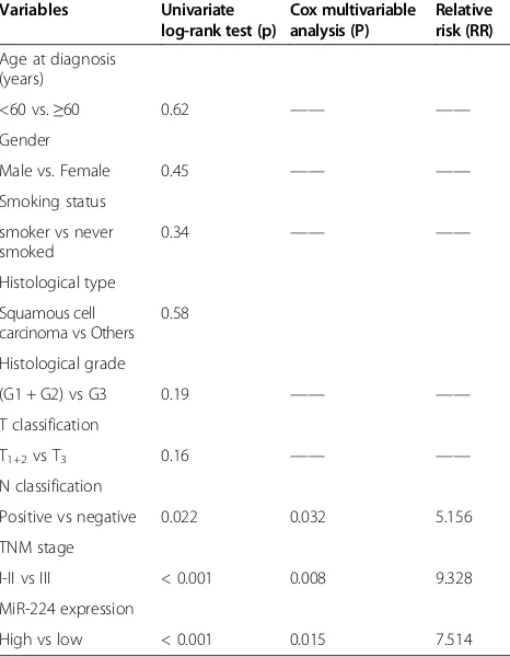
Table 2 Univariate and multivariate analysis of overall survival in 115 patients with non–small cell lung cancer
Variables Univariate log-rank test (p)
Cox multivariable analysis (P)
Relative risk (RR)
Age at diagnosis (years)
<60 vs.≥60 0.62 —— ——
Gender
Male vs. Female 0.45 —— ——
Smoking status
smoker vs never smoked
0.34 —— ——
Histological type
Squamous cell carcinoma vs Others
0.58
Histological grade
(G1 + G2) vs G3 0.19 —— ——
T classification
T1+2vs T3 0.16 —— ——
N classification
Positive vs negative 0.022 0.032 5.156
TNM stage
I-II vs III < 0.001 0.008 9.328
MiR-224 expression
High vs low < 0.001 0.015 7.514
Down-regulation of miR-224 confers poor prognosis in patients with NSCLC
We further evaluated whether miR-224 expression had prognostic potential for OS of NSCLC patients. Using the Kaplan–Meier method and logrank test, we found that the survival rate of patients with high miRNA-224 expression was higher than that of patients with low miRNA-224 ex-pression (P <0.001; Figure 2). Besides, the survival benefits were also found in those with negative N classification (P = 0.022) and early TNM stage (P <0.001; Table 2).
Multivariate Cox regression analysis enrolling above-mentioned significant parameters revealed that miR-224 expression (relative risk [RR] 7.514; P = 0.015), lymph node metastasis (RR 5.156; P = 0.032), and TNM stage (RR 9.328; P = 0.008) were independent prognostic markers for OS of NSCLC patients (Table 2).
Effects of miR-224 on the proliferation, apoptosis, invasion and migration of A549 cells
At last, we assessed the biological role of miR-224 in A549 cells. As shown in Figure 3A, the expression level of miR-224 in miR-224 mimics transfected cells was significantly higher compared with NC transfected cells (P <0.001). MTT assay showed that cell proliferation was significantly impaired after miR-224 mimics transfec-tion (Figure 3B). We also observed promoted cell apop-tosis in miR-224 mimics transfected cells (Figure 3C).
Transwell invasion and migration assays were per-formed to investigate whether miR-224 had a direct in-fluence on A549 cell migration and invasion. As shown in Figure 3D and E, up-regulation of miR-224 impeded cell invasion/migration compared with control. Scratch migration assay also confirmed the inhibitory effect of miR-224 on A549 cell migration (Figure 3F).
Discussion
Lung cancer is a malignant tumor that seriously threatens human health. It is of great significance to in-vestigate molecular and cellular mechanisms of lung cancer, and to identify novel genetic or protein markers for accurate diagnosis and prediction of prognosis. In the current study, we firstly observed that miR-224 was down-regulated in NSCLC compared with adjacent non-cancerous tissues. Then, decreased miR-224 expression
was significantly correlated with aggressive clinicopatho-logical features. Moreover, the Kaplan-Meier analysis revealed that NSCLC patients with low miR-224 ex-pression tend to have shorter OS. Multivariate Cox re-gression analysis identified miR-224 expression level as an independent prognostic factor for OS of NSCLC patients. Finally, in vitro functional assays demonstrated that up-regulation of miR-224 expression in A549 cells was able to reduce cell proliferation, invasion, and migration, and promote cell apoptosis. To the authors’knowledge, this is the first report regarding the clinical significance and functional attributes of miR-224 in NSCLC.
MiR-224 has been shown its tumor-suppressor func-tions in several cancers. Upraity et al. reported that miR-224 was downregulated in glioblastoma tumor tissues and cell lines [19]. Upregulation of miR-224 was found to reduce clonogenic potential of glioblastoma cells and enhance radiation sensitivity. Lower miR-224 expression showed significant correlation with poorer survival. Lin et al. revealed that reduced expression of miR-224 in prostate cancer was associated with metastasis, high PSA level, high Gleason scores, and poor biochemical recurrence-free survival [25]. Forced expression of miR-224 suppressed prostate cancer cell proliferation, inva-sion and migration, and promoted cell apoptosis [17,25]. MiR-224 expression has also been found to correlate in-versely with tumor stage and lymph node metastasis as well as survival times in colorectal cancer patients [26]. Metastatic colorectal cancer cells (SW620) transfected with miR-224 mimics had reduced migration and motil-ityin vitroand formed smaller tumors with fewer metas-tases in mice model. Furthermore, miR-224 upregulation enhances radiation sensitivity of medulloblastoma cells [27], and a 13-gene miRNA signature including increased miR-224 levels would predict good response of lung can-cer cells to EGFR inhibitor erlotinib treatment [28].
In contrast to the tumor-suppressive properties men-tioned above, miR-224 also acts as an oncogene in some other cancers. Overexpression of miR-224 in human he-patocellular carcinoma was associated with promoted cell migration and invasion and poorer patient survival [20,29]. In cervical cancer, miR-224 expression was sig-nificantly higher in the cancerous tissues of patients with poor differentiation, lymph node metastasis, vascular (See figure on previous page.)
invasion, advanced FIGO stage, and shorter overall survival [23]. In addition, the upregulation of miR-224 was also shown in breast cancer [30], clear cell renal cell carcinoma [31], pancreatic ductal adenocarcinoma [22], and bladder cancer [32]. So, miR-224 plays diverse func-tions in cancer pathogenesis and progression, and the role of miR-224 should be tumor specific and possibly dependent on its targets in different cancer types.
Previous research has identified many oncogenes or tumor suppressor genes as direct targets of miR-224, such as apoptosis inhibitor 5 (API5) [19], Homeobox D10 (HOXD10) [20], SMAD family member 4 (SMAD4) [33], SMAD family member 5 (SMAD5 [18], sarcolemma-associated protein (SLMAP) [18], Type 1 iodothyronine deiodinase (DIO1) [21], tumour protein D52 (TPD52) [17], tribbles homolog 1 (TRIB1) [25], chemokine (C-X-C motif ) receptor 4 (CXCR4) [34], hypoxia-inducible factor 1 (HIF1A) [35], Raf kinase inhibitor protein (RKIP) [30], and cell division control protein 42 (CDC42) [36]. It is now clear that miRNAs execute their oncogenic or tumor sup-pressive functions by regulating the expression of target genes. However, an average miRNA can have more than 100 targets [37], and more than one miRNA can converge on a single transcript target [38]. Therefore, the potential regulatory circuitry afforded by miR-224 is enormous, and the accurate mechanisms on how miR-224 influences NSCLC progression need further clarification.
Conclusion
In conclusion, our results revealed that miRNA-224 was down-regulated in NSCLC cell lines and clinical sam-ples. Decreased miRNA-224 expression was associated with aggressive progression and poor prognosis. Re-stored miR-224 expression in A549 cells exhibited anti-tumor effects in vitro. These findings demonstrate that miRNA-224 could not only be useful as a novel bio-marker but also serve as a potential target for gene ther-apy of NSCLC.
Competing interests
The authors declare that they have no competing interests.
Authors’contributions
Conceived and designed the experiments: DZ, HC. Performed the experiments: HC, XY, WC, LW. Analyzed the data: LW, JX, LY. Contributed reagents/materials/ analysis tools: JX, LY. Wrote the paper: DZ. All authors read and approved the final manuscript.
Received: 5 June 2014 Accepted: 7 October 2014
References
1. Jemal A, Siegel R, Xu J, Ward E:Cancer statistics, 2010.CA Cancer J Clin 2010,60:277–300.
2. Verdecchia A, Francisci S, Brenner H, Gatta G, Micheli A, Mangone L, Kunkler I, Group E-W:Recent cancer survival in Europe: a 2000–02 period analysis of EUROCARE-4 data.Lancet Oncol2007,8:784–796.
3. Osman A:MicroRNAs in health and disease–basic science and clinical applications.Clin Lab2012,58:393–402.
4. Zhao G, Cai C, Yang T, Qiu X, Liao B, Li W, Ji Z, Zhao J, Zhao H, Guo M, Ma Q, Xiao C, Fan Q, Ma B:MicroRNA-221 induces cell survival and cisplatin resistance through PI3K/Akt pathway in human osteosarcoma.PLoS One 2013,8:e53906.
5. Mendell JT, Olson EN:MicroRNAs in stress signaling and human disease. Cell2012,148:1172–1187.
6. Bartel DP:MicroRNAs: genomics, biogenesis, mechanism, and function. Cell2004,116:281–297.
7. Dieckmann KP, Spiekermann M, Balks T, Flor I, Loning T, Bullerdiek J, Belge G: MicroRNAs miR-371-3 in serum as diagnostic tools in the management of testicular germ cell tumours.Br J Cancer2012,107:1754–1760.
8. Takahashi M, Cuatrecasas M, Balaguer F, Hur K, Toiyama Y, Castells A, Boland CR, Goel A:The clinical significance of MiR-148a as a predictive biomarker in patients with advanced colorectal cancer.PLoS One2012, 7:e46684.
9. Liu X, Yu H, Cai H, Wang Y:The expression and clinical significance of miR-132 in gastric cancer patients.Diagn Pathol2014,9:57.
10. Meng W, Ye Z, Cui R, Perry J, Dedousi-Huebner V, Huebner A, Wang Y, Li B, Volinia S, Nakanishi H, Kim T, Suh SS, Ayers LW, Ross P, Croce CM, Chakravarti A, Jin VX, Lautenschlaeger T:MicroRNA-31 predicts the presence of lymph node metastases and survival in patients with lung adenocarcinoma.Clin Cancer Res2013,19:5423–5433.
11. Liu XH, Lu KH, Wang KM, Sun M, Zhang EB, Yang JS, Yin DD, Liu ZL, Zhou J, Liu ZJ, De W, Wang ZX:MicroRNA-196a promotes non-small cell lung cancer cell proliferation and invasion through targeting HOXA5.BMC Cancer2012,12:348.
12. Li Y, Jiang Q, Xia N, Yang H, Hu C:Decreased expression of microRNA-375 in nonsmall cell lung cancer and its clinical significance.J Int Med Res 2012,40:1662–1669.
13. Wang XC, Wang W, Zhang ZB, Zhao J, Tan XG, Luo JC:Overexpression of miRNA-21 promotes radiation-resistance of non-small cell lung cancer. Radiat Oncol2013,8:146.
14. Bian HB, Pan X, Yang JS, Wang ZX, De W:Upregulation of microRNA-451 increases cisplatin sensitivity of non-small cell lung cancer cell line (A549).J Exp Clin Cancer Res2011,30:20.
15. Scapoli L, Palmieri A, Lo Muzio L, Pezzetti F, Rubini C, Girardi A, Farinella F, Mazzotta M, Carinci F:MicroRNA expression profiling of oral carcinoma identifies new markers of tumor progression.Int J Immunopathol Pharmacol2010,23:1229–1234.
16. Iorio MV, Visone R, Di Leva G, Donati V, Petrocca F, Casalini P, Taccioli C, Volinia S, Liu CG, Alder H, Calin GA, Menard S, Croce CM:MicroRNA signatures in human ovarian cancer.Cancer Res2007,67:8699–8707. 17. Goto Y, Nishikawa R, Kojima S, Chiyomaru T, Enokida H, Inoguchi S,
Kinoshita T, Fuse M, Sakamoto S, Nakagawa M, Naya Y, Ichikawa T, Seki N: Tumour-suppressive microRNA-224 inhibits cancer cell migration and invasion via targeting oncogenic TPD52 in prostate cancer.FEBS Lett 2014,588:1973–1982.
18. Fellenberg J, Saehr H, Lehner B, Depeweg D:A microRNA signature differentiates between giant cell tumor derived neoplastic stromal cells and mesenchymal stem cells.Cancer Lett2012,321:162–168.
19. Upraity S, Kazi S, Padul V, Shirsat NV:MiR-224 expression increases radiation sensitivity of glioblastoma cells.Biochem Biophys Res Commun2014,448:225–230. 20. Li Q, Ding C, Chen C, Zhang Z, Xiao H, Xie F, Lei L, Chen Y, Mao B, Jiang M,
Li J, Wang D, Wang G:miR-224 promotion of cell migration and invasion by targeting Homeobox D 10 gene in human hepatocellular carcinoma. J Gastroenterol Hepatol2014,29:835–842.
21. Boguslawska J, Wojcicka A, Piekielko-Witkowska A, Master A, Nauman A: MiR-224 targets the 3′UTR of type 1 5′-iodothyronine deiodinase possibly contributing to tissue hypothyroidism in renal cancer.PLoS One2011, 6:e24541.
22. Mees ST, Mardin WA, Sielker S, Willscher E, Senninger N, Schleicher C, Colombo-Benkmann M, Haier J:Involvement of CD40 targeting miR-224 and miR-486 on the progression of pancreatic ductal adenocarcinomas. Ann Surg Oncol2009,16:2339–2350.
23. Shen SN, Wang LF, Jia YF, Hao YQ, Zhang L, Wang H:Upregulation of microRNA-224 is associated with aggressive progression and poor prognosis in human cervical cancer.Diagn Pathol2013,8:69. 24. Yanaihara N, Caplen N, Bowman E, Seike M, Kumamoto K, Yi M, Stephens
25. Lin ZY, Huang YQ, Zhang YQ, Han ZD, He HC, Ling XH, Fu X, Dai QS, Cai C, Chen JH, Liang YX, Jiang FN, Zhong WD, Wang F, Wu CL:MicroRNA-224 inhibits progression of human prostate cancer by downregulating TRIB1. Int J Cancer2014,135:541–550.
26. Yuan K, Xie K, Fox J, Zeng H, Gao H, Huang C, Wu M:Decreased levels of miR-224 and the passenger strand of miR-221 increase MBD2, suppressing maspin and promoting colorectal tumor growth and metastasis in mice. Gastroenterology2013,145:853–864 e859.
27. Gokhale A, Kunder R, Goel A, Sarin R, Moiyadi A, Shenoy A, Mamidipally C, Noronha S, Kannan S, Shirsat NV:Distinctive microRNA signature of medulloblastomas associated with the WNT signaling pathway.J Cancer Res Ther2010,6:521–529.
28. Bryant JL, Britson J, Balko JM, Willian M, Timmons R, Frolov A, Black EP:A microRNA gene expression signature predicts response to erlotinib in epithelial cancer cell lines and targets EMT.Br J Cancer2012,106:148–156. 29. Wang Y, Ren J, Gao Y, Ma JZ, Toh HC, Chow P, Chung AY, Ooi LL, Lee CG:
MicroRNA-224 targets SMAD family member 4 to promote cell proliferation and negatively influence patient survival.PLoS One2013, 8:e68744.
30. Huang L, Dai T, Lin X, Zhao X, Chen X, Wang C, Li X, Shen H, Wang X: MicroRNA-224 targets RKIP to control cell invasion and expression of metastasis genes in human breast cancer cells.Biochem Biophys Res Commun2012,425:127–133.
31. Wotschofsky Z, Busch J, Jung M, Kempkensteffen C, Weikert S, Schaser KD, Melcher I, Kilic E, Miller K, Kristiansen G, Erbersdobler A, Jung K:Diagnostic and prognostic potential of differentially expressed miRNAs between metastatic and non-metastatic renal cell carcinoma at the time of nephrectomy.Clin Chim Acta2013,416:5–10.
32. Han Y, Chen J, Zhao X, Liang C, Wang Y, Sun L, Jiang Z, Zhang Z, Yang R, Chen J, Li Z, Tang A, Li X, Ye J, Guan Z, Gui Y, Cai Z:MicroRNA expression signatures of bladder cancer revealed by deep sequencing.PLoS One 2011,6:e18286.
33. Zhang GJ, Zhou H, Xiao HX, Li Y, Zhou T:Up-regulation of miR-224 promotes cancer cell proliferation and invasion and predicts relapse of colorectal cancer.Cancer Cell Int2013,13:104.
34. Zhu S, Sachdeva M, Wu F, Lu Z, Mo YY:Ubc9 promotes breast cell invasion and metastasis in a sumoylation-independent manner. Oncogene2010,29:1763–1772.
35. Lichner Z, Mejia-Guerrero S, Ignacak M, Krizova A, Bao TT, Girgis AH, Youssef YM, Yousef GM:Pleiotropic action of renal cell carcinoma-dysregulated miRNAs on hypoxia-related signaling pathways.Am J Pathol2012, 180:1675–1687.
36. Ke TW, Hsu HL, Wu YH, Chen WT, Cheng YW, Cheng CW:MicroRNA-224 suppresses colorectal cancer cell migration by targeting Cdc42. Dis Markers2014,2014:617150.
37. Brennecke J, Stark A, Russell RB, Cohen SM:Principles of microRNA-target recognition.PLoS Biol2005,3:e85.
38. Krek A, Grun D, Poy MN, Wolf R, Rosenberg L, Epstein EJ, MacMenamin P, da Piedade I, Gunsalus KC, Stoffel M, Rajewsky N:Combinatorial microRNA target predictions.Nat Genet2005,37:495–500.
doi:10.1186/s13000-014-0198-4
Cite this article as:Zhuet al.:Decreased microRNA-224 and its clinical significance in non-small cell lung cancer patients.Diagnostic Pathology
20149:198.
Submit your next manuscript to BioMed Central and take full advantage of:
• Convenient online submission
• Thorough peer review
• No space constraints or color figure charges
• Immediate publication on acceptance
• Inclusion in PubMed, CAS, Scopus and Google Scholar
• Research which is freely available for redistribution