L E T T E R T O T H E E D I T O R
Open Access
Memory T cells skew toward terminal
differentiation in the CD8+ T cell
population in patients with acute myeloid
leukemia
Ling Xu
1†, Danlin Yao
1†, Jiaxiong Tan
1, Zifan He
1, Zhi Yu
1, Jie Chen
1, Gengxin Luo
1, Chunli Wang
1, Fenfang Zhou
1,
Xianfeng Zha
2, Shaohua Chen
1and Yangqiu Li
1*Abstract
Stem cell memory T (T
SCM) and central memory T (T
CM) cells can rapidly differentiate into effector memory (T
EM)
and terminal effector (T
EF) T cells, and have the most potential for immunotherapy. In this study, we found that the
frequency of T
SCMand T
CMcells in the CD8+ population dramatically decreased together with increases in T
EMand
T
EFcells, particularly in younger patients with acute myeloid leukemia (AML) (< 60 years). These alterations persisted
in patients who achieved complete remission after chemotherapy. The decrease in T
SCMand T
CMtogether with the
increase in differentiated T
EMand T
EFsubsets in CD8+ T cells may explain the reduced T cell response and subdued
anti-leukemia capacity in AML patients.
Keywords:
Stem cell memory T cells, Central memory T cells, Effector memory T cells, CD8+ T cells, Acute myeloid
leukemia, Bone marrow, Peripheral blood
To the editor
Clinical applications of immunotherapy for AML lag
be-hind those for solid tumors and lymphocytic leukemia
[
1
–
3
]. Recently, a new memory T cell subset, stem cell
memory T (T
SCM), which has stem cell-like capacity, has
been discovered [
4
–
6
]. However, little is known about
the role of these cells in AML. In this study, we assessed
the distribution of CD4+ and CD8+ T
SCM, central
mem-ory T (T
CM), T effector memory (T
EM), and T terminal
effector (T
EF) cells in peripheral blood (PB) and bone
marrow (BM) from patients with AML and those with
AML in complete remission (AML-CR) by multicolor
flow cytometry. The gating strategy used in this study
followed a published protocol [
7
]. The CD4+ and CD8+
T cells were divided into four subgroups according to
the CCR7 and CD45RO expression pattern: naïve and
T
SCMcells
(CCR7+CD45RO
−
),
T
CMcells
(CCR7
+CD45RO+), T
EMcells (CCR7
−
CD45RO+), and T
EFcells (CCR7
−
CD45RO
−
). The T
SCMpopulation was
de-fined by double positive CD95 and CD28 expression.
The percentages of the T
SCM, T
CM, T
EM, and T
EFcells
in the CD4+ and CD8+ populations were analyzed in 20
cases with AML (17 cases in newly diagnosed and 3
cases with AML relapse) (Fig.
1a, d
) [
8
,
9
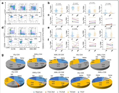
]. The CD8+
T
SCMand CD8+ T
CMcells significantly decreased in the
PB of these patients (Fig.
1e, g
), whereas there was no
significant change in the CD4+ population (Fig.
1b,
g
). Thus, the changes in the memory T cell subsets
appeared to mainly involve CD8+ T cells. The shift
from T
SCMand T
CMcells to a higher ratio of
differ-entiated T
EMand T
EFcells is thought to be due to
the constant exposure of T cells to AML cells and
the leukemia environment, leading to T cell
exhaus-tion and/or dysfuncexhaus-tion [
3
].
To study the influence of the tumor
microenviron-ment on the memory T cell distribution and function in
leukemia patients, we collected seven pairs of PB and
BM samples from AML patients at the time of diagnosis
* Correspondence:yangqiuli@hotmail.com
†Ling Xu and Danlin Yao contributed equally to this work.
1
Department of Hematology, First Affiliated Hospital, Institute of Hematology, School of Medicine; Key Laboratory for Regenerative Medicine of Ministry of Education, Jinan University, No.601 West of Huangpu Avenue, Guangzhou 510632, China
Full list of author information is available at the end of the article
and compared the distributions of memory T cell
sub-sets. The differences in each subset appeared to vary
widely (Fig.
1
c, f ). A low percentage of CD4+ T
CMcells
and a corresponding high percentage of CD4+ T
EMand
T
EFcells were observed in the BM compared with PB
(Fig.
1
c). In the CD8+ population, the changes appeared
to be specific to each individual, and lower CD8+ T
SCMand CD8+ T
CMpercentages were observed in the BM in
half of the patients, whereas there were high percentages
of CD8+ T
SCMand CD8+ T
CMcells in the BM
compared with PB in the remaining samples. It has been
reported that T cells in normal BM mainly possess a
memory phenotype, particularly for CD8+ T
CMcells
[
10
], suggesting that alterations in the leukemic BM
niche in different AML individuals and AML subtypes
may have different impact on T
CMhoming.
Next, we compared the distribution of memory T cells
in AML patients younger (AMLy) and older (AMLo)
than 60 years [
11
]. Unlike healthy individuals (HIs), the
memory T cell subset distribution in the AMLy cohort
Fig. 1Gating strategy for identifying the CD4+ and CD8+ T cells and the percentage of memory T cell subsets in the patients with AML and healthy individuals.a,dCD4+ (a) and CD8+ T (d) cells were differentiated into four subsets based on the expression of CCR7 and CD45RO in one HI-PB, one AML-PB, and one AML-BM patient: central memory T cells (CCR7+CD45RO+), effector memory T cells (CCR7−CD45RO+), and effector T cells (CCR7−CD45RO−). In the CCR7+CD45RO−subset, the expression of CD28 and CD95 was used to identify naïve T cells (CD28+CD95−) and TSCMcells (CD28+CD95+).b,eFrequency of the TSCM, TCM, TEM, and TEFsubsets in the CD4+ (b) and CD8+ (e) T cell populations from 27 HIs and
was strikingly different than that in younger HIs (HIy)
and tended to have a similar distribution pattern as that
detected in the HIo and AMLo groups with a more
ob-vious difference in the CD8+ population (Figs.
1g
and
2a, b
). These findings indicate that the leukemia
micro-environment might drive T cell differentiation in AMLy.
Whether such a skewed T cell distribution in AMLy
truly represents T cell senescence remains an open
ques-tion [
8
]; however, T cells in AMLo patients may not be
able to further differentiate due to inherent T cell
senes-cence, which may be an immune factor underlying the
inferior prognosis of AMLo patients. Together, these
data may suggest that T cell exhaustion and senescence
are involved in T cell immune impairment, leading to an
inefficient anti-tumor response.
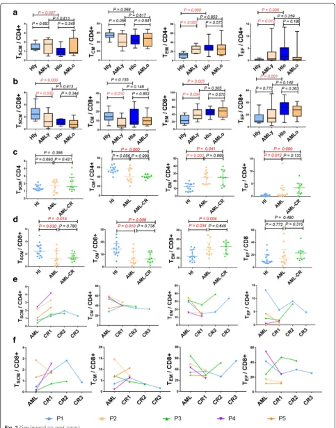
We next compared differences in the distribution of
memory T cell subsets between the AMLy, AML-CR,
and HIy groups. A persistent, skewed memory T cell
dis-tribution was demonstrated for AML patients who
achieved CR after chemotherapy (Fig.
2c, d
). CD4+ and
CD8+ T
SCMcells were predominantly increased at
dif-ferent time points after CR, while the change in other
memory T cell subsets was relatively different (Fig.
2e,
f
). Overall, with the exception of incomplete recovery of
the T
SCMcells, the reduction in T
CMcells and
corre-sponding excessive accumulation of T
EMand T
EFcells
were more evident in AML patients with CR (Fig.
1g
),
which may be related to the immune suppression of
chemotherapy.
Abbreviations
AML:Acute myeloid leukemia; BM: Bone marrow; CML: Chronic myeloid leukemia; CR: Complete remission; HSCT: Hematopoietic stem cell transplantation; PB: Peripheral blood; PBMCs: Peripheral blood mononuclear cells; TCM: Central memory T cells; TEF: Terminal effector T cells; TEM: Effector
memory T cells; TSCM: Stem cell memory T cells
Acknowledgements
We want to thank the flow facility of the Biological Translational Research Institute of Jinan University as well as Yanqiong Jia, a research assistant from the Translational Research Institute of Jinan University. We also would like to thank the volunteers who donated blood for this project.
Funding
This study was supported by grants from the National Natural Science Foundation of China (Nos. 91642111, 81770152, and 81570143), the Guangdong Provincial Basic Research Program (No. 2015B020227003), the Guangdong Provincial Applied Science and Technology Research & Development Program (No. 2016B020237006), the Guangzhou Science and Technology Project (Nos. 201510010211, 201807010004, and 201803040017), and Special Funds for the Cultivation of Guangdong
College Students’Scientific and Technological Innovation (No. pdjh2017b0065).
Availability of data and materials
The datasets used and/or analyzed during the current study are available from the corresponding author on reasonable request.
Authors’contributions
YQL contributed to the concept development and study design. LX coordinated the study. LX, DLY, JXT, ZFH, SHL, XFZ, and SHC performed the laboratory studies. ZY, JC, GXL, CLW, and FFZ collected the clinical data. DLY contributed to figure preparation. YQL, XL, and DLY drafted the manuscript. All authors read and approved the final manuscript.
Ethics approval and consent to participate
This study was approved by the ethics committee of The First Affiliated Hospital of Jinan University.
Consent for publication
Not applicable.
Competing interests
The authors declare that they have no competing interests.
Publisher
’
s Note
Springer Nature remains neutral with regard to jurisdictional claims in published maps and institutional affiliations.
Author details
1Department of Hematology, First Affiliated Hospital, Institute of Hematology,
School of Medicine; Key Laboratory for Regenerative Medicine of Ministry of Education, Jinan University, No.601 West of Huangpu Avenue, Guangzhou 510632, China.2Department of clinical laboratory, First Affiliated Hospital, Jinan University, Guangzhou 510632, China.
Received: 12 March 2018 Accepted: 29 June 2018
References
1. Lichtenegger FS, Krupka C, Haubner S, Kohnke T, Subklewe M. Recent developments in immunotherapy of acute myeloid leukemia. J Hematol Oncol. 2017;10(1):142.
2. Li Y, Yin Q, Yang L, Chen S, Geng S, Wu X, et al. Reduced levels of recent thymic emigrants in acute myeloid leukemia patients. Cancer Immunol Immunother. 2009;58(7):1047–55.
3. Tan J, Chen S, Lu Y, Yao D, Xu L, Zhang Y, et al. Higher PD-1 expression concurrent with exhausted CD8+ T cells in patients with de novo acute myeloid leukemia. Chin J Cancer Res. 2017;29(5):463–70.
4. Zhang Y, Joe G, Hexner E, Zhu J, Emerson SG. Host-reactive CD8+ memory stem cells in graft-versus-host disease. Nat Med. 2005;11(12):1299–305. 5. Gattinoni L, Lugli E, Ji Y, Pos Z, Paulos CM, Quigley MF, et al. A human
memory T cell subset with stem cell-like properties. Nat Med. 2011; 17(10):1290–7.
6. Xu L, Zhang Y, Luo G, Li Y. The roles of stem cell memory T cells in hematological malignancies. J Hematol Oncol. 2015;8:113. 7. Lugli E, Gattinoni L, Roberto A, Mavilio D, Price DA, Restifo NP, et al.
Identification, isolation and in vitro expansion of human and nonhuman primate T stem cell memory cells. Nat Protoc. 2013;8(1):33–42. 8. Saule P, Trauet J, Dutriez V, Lekeux V, Dessaint JP, Labalette M.
Accumulation of memory T cells from childhood to old age: central and (See figure on previous page.)
Fig. 2Memory T cell subset distribution in CD4+ and CD8+ T cells in patients younger or older than 60 years with AML and AML-CR.a,bTSCM,
TCM, TEM, and TEFsubsets within the CD4+ (a) and CD8+ (b) populations in the HIy, AMLy, HIo, and AMLo groups. HIy (n= 13), AMLy (n= 10), HIo
(n= 14), and AMLo (n= 10).c,d: Frequency of TSCM, TCM, TEM, and TEFcells within the CD4+ (c) and CD8+ (d) T cell populations in age matched
HI, AML and AML-CR cohorts. HIs (n= 13), AML (n= 10), and AML-CR (n= 10).e,fFive AML patients were dynamically assayed for the TSCM, TCM,
TEM, and TEFsubsets in the CD4+ (e) and CD8+ (f) T cell populations at different time points. AML-CR, AML patients who achieved complete
effector memory cells in CD4(+) versus effector memory and terminally differentiated memory cells in CD8(+) compartment. Mech Ageing Dev. 2006;127(3):274–81.
9. Yao DL, Xu L, Tan JX, Zhang YK, Lu S, Li MD, et al. Re-balance of memory T cell subsets in peripheral blood from patients with CML after TKI treatment. Oncotarget. 2017;8(47):81852–9.
10. Mazo IB, Honczarenko M, Leung H, Cavanagh LL, Bonasio R, Weninger W, et al. Bone marrow is a major reservoir and site of recruitment for central memory CD8+ T cells. Immunity. 2005;22(2):259–70.