Volume-6 Issue-1
International Journal of Intellectual Advancements
and Research in Engineering Computations
Detection of cervical cancer by using thresholding,
watershed segmentation and SVM classifier in image
processing
Mrs.S.YAMUNA DEVI
#1,Assistant Professor, Department of Biomedical Engineering,
MONIKA M
#2, SAKITHYA N
#3, SUKANYA E.P
#4, VIJAY S
#5DEPARTMENT OF BIOMEDICAL ENGINEERING,
VELALAR COLLEGE OF ENGINEERING AND TECHNOLOGY,
1
[email protected]
2
[email protected]
3
[email protected]
4
[email protected]
5
[email protected]
Abstract - Cervical cancer is an abnormal growth of cervix tissue. We use algorithm to detect cervical cancer. Noise removal is applied on the image to remove the noise. Image sharpening is applied to detect boundary of the image. Segmentation used to divide the image into regions. Two types of segmentation are used they are thresholding and watershed segmentation. Thresholding value convert grayscale into binary images. Here main selection is the threshold value. Watershed segmentation is the best method to group the pixels on the basis of intensity. After watershed segmentation apply morphological operation. It’s main aim to separate tumor parts from an image. Next phase of the proposed system is multiclass classification that is based on these extracted features. This method uses multiclass Support Vector Machine (SVM) to classify the tumor. After classifying the tumor as stage IA,IB,IIA,IIB,IIIA,IIIB,IVA,IVB, using SVM classifier the overall accuracy is found to be 80 – 90%. The fine detection of tumourous area is a challenging task in medical image science. Using this proposed method it is possible to detect infected area.
I.INTRODUCTION
The lower part of the uterus is the cervix or cervix uteri. Approximately 14,000 women has a major health disease like cervical cancer in less developed countries due to not proper vaccination programs. It’s a major disease in women. It is a method for conformal and stereotactic radiation therapy. Smear screening used to detect presents of abnormality. Image processing is a good technique to extract the information. It is used to measure cervical cancer size and uterine extension.
A. Cancer:
Cancer is a group of diseases involving abnormal cell growth with the potential to invade or spread to other parts of the body.
B. Cervical Cancer:
Cervical cancer is a cancer arising from the cervix. It is due to the abnormal growth of cells that have the ability to invade or spread to other parts of the body.
Staging is a technique used to give better treatment for cancer and to detect the stages of cancer. Higher priority given to staging. It is used to detect the difference between cancer and helping to select treatment. At stage 0 it is consider as non invasive cancer, up to stage 4 is called metastatic stage.
a)Stage 0:At this stage it is used to find cancer cells only on the lining of the cervix not on the deeper tissues.
b)Stage IA: It is the invasive cervical cancer, the stage is identified only by microscope not by naked eye.
Stage IA1 - The invasion area is not more than 3 mm deep and not more than 7 mm wide.
Stage IA2 - The invasion area is greater than 3 mm but not more than 5 mm deep, and not more than 7 mm wide. c)Stage IB: The tumor is slightly greater than stage IA. It cannot be seen by microscope.
Stage IB1 - These tumors can only be seen under the microscope. The invasion is not more than 5 mm deep and not more than 7 mm wide, or they can be seen under the microscope but are less than 4 cm in size.
Stage IB2 - These tumors can be seen without a microscope and are larger than 4 cm in size.
d)Stage II: It spread beyond the cervix and not on the pelvic wall or lower third of the vagina.
e)Stage IIA: Cancer spread beyond the cervix to upper two third of the vagina and not spread around uterus.
Stage IIA1 - The tumor can be seen without a microscope but is not more than 4 cm in size.
Stage IIA2 - The tumor can be seen without a microscope and is more than 4 cm in size.
f)Stage IIB: Cancer spread to the tissues around the uterus and upper two third of the vagina but not to the vagina.
g)Stage III: In stage III, in addition to spread as noted above to the upper 2/3 of the vagina and to the tissues around the uterus, these cancers may have
spread to the lower 1/3 of the vagina, to the pelvic wall, and/or may involve the kidneys.
h) Stage IIIA: These cancers may have spread to the lower 1/3 of the vagina, but not to the pelvic wall.
i)Stage IIIB: There are a few reasons why a cervical cancer would be classified as stage IIIB. One is if it has invaded the pelvic wall. The other is if it has blocked one or both uterus (the tubes which travel from the kidney to the bladder) such that it has caused the kidneys to become enlarged or stop working as well as usual.
j)Stage IV: In stage IV cervical cancer the tumor has spread beyond the region of the cervix to involve the wall of the bladder or rectum or has spread to other regions of the body.
k)Stage IVA:
These cancers have spread so that they have invaded either the bladder or rectum or both (spread to adjacent pelvic organs.)
l)Stage IVB: These cancers have spread to distant regions of the body, for example, lymph nodes in a distant region of the body, or the lungs, liver, or bones.
II. LITERATURE REVIEW
The main aim of implementing SVM classifier algorithm is to detect and classify the various types of cervical cancer. Within the field of cervical cancer imaging, this involves a huge challenge. Since large variability is observed in the image acquisition process and a natural variation is reported in the appearance of cervix. The proposed method utilizes the detection and classification of cervical cancer. The following paper supports this idea.
2)M. Anousouya Devi, S. Ravi and J. Vaishnavi S. Punitha (2016)[3], proposed a method in which the different types of cervical cancer are classified. It is a multi-stage system for cervical cancer diagnosis and tumor region extraction. In this, they have used multiclass support vector machine, fuzzy based technique, textural classification, Hierarchical Clustering to classify the tumor.
3)N. Sakthi Priya (2013)[5], proposed a novel approach to classify the various malignancies in cervical images using acoustic shadowing. For classification we have used SVM classifier that would help us to classify the stages of the cancer and help the pathologist detect the cancer better. The proposed image has been tested with a set of images and has proved to be efficient.
4)Saif D. Salman &Ahmed A.Bahrani (2013)[6], proposed a method to segment a tumor tissue in gray medical images using watershed segmentation method. Edge detection used to detect involved region like tumor in gray level. Watershed theory used to detect the minimum region from other values in the same image. Finally superimposed between original image and the obtain by threshold algorithm to get the final image differentiable and colored.
III.PROPOSED METHODOLOGY A. Image Acquisition:
Images are acquired by using MRI scan and they are displayed in a two dimensional matrices having pixels as its elements. Images are stored in database and displayed in the form of grayscale image. The range of grayscale image from 0 to 255.0 represents a black and 255 represents a white color. B. Pre-Processing:
Enhancement and noise detection are most commonly used techniques. Enhancement is used to detect edges and it gives a sharpened image, noise and blurring effect will be reduced. Edge detection will help to find the exact location of tumor.
The obtained scanned image is stored in database and it is converted into gray scale image. To remove noise image is processed. The high pass filter operate the noiseless, high quality image for sharpening and edge detection. This image is added to original image for further enhancement.
a)Noise removal: Median filter is used to remove noise from the images. On removing noise its gives better image clarity.
b)Image sharpening: It can be achieved by using different high pass filter. It helps in detecting the boundary of the images.
C. Processing:
It involves segmenting the images.
a)SEGMENTATION: It is based on the division of the image into groups. Division is based on the attributes of similarities. The Segmentation’s basic purpose is to extraction of important features from the image. The Image will give information. D. Post Processing:
Postprocessing is done by the following methods.
a)Threshold Segmentation: It is one of the simplest method. The input is grayscale image and is convert into a binary image. The base of this method is threshold value.
b)Watershed Segmentation: It is an algorithm method and it is based on the intensity to group pixel of an image. It usually suffers from over segmentation and under segmentation.
In this two methods are used to segment the cervical cancer from the original image.
i)It choose marker as a computed local minima of the image.
ii)The marker positions a defined either by user or by using morphological tools.
c)Morphological Operators: After the completion of binary conversion, some morphological operations are applied into the cervical image. The purpose is to separate the cancer part of the cervical image. The cancer part is shown white in color and it has high intensity.
D. Feature Extraction:
It is defined as the transforming the input data into the set of features. It detects the various desired position, parameters or shapes of the image by using various algorithm. The region properties extract the features of the nucleus. The normal cells and abnormal cells of the nucleus area can be calculated.
E.SVM Classification:
methods. The vector near the hyper plane are the support vector.
IV.RESULTS
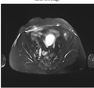
First of all input image is shown here, fig 1 shows input image which has brain tumor.
Fig 1.Original MRI Image
Edge detection is applied on this original image. The results are shown in fig2.
Fig2.Edge Detection
Threshold segmentation is applied on these images. The results are shown in fig 3.
Fig 3.Threshold image
Gradient magnitude is applied on the images. The results are shown in fig 4.
Fig 4.Gradient Magnitude
After that Watershed segmentation is applied on the image. The results are shown in fig5 .
Fig 5.Watershed Segmentation
Finally morphological operator is applied to the image. This will separate the cancerous area from the original image. The results are shown in fig 6.
Fig 6.Morphological Operator
cervix MRI Image
edge detection
Threshold Image
Gradient magnitude (gradmag)
Watershed transform of gradient magnitude (Lrgb)
V.CONCLUSION
In this method we use an efficient algorithm to detect cervical cancerous area with accurate dimension. The MRI image is preprocessed by using edge detection in image processing. Then it is segmented by using hybrid segmentation. Morphological operators are used to extract the cancerous area.SVM classification is used to classify the stages of cervical cancer. It is concluded from results that the affected timorous area is well defined and classify by using hybrid segmentation and SVM classifier.
VI. REFERENCES
[1] How many women get cancer of the cervix? Atlanta, GA, Cancer Soc., 2010(online). Available:http://www.cancer.org/cancer/Cervical Cancer /DetailedGuide
[2]Anam Mustaqeem, Ali Javed,Tehseen Fatima,”An Efficient Brain Tumor Detection algorithm using
Watershed and Thresholding based
segmentation”,I.J.Image Graphics and signal processing, September 2012,10,34-39
[3]M.Anousouya Devi,S.Ravi,J.Vaishnavi S.Punitha,”Detection of Cervical cancer using the
Image Classification
Algorithm”,IJCTA,9(3),2016,pp.153-166
[4]S.Athinarayanan,M.V.Srinath,”Classification of cervical cells in pap smear screening test”,DOI:10.21917/ijivp.2016.0179
[5] N.Sakthi Priya,” Cervical Cancer Screening and Classification Using Acoustic Shadowing”, Volume 1, Issue 8, October 2013.
[6]SaifD.Salman&AhmedA.Bahrani,”Segmentation of tumor tissue in gray medical images using
watershed transformation
method”,doi:10.4156/ijact.vol2.issue4.13
[7]Nikita V. Chavan, Jadhav, B D. and Patil, P M. (2015) ‘Detection and Classification of Brain Tumors’, International Journal of Computer Applications, Vol.112, No. 8, pp.48-53.
[8] Jahanavi, M S . and Sree Priya Kurup (2016) ‘Hybrid Technique to Detect Brain Tumor Using SVM Classifier’, International Journal of Advanced Research in Electrical, Electronics and Instrumentation Engineering, Vol. 5, No. 6, pp.48-52. [9]SetuGarg,Shabana Urooj,Ritu Vijay (2015),”Detection of cervical cancer by using thresholding and Watershed Segmentation”,978-9-3805-4416-8/15