Effect of laser photocoagulation and bevacizumab intravitreal
in proliferative diabetic retinopathy: review on biomarkers of
oxidative stress
Abstrak
Latar belakang: Penelitian ini bertujuan membandingkan pengaruh fotokoagulasi laser dan bevacizumab intravitreal (BIV) terhadap penanda biologis stres oksidatif, antara lain aktivitas aldehid dehidrogenase (ALDH) plasma, kadar malondialdehid (MDA), aktivitas superoxide dismutase (SOD) dan kadar vitreal vascular endothelial growth factor (VEGF) pada penyandang retinopati diabetik (RD) proliferatif.
Metode: Penelitian ini merupakan uji klinis, prospektif, acak, tersamar tunggal di Rumah Sakit Cipto Mangunkusumo antara Februari 2011 - Juni 2013. Sebanyak 72 mata dari 69 penyandang RD proliferatif dirandomisasi menjadi 4 kelompok: 1) kontrol yang langsung menjalani vitrektomi sesuai indikasi (n = 18); 2) fotokoagulasi laser vitrektomi (n = 18); 3) BIV pra-vitrektomi (n = 18); dan 4) kombinasi BIV dan fotokoagulasi laser pra-vitrektomi (n = 18). One-way ANOVA digunakan untuk perbandingan parameter stres oksidatif.
Hasil: Rerata aktivitas ALDH plasma pada kelompok 1, 2, 3 dan 4 masing-masing 0,034 ± 0,02; 0,027 ± 0,02; 0,025 ± 0,02 dan 0,031 ± 0,11 IU/mg protein (p = 0,66), kadar MDA vitreus 1,661 ± 1,21; 1,557 ± 1,32; 1,717 ± 1,54 dan 1,501 ± 1,09 nmol/mL (p = 0,96), dan aktivitas SOD 0,403 ± 0,50; 0,210 ± 0,18; 0,399 ± 0,49 dan 0,273 ± 0,32 U/mL (p = 0,38). Sedangkan rerata kadar VEGF vitreus adalah 0,356 ± 0,60; 0,393 ± 0,45; 0,150 ± 0,24 dan 0,069 ± 0,13 pg/mL menunjukkan perbedaan bermakna (p = 0,05). VEGF pada kelompok 4 lima kali lebih rendah dibandingkan kelompok kontrol (p = 0,05).
Kesimpulan: Kombinasi BIV dan fotokoagulasi laser berpengaruh terhadap MDA dan VEGF vitreus, namun tidak terhadap aktivitas ALDH plasma dan SOD vitreus. Kombinasi BIV dengan fotokoagulasi laser perlu dilakukan pada RD proliferatif.
Abstract
Background: This study was aimed to compare the effect of laser photocoagulation (LF), intravitreal bevacizumab (IVB) and combined treatments on biomarkers of oxidative stress such as aldehhyde dehidrogenase (ALDH), malondialdehyde (MDA) level, superoxide dismutase (SOD) activities, and vitreal vascular endothelial growth factor (VEGF) on proliferative diabetic retinopathy (DR) patients.
Methods: In this single blind randomized clinical trial, 72 eyes from 69 cases of proliferative DR in Cipto Mangunkusumo Hospital between February 2011 - June 2013 were randomized into 4 groups : 1) control (n = 18); 2) LF pre-vitrectomy (n = 18); 3) IVB pre-vitrectomy (n = 18); and 4) combined IVB and LF pre-vitrectomy (n = 18). One-way ANOVA was used to compare oxidative stress parameters in the four groups.
Results: There were no statistically significant differences in the average plasma ALDH activity (0.034 ± 0.02; 0.027 ± 0.02; 0.025 ± 0.02; 0.031 ± 0.11 IU/mg protein; p = 0.66), vitreal MDA level (1.661 ± 1.21; 1.557 ± 1.32; 1.717 ± 1.54; 1.501 ± 1.09 nmol/mL; p = 0.96) and SOD activity) (0.403 ± 0.50; 0.210 ± 0.18; 0.399 ± 0.49; 0.273 ± 0.32 U/mL; p = 0.38) among these four groups, respectively. However, the VEGF vitreal level (pg/mL) showed a statistically significant difference (0.356 ± 0.60; 0.393 ± 0.45; 0.150 ± 0.24; 0.069 ± 0.13; p = 0.05). The VEGF level in combination group was five times lower than the control group (p = 0.05).
Conclusion: Combined treatments of DR by IVB and LF were correlated with lower vitreal MDA and plasma VEGF level, but did not have any effect on plasma ALDH and vitreal SOD in proliferative DR. Combined treatments with IVB and LF are recommended for the management of proliferative DR patients.
Keywords: bevacizumab, diabetic retinopathy, laser photocoagulation, oxidative stress
pISSN: 0853-1773 • eISSN: 2252-8083 • http://dx.doi.org/10.13181/mji.v23i2.756 • Med J Indones. 2014;23:79-86
Correspondence author: Andi A. Victor, arvimadao@yahoo.com
Clinical Research
Copyright @ 2014 Authors. This is an open access article distributed under the terms of the Creative Commons Attribution-NonCommercial-ShareAlike 4.0 International License (http://creativecommons.org/licenses/by-nc-sa/4.0/), which permits unrestricted non-commercial use, distribution, and reproduction in any medium, provided the original author and source are properly cited.
Andi A. Victor,1 Tjahjono D. Gondhowiardjo,1 Sarwono Waspadji,2 Septelia I. Wanandi,3 Adang Bachtiar,4
Franciscus D. Suyatna,5 Habibah Muhiddin,6
1 Department of Ophtalmology, Faculty of Medicine, Universitas Indonesia, Jakarta, Indonesia 2 Department of Internal Medicine, Faculty of Medicine, Universitas Indonesia, Jakarta, Indonesia
3 Department of Biochemistry and Molecular Biology, Faculty of Medicine, Universitas Indonesia, Jakarta, Indonesia 4 Department of Administration and Health Policy, Faculty of Public Health, Universitas Indonesia, Jakarta, Indonesia 5 Department of Pharmacology, Faculty of Medicine, Universitas Indonesia, Jakarta, Indonesia
http://mji.ui.ac.id
Diabetic retinopathy (DR) is a retinal vascular complication in patients with diabetes mellitus (DM) due to long term uncontrolled blood glucose levels.1,2
Diabetic retinopathy is the most common retinal vascular disorder found in Cipto Mangunkusumo Hospital. Between 2004-2009, among 3988 DR patients in RSCM, 61.7% were having non proliferative DR while the other 38.3% were having proliferative DR. In 2010-2012 the percentage of proliferative DR patients increased to 47.9%.3
In DR, retinal ischemia occurs due to microvascular occlusion and capillary nonperfusion.2 In this ischemic
state, retina underwent oxidative stress and formed a compound that will stimulate free radical reactions and pathological lipid peroxidation,4 forming an aldehyde
compound that is highly reactive and cytotoxic such as malondialdehyde (MDA).5-7 Superoxide dismutase
(SOD) is an antioxidant enzyme that acts to protect cells from the effects of oxidative stress. However, the low concentrations of SOD were found in people with DM.8
Free radicals and aldehyde compounds will increase the activity of the aldehyde dehydrogenase (ALDH) enzyme.9 Gondhowiardjo10 reported ALDH activity
in the cornea, lens, aqueous humor and retina. Hasibuan11 further reported the differences in
diabetic patient plasma ALDH activity. ALDH activity in plasma of diabetic patients without diabetic retinopathy was higher than patients with diabetic retinopathy. ALDH activity in plasma is
influenced by many abnormalities in other organs. Nevertheless, it will be very beneficial to know
more about how ALDH activity in plasma level and its association with vitreal MDA as a substrate of ALDH activity in the vitreous, vitreal SOD and vitreal vascular endothelial growth factor (VEGF) in patients with intravitreal bevacizumab (IVB), an anti-VEGF agent, administration and/or laser photocoagulation (LF) as the treatments for proliferative diabetic retinopathy.
METHODS
The protocol of this study has been approved by the Ethic Research Committee, Faculty of Medicine Universitas Indonesia with letter number 352/PT02. FK/ETIK/2011 dated 16 June 2011.
The study was designed as a prospective, single-blind randomized clinical trial. Between February 2011 and June 2013, type 2 DM patients with
proliferative diabetic retinopathy were considered for enrollment into the study. Patients enrolled in this study underwent systemic evaluation which include anamnesis (duration of DM and DM medication), measurement of blood pressure, and serum levels of HbA1C and blood glucose level as well as ocular inspection including best corrected visual acuity (BCVA) using the Snellen Chart (converted to logMAR), intraocular pressure, fundus photography and central macular thickness (CMT) as measured by Ocular Computed Tomography (OCT). An
improvement in visual acuity was defined as two or
more improved lines of BCVA.
Inclusion and exclusion criteria
Inclusion criteria for this study were patients with proliferative diabetic retinopathy who are willing to participate by signing informed consent, and agree to follow the steps of the study. Exclusion criteria were patients with systemic disease such as uncontrolled hypertension (systolic > 180 mmHg or diastolic > 110 mmHg), hemostatic disorder or using anticoagulant therapy, impaired renal function (hemodialysis patients), history of stroke and congestive heart failure, patients who ever had previous vitrectomy or got IVB or intravitreal triamcinolone or LF. Drop out criteria was patient who did not come for follow-up at 2-4 weeks after LF or IVB treatment and refused to undergo vitrectomy.
Patient enrollment
Seventy two eyes of 69 proliferative diabetic retinopathy patients obtained from Vitreoretina Division, Department of Ophthalmology, Faculty of Medicine-RSCM were randomized into 4 groups. Control group (1) (n = 18) underwent direct vitrectomy according to clinical indication, LF group (2) (n = 18) underwent a pre-vitrectomy laser photocoagultion, IVB group (3) (n = 18) received intravitreal bevacizumab (IVB) pre-vitrectomy and the group 4 received combination of LF and IVB pre-vitrectomy (n = 18). Informed consent form was signed by all study participants after explanation of the nature and possible consequences of the study.
Biomarkers of oxidative stress measurement
Plasma ALDH activity9 and MDA level12-14 were
measured using spectophotometry method. The
determined using RanSOD® kit.15 VEGF level
measurement was determined using ELISA system kit.16
Follow-up protocol
Follow-up visits were scheduled at 2nd, 4th and 12th
weeks after surgery. Follow-up evaluation includes BCVA and OCT examination. At each visit, patients were also asked about the perceived side effects.
Outcome measures
The primary outcomes measured were the value of plasma ALDH, VEGF, MDA and SOD in the vitreous body of the four groups. Secondary outcomes measured include the proportion of the increase in visual acuity and reduction of the proportion of CMT measured by OCT 2 weeks, 4 weeks, and 12 weeks after intervention.
Statistical analysis
Results were analyzed using computerized statistic program. Visual acuity was converted to logMar before statistical analysis. Statistical test comparing two mean ALDH activity, MDA level, SOD activity, VEGF level, central macular thickness and visual acuity in each group were performed by one-way ANOVA. Multivariate analysis performed by two-way ANOVA/multi regression analysis.
Group
Control LF IVB IVB + LF
Age (years) ± SD 51.00 ± 6.95 51.00 ± 6.62 50.44 ± 9.87 48.89 ± 9.53
Male/Female 12/6 7/11 10/8 6/12
DM duration (years) ± SD 9.83 ± 6.25 9.11 ± 4.77 8.44 ± 6.88 9.67 ± 5.122
HbA1c (%) ± SD 8.13 ± 1.54 8.45 ± 2.26 6.87 ± 1.16 7.23 ± 1.45
Anti-hyperglycemic drug usage
Insulin 3 6 1 2
SU and insulin 2 2 1 3
Non-SU and insulin 2 2 1 3
SU 9 7 11 7
Non-SU 2 1 4 3
CMT (µm) ± SD 360.20 ± 191.35 452.10 ± 327.57 464.22 ± 297.77 519.00 ± 319.19
Pre-vitrectomy CMT (µm) ± SD 428.44 ± 226.77 452.83 ± 362.17 409.17 ± 231.60 480.39 ± 264.20
Visual acuity (logMAR) ± SD 1.58 ± 0.55 1.79 ± 0.26 1.83 ± 0.49 1.84 ± 0.32
Pre-vitrectomy visual acuity (logMAR) ± SD 1.61 ± 0.52 1.79 ± 0.24 1.80 ± 0.37 1.87 ± 0.28
Table 1. Baseline characteristics of diabetic retinophathy patients in the four groups
SU: Sulphonylurea, CMT: Central macular thickness, DM: Diabetes mellitus, IVB: Intravitreal bevacizumab, LF: Laser photocoagulation RESULTS
In this study, male and female subjects ratio was 51.4% : 48.6%. The age range of the subjects was 26 - 67 year old with the average age was 50.33 ± 8.236 year old. A total of 70.8% of these study subjects had been diagnosed with type 2 DM for less than 10 years, while the rest was diagnosed as type 2 DM for more than 10 years.
There was no statistically significant difference in
the baseline clinical features in these four groups, with the exception of the average glycemic control
status. There was statistically significant difference
between group 3 and 4 and group 1 and 2 of the average glycemic control status of the subjects. The
other variables showed no statistically significant
differences (Table 1).
Primary outcomes: biomarkers values
http://mji.ui.ac.id
The activity of plasma ALDH in the IVB group
showed no statistically significant differences among
all groups (p = 0.66). The highest plasma ALDH activity was found in the patients with bad glycemic control status, while the lowest was found in the good glycemic control status patients (Table 3).
In the LF group, the vitreal MDA level was lower than the control group. The vitreal MDA level in the combination group was the lowest among all groups. However, the differences of vitreal MDA
level in all groups were not statistically significant
(p = 0.96).
The activity of vitreal SOD showed no statistically
significant differences (p = 0.38). The highest SOD
activity was found in the patients with good glycemic control status, while the lowest was found in the bad glycemic control status (Table 3).
The lowest VEGF level was found in the combination group (0.069 pg/mL). The VEGF level in combination group was five times lower than the control group and statistically significant. In the IVB group the VEGF level was also low (0.150 pg/mL), twice lower than the control group. The VEGF level of LF group and the control group were comparable. The differences of vitreal VEGF level in all groups were statistically significant (p = 0.05).
Group
p*
Control LF IVB IVB + LF
Plasma ALDH activity (U/mg protein) ± SD 0.034 ± 0.02 0.027 ± 0.02 0.025 ± 0.02 0.031 ± 0.11 0.66 Vitreal MDA level (nmol/mL) ± SD 1.661 ± 1.21 1.557 ± 1.32 1.717 ± 1.54 1.501 ± 1.09 0.96 Vitreal SOD activity (U/mL) ± SD 0.403 ± 0.50 0.210 ± 0.18 0.399 ± 0.49 0.273 ± 0.32 0.38 Vitreal VEGF level (pg/mL) ± SD 0.356 ± 0.60 0.393 ± 0.45 0.150 ± 0.24 0.069 ± 0.13 0.05
Table 2. Comparison of biomarkers values among the four groups
*ANOVA analysis, ALDH: Aldehyde dehydrogenase, MDA: Malondialdehyde, SOD: Superoxide dismutase, VEGF: Vascular endothelial growth factor, LF: Laser photocoagulation, IVB: Intravitreal bevacizumab
Group
p*
Control LF IVB IVB + LF
Plasma ALDH activity (U/mg protein) ± SD 0.034 ± 0.02 0.027 ± 0.02 0.025 ± 0.02 0.031 ± 0.11 0.66 Vitreal MDA level (nmol/mL) ± SD 1.661 ± 1.21 1.557 ± 1.32 1.717 ± 1.54 1.501 ± 1.09 0.96 Vitreal SOD activity (U/mL) ± SD 0.403 ± 0.50 0.210 ± 0.18 0.399 ± 0.49 0.273 ± 0.32 0.38 Vitreal VEGF level (pg/mL) ± SD 0.356 ± 0.60 0.393 ± 0.45 0.150 ± 0.24 0.069 ± 0.13 0.05
Table 3. Associations between biomarkers and glycemic control status (HbA1c) (n = 72)
*Using Kruskal-Wallis analysis, ALDH: Aldehyde dehydrogenase, MDA: Malondialdehyde, SOD: Superoxide dismutase, VEGF: Vascular endothelial growth factor, LF: Laser photocoagulation, IVB: Intravitreal bevacizumab
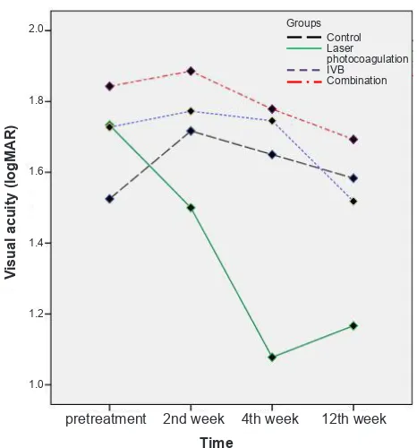
Visual acuity
Visual acuity in the LF group was improved compared to the other groups. However the improvement occurred only until the 4th week. The visual acuity in
this group decreased when measured in the 12th week.
However in the 12th week the visual acuity in this
group was still better than the other groups (Figure 1).
Figure 1. The changes of visual acuity among the four groups in 2nd, 4th, and 12th week
Groups Control Laser photocoagulation IVB
Combination
pretreatment 2nd week 4th week 12th week
Time
Visual acuity (logMAR)
http://mji.ui.ac.id
This study also showed that ALDH activity less than 0.024 IU was associated with improved visual acuity
with a sensitivity of 0.54 and specificity of 0.45.
Central macular thickness
Central macular thickness was reduced in control, LF and combination groups until week-4, and it increased again when measured in week-12. The exception was in the IVB group, in which the CMT is continuously reduced until week-12. However, the biggest reduction in CMT was found in the combination group (Figure 2).
It was also shown that the changes in CMT were not associated with the level of vitreal MDA, the activity of vitreal SOD and the level of vitreal VEGF with the p value respectively 0.09, 0.64, and 0.13.
The plasma ALDH activity below 0.024 IU in this study was associated with reduced CMT with sensitivity
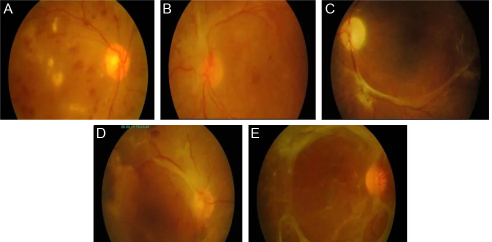
of 0.54 and specificity of 0.53. From the OCT, this study also found a method to grade the fibrovascular membrane. This finding is shown in figure 3.
Complications
In this study, 11 cases (15.3%) of recurrent vitreal Figure 2. The changes of CMT among the four groups in 2
nd, 4th, and 12th Week
Grade 0
No fibrovascular membrane in
every quadrant.
Grade 1
Fibrovascular membrane in 1
quadrant.
Grade 2
Fibrovascular membrane in 2
quadrants.
Grade 3
Fibrovascular membrane in 2
quadrants.
Grade 4
Fibrovascular membrane in all
quadrants.
hemorrhage and hyphema post vitrectomy were found. Other complication was retinal detachment which was found in 5 cases (6.9%). Fifteen of 72 (20.8%) subjects developed increase in lens opacities. Other complication comprised glaucoma in 5 cases (6.9%) and optic nerve atrophy in 1 case (1.4%). The number of complications occurred in each group is shown on table 4.
Figure 3. Grading of fibrovascular membrane: A) Grade 0 no fibrovascular membrane in every quadrant; B) Grade 1 fibrovascular mem
-brane in 1 quadrant; C) Grade 2 fibrovascular mem-brane in 2 quadrants; D) Grade 3 fibrovascular mem-brane in 2 quadrants; E) Grade 4 fibrovascular membrane in all quadrants
Groups Control Laser photocoagulation IVB
Combination
12th week pretreatment 2nd week 4th week
Time Central macular thickness300
400 500 600
200
Grade 0
No fibrovascular membrane in
every quadrant.
Grade 1
Fibrovascular membrane in 1
quadrant.
Grade 2
Fibrovascular membrane in 2
quadrants.
Grade 3
Fibrovascular membrane in 2
quadrants.
Grade 4
Fibrovascular membrane in all
quadrants.
Grade 0
No fibrovascular membrane in
every quadrant.
Grade 1
Fibrovascular membrane in 1
quadrant.
Grade 2
Fibrovascular membrane in 2
quadrants.
Grade 3
Fibrovascular membrane in 2
quadrants.
Grade 4
Fibrovascular membrane in all
quadrants.
Grade 0
No fibrovascular membrane in
every quadrant.
Grade 1
Fibrovascular membrane in 1
quadrant.
Grade 2
Fibrovascular membrane in 2
quadrants.
Grade 3
Fibrovascular membrane in 2
quadrants.
Grade 4
Fibrovascular membrane in all
quadrants.
Grade 0
No fibrovascular membrane in
every quadrant.
Grade 1
Fibrovascular membrane in 1
quadrant.
Grade 2
Fibrovascular membrane in 2
quadrants.
Grade 3
Fibrovascular membrane in 2
quadrants.
Grade 4
Fibrovascular membrane in all
quadrants.
A
B
C
http://mji.ui.ac.id DISCUSSION
Intravitreal bevacizumab and LF is the current treatment used in the proliferative DR patients as the adjuncts for the vitrectomy. Various studies were done especially on IVB to measure the clinical outcomes of the therapies on these proliferative DR patients. The vitrectomy on those studies appeared safe and effective on reducing the recurrent vitreal
hemorrhage, albeit without the benefit of visual
acuity.17 Our study was aim to measure the effect of
these therapies on the biomarkers of oxidative stress. Oxidative stress is the underlying mechanism of DR, thus by measuring it; the effectivity of the therapies can be accurately measured.
Laser photocoagulation in DR is aimed at ischemic retinal tissue to turn them into scar tissue thus decreasing the oxidative stressed caused by the high oxygen demand. However before these effect can be achieved, LF causes a physical trauma due to its thermal effect which in turn worsen the oxidative stress and increase the aldehyde level.18
The administration of IVB as anti-VEGF influenced
the biochemical processes that happened in DR patients. Intravitreal bevacizumab will decrease the VEGF level and consequently restore the retinal vascular permeability.19,20 These processes will
inhibit the oxidative stress and aldehyde formation.4
However IVB cannot reduce the ischemic retinal area. Thus its effect is limited by its duration of action.21
The ALDH activity in all groups showed no
statistically significant and clinically important results. These finding was due to biochemical
processes that happened because of LF, IVB, and combination of both of them were only a small proportion of the whole biochemical processes in the body. However, Gondhowiardjo10 found that plasma
ALDH activity in type 2 DM patients without DR
Complications n (%)
Control LF IVB IVB + LF Total
Recurrent vitreous hemorrhage and / or hyphema 2 (2.8) 1 (1.4) 3 (4.2) 5 (6.9) 11 (15.4)
Retinal detachment 1 (1.4) 1 (1.4) 0 3 (4.2) 5 (6.9)
Lens opacity 5 (6.9) 4 (5.6) 2 (2.8) 4 (5.6) 15 (20.8)
Glaucoma 1 (1.4) 3 (4.2) 1 (1.4) 0 5 (6.9)
Optic nerve atrophy 0 1 (1.4) 0 0 1 (1.4)
Table 4. Complications of the treatments with laser photocoagulation, intravitreal bevacizumab, or combination (n = 72)
is higher than plasma ALDH activity in type 2 DM patients with DR. Plasma ALDH activity decrease as the duration of type 2 DM increase and is associated with blood glucose level and the use of anti-diabetic drugs.10
The MDA level in the combination group was lower than the other groups while in the IVB group the value was higher than the other groups. Intravitreal bevacizumab is given to inhibit the formation of VEGF which can cause microvascular occlusion, telangiectasia and retinal ischemia.2 Through
inhibition of VEGF formation, the retinal ischemia can be minimized and so do the oxidative stress. Subsequently, the end products of aldehyde such as MDA will not be formed. However, the IVB effect is limited by the duration of its action21 and
also the ischemic retina cannot be treated as in the LF. Therefore the vitreal MDA level will increase because of the retinal ischemia that is not resolved.
The activity of vitreal SOD in the combination group was not lower than the control or IVB groups. However, high SOD activity will overcome the oxidative stress22 and in turn will lower the formation
of VEGF.4 Nevertheless, this is consistent with the
finding in this study that the high activity of SOD
will be followed by lower VEGF level.
Laser photocoagulation is known to increase the oxidative status of the retina. In this study, the activity of SOD in the LF group was lower than the control group, even though the difference was not
statistically significant. The low SOD activity can be
caused by the bad glycemic control status in the LF group.
In this study, the lowest vitreal VEGF level was found in the combination group and is consistent with Jing, et al23 study. The IVB group also showed low VEGF
level. The effect of LF in decreasing VEGF levels was twice lower than the IVB.
Visual acuity in the LF group was improved better compared to the other groups even though it only occurred until the 4th week. The visual acuity in this
group decreased when measured in the 12th week.
However in the 12th week the visual acuity in this
group was still better than the other groups (Figure 1). Nevertheless, visual acuity should not be used as a sole indicator of the success of the therapies because it is affected by many factors. It was showed that in the combination group the visual acuity was the worst before any intervention was made, thus the visual acuity after the treatment could not be expected to improve very much.
In this study, CMT was reduced in all group, with the biggest reduction occurred in the combination group
(Figure 2). This finding showed that both of these
therapies have a synergistic effect in reducing the CMT in proliferative DR patients and is consistent with Stefansson, et al24 study.
This study showed that plasma ALDH activity below 0.024 IU was associated with improved
CMT with sensitivity of 0.54 and specificity of
0.53. Similarly, the ALDH activity less than 0.024 IU was associated with improved visual acuity
with a sensitivity of 0.54 and specificity of 0.45.
Thus proliferative DR patients with plasma ALDH activity below 0.024 IU post vitrectomy will show a better improvement in visual acuity and CMT.
However the sensitivity and specificity of this test
as a prognostic factor is not high.
The correlation between MDA and VEGF showed negative correlation in which the high MDA level is associated with low VEGF level. This negative correlation is not found in the control group. However the correlations in all of these groups were weak and
were not statistically significant. These showed that the
negative correlation was caused by the intervention.
The correlation between VEGF and SOD in this study showed a positive correlation, in which high VEGF levels was associated with high SOD activity, with the exception in the IVB group. These correlations, however, were weak and were not statistically
significant.
Theoretically, oxidative stress will promote the formation of superoxide radical (O2-)4 which in turn
will cause the lipid peroxidation of cell membrane25
and cause the formation of MDA and then VEGF.4,26
Superoxide dismutase acts to eliminate O2- so
MDA will not be formed.22 Consequently the stress
oxidative and retinal ischemia is reduced and the formation of VEGF is inhibited.
It was also shown that the changes in CMT were not associated with the level of vitreal MDA, VEGF and the activity of vitreal SOD. The changes in
CMT could be influenced by the activity of another
enzyme or biomarkers such as Na+-K+-ATPase
or prostaglandin-E2.27 The reduction of Na+-K+
-ATPase will disrupt the function of retinal epithelial barrier that can lead to macula oedema. The levels of vitreal MDA and VEGF and vitreal SOD activity are dynamic and constantly changing while the change in CMT is the result of a long processes.
In conclusion, combination of LF and IVB did not
influence the activities of plasma ALDH and vitreal SOD. However, these combination was significantly
associated with lower vitreal VEGF level. These combination was also associated with lower vitreal MDA level even though the association was not
statistically significant. These finding showed that
the combination of both therapies decreased the oxidative stress that occurs on proliferative DR. Thus, combination of both IVB and LF should be used in the management of proliferative DR patient. Moreover, the measurement of plasma ALDH can also be used as a prognostic factor for determining the visual acuity
and CMT although the sensitivity and specificity is
not high. However, more study with more sensitive measurement of plasma ALDH should be conducted to get a more accurate ALDH measurement.
Conflict of interest
The authors hereby affirm that there is no conflict of
interest in this study.
REFERENCES
1. Chew EY, Ferris III FL. Nonproliferative diabetic retinopathy. In: Ryan SJ, editor. Retina. 5th ed. St. Louis: Mosby; 2006. p.1271-84.
2. Frank RN. Etiologic mechanism in diabetic retinopathy. In: Ryan SJ, editor. Retina. 5th ed. St. Louis: Mosby; 2006. p.1241-70.
3. Vitreoretinal Division. Department of Ophthalmology Faculty of Medicine Universitas Indonesia. (2013). [Prevalence of diabetic retinopathy]. Unpublished raw data. 4. Kowluru RA, Chan P-S. Oxidative stress and diabetic
retinopathy. Exp Diabet Res. 2007;2007:1-12.
http://mji.ui.ac.id
6. Atamer Y, Koçyiğit Y, Atamer A, Mete N, Canoruç N,
Toprak G. Alterations of erythrocyte and plasma lipid peroxides as well as antioxidant mechanism in patients with type II diabetes mellitus (NIDDM). Tr J Medical Sciences. 1998;28:143-8.
7. Mancino R, Di Pierro D, Varesi C, Cerulli A, Feraco A, Cedrone C, et al. Lipid peroxidation and total antioxidant capacity in vitreous, aqueous humor, and blood samples from patients with diabetic retinopathy. Mol Vis. 2011;17:1298-304.
8. Samuel TV, Murthy DSJ, Dattatreya K, Babu PS, Johncy SS. Impaired antioxidant defence mechanism in diabetic retinopathy. JCDR. 2010;4(6):3429-36.
9. Gondhowiardjo TD. Characterization of Human Corneal Aldehyde Dehydrogenase [dissertation]. Netherlands: The Netherlands Ophthalmic Research Institute; 1992.
10. Gondhowiardjo TD. The role of enzymatic exploration in ocular diabetics. In: Kadarisman RS, editor. Understanding ocular diabetics. Jakarta: Department of Ophthalmology Faculty of Medicine Universitas Indonesia; 1999. p. 32-40. 11. Hasibuan H. Aktivitas Enzim ALDH pada Retinopati Diabetik [thesis]. Mount Pleasant (MI): Universitas Indonesia; 1997. Indonesian.
12. Suyatna FD, Priyanto, Istiantoro J, Sadikin M. Oxidant and
antioxidant status of police officer in the city and rural area.
Med J Indones. 2004;13(2):77-80.
13. Oen LH, Utomo H, Suyatna FD, Hanafiah A, Asikin N.
Plasma lipid peroxides in coronary heart disease. Int J Clin Pharmacol Ther Toxicol 1992;30(3):77-80.
14. Wills ED. Mechanisms of lipid peroxide formation in animal tissues. Biochem J. 1966;99(3):667-76.
15. Randox Laboratories [Internet]. Ransod superoxide dismutase manual. Available from: http://www.randox.com. 16. Ishizaki E, Takai S, Ueki M, Maeno T, Maruichi M,
Sugiyama T, et al. Correlation between angiotensin-converting enzyme, vascular endothelial growth factor, and matrix metalloproteinase-9 in the vitreous of eyes with diabetic retinopathy. Am J Ophthalmol. 2006;141(1):129-34.
17. Sinawat S, Rattanapakorn T, Sanguansak T, Yospaiboon Y, Sinawat S. Intravitreal bevacizumab for proliferative
diabetic retinopathy with new dense vitreous hemorrhage after full panretinal photocoagulation. Eye (Lond). 2013;27(12):1391-6.
18. Galetović D, Bojić L, Bućan K, Karlica D, Lesin M,
Znaor L. The role of oxidative stress after retinal laser photocoagulation in nonproliferative diabetic retinopathy. Coll Antropol. 2011;35(3):835-40.
19. Lam DS, Lai TY, Lee VY, Chan CK, Liu DT, Mohamed
S, et al. Efficacy of 1.25 mg versus 2.5 mg intravitreal
bevacizumab for diabetic macular edema. six month results of a randomized controlled trial. Retina. 2009;29(3):292-9. 20. Cho WB, Oh SB, Moon JW, Kim HC. Panretinal photocoagulation combined with intravitreal bavacizumab in high risk proliferative diabetic retinopathy. Retina. 2009;29(4):516-22.
21. Krohne TU, Eter N, Holz FG, Meyer CH. Intraocular pharmacokinetics of bevacizumab after a single intravitreal injection in humans. Am J Ophthalmol. 2008;146(4):508-12.
22. Green K, Brand MD, Murphy MP. Prevention of mitochondrial oxidative damage as a therapeutic strategy in diabetes. Diabetes. 2004;53(suppl1):S110-8.
23. Qian J, Lu Q, Tao Y, Jiang YR. Vitreous and plasma concentrations of apelin and vascular endothelial growth factor after intravitreal bevacizumab in eyes with proliferative diabetic retinopathy. Retina. 2011;31(1):161-8.
24. Stefánsson E, Novack RL, Hatchell DL. Vitrectomy prevents retinal hypoxia in branch retinal vein occlusion. Invest Ophthalmol Vis Sci. 1990;31(2):284-9.
25. Durmuş M, Yilmaz HR, Uz E, N Özçelik. The effect of
caffeic acid phenethyl ester (CAPE) treatment on levels of MDA, NO, and antioxidant enzymes activities in retinas of streptozotocin-induced diabetic rats. Turk J Med Sci. 2008;38(6):525-30.
26. Halliwell B, Gutteridge JM. Lipid peroxidation, oxygen radicals, cell damage and antioxidant therapy. Lancet. 1984;1(8391):1396-7.