B.Vani et al JMSCR Volume 07 Issue 11 November 2019 Page 517
Original Article
Spectrum of histopathological study of colonoscopic biopsies in a tertiary
care hospital
Authors
B.Vani
1, Swathi Cheruku
2*1, 2
Assistant Professor, Department of Pathology, Malla Reddy Institute of Medical Sciences, Hyderabad, Telangana State, India
*Corresponding Author
Dr Swathi Cheruku
Assistant Professor, Department of Pathology, Malla Reddy Institute of Medical Sciences, Hyderabad, Telangana State, India
Abstract
Introduction: Colorectal cancer ranks second among the most common tumours of the world according
to the World Cancer Report of 2000. The incidence in India is about 7/1,00,000. The increasing role of colonoscopy and polypectomy in the diagnosis and management of colorectal adenomas during the past decade has provided the histopathologist with an abundance of valuable material for study.
Materials & Methods: This was a prospective study carried out over a period of 26 months on all the colonoscopic biopsy specimens received in the Department of Pathology. All the colonoscopic biopsies received in Dept of Pathology were considered in the study. The resected specimens of large intestine were excluded from the study. All the clinical findings and radiological diagnosis were also considered.
Results: In the present study, total of 76 cases were considered. Among them 24 cases were inflammatory, 19 benign and 33 malignant lesions. Among the 76 patients, 45 were males and 31 females with a male to female ratio of 1.5:1. Age group between 41 to 50 years were most commonly affected.
Conclusion: Colonoscopic examination with biopsy may be used for follow up of Inflammatory bowel diseases and to define epithelial dysplasia in cases of chronic colitis and thus help in the early detection of carcinoma in such cases. Hence, colonoscopic biopsy has improved the role of pathologists in the diagnosis and management of large bowel diseases while underscoring a constant correlation with clinicians, but a correct interpretation still proves to be an exciting diagnostic challenge for pathologists because of small size of the specimens.
Keywords: Inflammatory bowel diseases, Papilloma, Adenocarcinoma.
Introduction
The study of colonoscopic biopsies is fascinating; among these lesions intestinal neoplasms constitute a considerable share(1). The colon and rectum can be sites for infections, idiopathic inflammatory bowel disease (IBD), vascular disorders, motor and mechanical conditions, toxic
and physical reactions, other inflammatory conditions and various neoplasms.
The increasing role of colonoscopy and polypectomy in the diagnosis and management of colorectal adenomas during the past decade has provided the histopathologist with an abundance of valuable material for study. This material is
http://jmscr.igmpublication.org/home/ ISSN (e)-2347-176x ISSN (p) 2455-0450
B.Vani et al JMSCR Volume 07 Issue 11 November 2019 Page 518 useful because, in most patients subjected to
colonoscopy, the entire colorectum has been visualised at one examination and all the polyps removed for histological examination. Colonoscopies biopsies are now performed not only for the diagnosis of disease but also for the monitoring of the course of a wide variety of conditions and for the early detection of complications.
Colonoscopic biopsies are now performed not only for the diagnosis of disease but also for the monitoring of the course of a wide variety of conditions and for the early detection of complications. The colon and rectum can be sites for infections, idiopathic inflammatory bowel disease (IBD), vascular disorders, motor and mechanical conditions, toxic and physical reactions, other inflammatory conditions and various neoplasms. This material is useful because, in most patients subjected to colonoscopy, the entire colorectum has been visualised at one examination and all the polyps removed for histological examination.
Materials and Methods
This is a prospective study carried out over a period of 26 months from August 2013 to September 2015. The colonoscopic biopsy specimens were received to the Department of Pathology. All the cases irrespective of age and sex who presented to the Department of Gastroenterology and Surgery with various gastrointestinal symptoms who were subjected to colonoscopy for colorectal pathology were taken to study. All the colonoscopic biopsies received in Dept of Pathology, were considered in the study. The resected specimens of large intestine are excluded from the study.
Relevant clinical data was collected from patients through questionnaire, clinical examination, requisition forms and from case sheets. All colonoscopic biopsies received in Dept of Pathology were subjected to histopathological examination. The biopsy material, after 24 hours of fixation in 10% formalin is processed for
paraffin embedding. Sections are cut at 5-6 mm thickness, cleared in xylene, hydrated and dehydrated in graded alcohols and stained with haematoxylin & eosin stain. Special stains like PAS, Alcian Blue and Congo red are carried out wherever required along with routine haematoxylin and eosin staining. The clinical details of the cases like age, clinical history, clinical diagnosis was collected from the patients requisition forms. The histopathological findings were noted and the sections were submitted for special stains and results were noted accordingly.
Results
The present study was conducted from August 2013 to September 2015 in the Department of pathology. A total of 76 biopsies were studied. Among them 24 cases were non-neoplastic, 52 cases were neoplastic. Out of all neoplastic 19 were benign and 33 were malignant neoplasms. The present study involved 76 cases and colonoscopic biopsy was performed on patients of all age groups, ranging from a 5year old male child to an 81 years old female. The majority of cases were males 45 (59.2%) and females 31 (40.8%). [Table 1]
Table 1: Sex distribution of all colorectal lesions
Sex Number of cases Percentage
Male 45 59.2%
Female 31 40.8%
Total 76 100%
Maximum numbers of cases were observed in the age group of 40-49 years (23.7%), followed by 30-39 years (22.4%) and 50-59 years (19.7%) and minimum number of cases were seen in the age group 80-89 years (1.3%). The mean age of the study was 42.89.
B.Vani et al JMSCR Volume 07 Issue 11 November 2019 Page 519
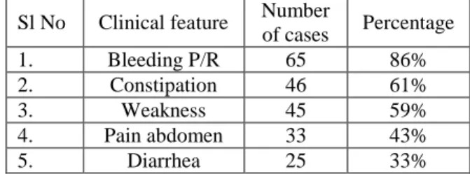
Table - 2: Clinical features of all cases
Sl No Clinical feature Number
of cases Percentage
1. Bleeding P/R 65 86%
2. Constipation 46 61%
3. Weakness 45 59%
4. Pain abdomen 33 43%
5. Diarrhea 25 33%
In the present study of 76 colorectal biopsies: 24 cases were diagnosed as Non-neoplastic lesions, 19 cases were diagnosed as benign neoplasms and 33 cases were diagnosed as malignant neoplasms. [Table 3]
Table - 3: Distribution of all lesions
Sl.no Diagnosis Number of
cases Percentage
1 Non-neoplastic 24 31.5%
2
Neoplastic-benign 19 25%
3
Neoplastic-malignant 33 43.4%
Total 76 100%
In the present study, the range of age distribution of all non-neoplastic lesions was 5years to 62 years. There was clustering of cases between 20-59 years, with maximum cases seen in 30-39 years. Mean age of non-neoplastic lesions was 34.91. There were 24 cases of benign neoplasms, out of which 15 cases were seen in males (62.5%) and 9 cases were seen in females (37.5%). Male: female ratio was 1.7:1.
In the present study of 76 colonoscopic biopsies, 24 cases were non-neoplastic lesions, out of these 6 cases were of Ulcerative colitis, 4 cases were of Tuberculous colitis, 5 cases were of Crohn’s colitis, 3 cases were of chronic non-specific colitis, 2 cases of Solitary rectal ulcer, 2 cases of Amyloidosis and 1 case each of Acute colitis and Eosinophilic colitis.
Out of 19 benign neoplasms, 7 cases were of Hyperplastic polyps, 3 cases were of Juvenile polyps, 3 cases were of Tubular adenoma, 3 cases were of villous adenoma, there were 2 cases of Tubulovillous adenoma and 1 case of Lipoma. Majority of cases were seen in 40-49 years (26.3%) age group followed by 3rd, 4th and 6th decades (15.7% each). Mean age of benign
neoplasms was 31.36.
In the present study, malignant neoplastic lesions were distributed between 20 – 89 years with clustering of cases seen between 30 – 79 years, with predominance in 6th decade. Mean age of malignant neoplasms was 55.36. Rectum was the commonest site in large intestinal tumours with 10 (30.3%) cases followed by ascending colon with 6 cases (18.2%).
The predominant histological variant among all colorectal malignancies is Adenocarcinoma with 27 cases (81.8%). 4 cases (12.1%) of Mucinous adenocarcinomas were diagnosed. 1 case of signet-ring adenocarcinoma was diagnosed (3.03%) and characterised by tumour cells arranged in sheets in lamina propria and infiltrating the sub-mucosa. The tumour cells had abundant intracellular mucin which was PAS positive with eccentric hyperchromatic nuclei. Out of 27 cases of Adenocarcinoma, 11 cases were graded as well differentiated adenocarcinoma (40.7%) and were characterised by malignant glands invading the sub-mucosa. The glands were well formed and lined by tall-columnar cells with hyperchromatic nuclei and only mild nuclear stratification [Table 4]
Table 4: Histological types of malignant
colorectal neoplasms
Microscopic appearance Number
of cases Percentage
Adenocarcinoma 27 81.8%
Mucinous adenocarcinoma 4 12.1%
Signet ring cell carcinoma 1 3.03%
GIST 1 3.03%
Total 33 100%
B.Vani et al JMSCR Volume 07 Issue 11 November 2019 Page 520 and scanty cytoplasm. The tumour cells were PAS
positive.
Ulcerative colitis (Transverse colon)- Inflammatory infiltrate in lamina propria, crypt distortion & crypt abscess formation (H&E;10x)
Tuberculous colitis- large confluent granulomas, caseating necrosis (H&E;40x)
Hyperplastic polyp (Sigmoid colon)- showing tufting of epithelium composed predominantly of goblet cells (H&E;10x)
Tubulovillous adenoma (Ascending colon) -admixture of villous & glandular structures (H&E;10x)
Well-differentiated adenocarcinoma (Descending colon) with >95% glandular elements (H&E;10x)
Signet ring cell carcinoma (Ascending colon)- showing signet ring cells with prominent intracytoplasmic mucin with nucleus pushed to periphery (H&E; 40x)
Discussion
B.Vani et al JMSCR Volume 07 Issue 11 November 2019 Page 521 conditions like infections, IBD, polyps and
colorectal tumours are important lesions which often require colonic biopsy for their conclusive diagnosis.
A rising trend in the incidence and prevalence of inflammatory bowel disease (IBD) in Asia has been recognized for the past two decades. The highest incidence rates are recorded from India, Japan and the Middle East and there exists a genetic predisposition of South Asians (Indians, Pakistanis and Bangladeshis) to ulcerative colitis (UC) (2).
Colorectal cancer (CRC) is a formidable health problem worldwide. As per the GLOBOCAN project undertaken by WHO in 2008, It is the third most common cancer in men (663000 cases, 10.0% of all cancer cases) and the second most common in women (571000 cases, 9.4% of all cancer cases)(3). Worldwide, an estimated 1.2 million cases of colorectal cancer occurred in 2008(3). About 608,700 deaths from colorectal cancer occurred in 2008 worldwide, accounting for 8% of all cancer deaths(3).
In India, the annual incidence rates (AARs) for colon cancer and rectal cancer in men are 4.4 and 4.1 per 100000, respectively. The AAR for colon cancer in women is 3.9 per 100000(4). In the 2013 report, the highest AAR in men for CRCs was recorded in Thiruvananthapuram (4.1) followed by Bangalore (3.9) and Mumbai (3.7). The highest AAR in women for CRCs was recorded in Nagaland (5.2) followed by Aizwal (4.5)(4)
The current recommendation for colorectal cancer screening in most countries is to begin screening at age 50 for men and women who are at average risk for developing colorectal cancer. While colonoscopy is a highly sensitive test, it requires a skilled examiner, involves greater cost, and is less convenient and has more risk for the patient (5). in contrast to the study of R.H.Teague et al, Sidney. J et al and Azar Qayyam et al, where non-neoplastic lesions were dominant followed by neoplastic lesions, in our study, out of 76 cases, 24 (31.5%) cases were non-neoplastic and 33 (43.4%) cases were neoplastic malignant. Benign
neoplastic lesions were 19 accounting to 25%, which matched with the study of R.H.Teague et al (1973) (6)
Dickinson et al(7) in their study of 74 cases described 15 cases (20.3%) of Acute non-specific colitis.
Tablot and price(8) described acute non-specific inflammation of the colon as displaying focal polymorphic exudate in the lamina propria, infiltrating crypts and surface epithelium with an arbitrary involvement of 3-4 crypts, not necessarily contagiously; accompanied by mild increase in lymphocytes and plasma cells; straight crypts and mild decrease to a normal number of goblet cells.
In the present study, there was one case of Acute colitis (4.2%), characterised by neutrophilic and mononuclear cell infiltrate in the lamina propria. No crypt abscesses or branching or decrease in goblet cells were observed.
Dickinson et al described 20 cases (27%) of chronic colitis in their study of 74 cases.
Talbot and Price(8), Carpenter and Tally in 2000(9), diagnosed cases showing a marked increase in plasma cells and lymphocytes in the lamina propria, focal or diffuse with intact and well-aligned crypts with/without some goblet cell depletion as ‘chronic non-specific inflammation of the colon’.
The present study showed 3 cases of chronic non-specific colitis with predominantly lymphoplasmacytic infiltrate in the lamina propria with well-preserved mucosal glands and normal goblet cells.
The present study was compared with the study series of Hassan Abdulla Al-aquli et al(10). Of the 31 cases (11.75%) which included 21 (67.7%) were of Juvenile polyps, 6 cases (19.3%) were of Hyperplastic polyps and 4 cases (13%) were of Hamartomatous polyps. In the present study Hyperplastic polyps were predominant, followed by Juvenile polyps.
B.Vani et al JMSCR Volume 07 Issue 11 November 2019 Page 522 al (11).
In the present study, the incidence of colorectal cancers was more in males than in females, with a male: female ratio of 1.2:1. This finding was consistent with sex incidence in the studies done by Mostafa G et al and Newland RC et al(12). Most colorectal carcinomas are located in the sigmoid colon and rectum, but there is evidence of changing distribution in recent years, with an increasing proportion of more proximal carcinomas (13)
In high-risk countries, colorectal carcinoma most commonly arises in the rectosigmoid region, but though we are in low risk countries we encountered more lesions in the distal region whether it was due to environmental factors, or easy approachability detected them at early stage, as many of these were well differentiated adenocarcinomas.
Conclusion
A variety of both Non-neoplastic and Neoplastic lesions were reported in the present study across a wide age distribution and the findings correlated well with that of similar studies. Much difficulty was encountered in correct interpretation, because of the small size of the biopsy specimens received. A greater awareness of the disease and understanding of pathogenesis on the part of the pathologist was felt to be necessary for a better improved diagnosis.
Histopathological diagnosis correlated well with the colonoscopic diagnosis offered especially in malignant neoplasms. Therefore, colonoscopic biopsy is an important tool in the diagnosis of large bowel diseases. The histopathological study of colonoscopic biopsy specimens should be comprehensive by constant correlation with the clinical details and colonoscopic features. Colonoscopic examination with biopsy may be used for follow up of Inflammatory bowel diseases and to define epithelial dysplasia in cases of chronic colitis and thus help in the early detection of carcinoma in such cases.
Hence, colonoscopic biopsy has improved the role of pathologists in the diagnosis and management of large bowel diseases while underscoring a constant correlation with clinicians, but a correct interpretation still proves to be an exciting diagnostic challenge for pathologists because of small size of the specimens.
References
1. Ferlay J, Shin HR, Bray F, Forman D, Mathers C, Parkin DM. Estimates of worldwide burden of cancer in 2008: GLOBOCAN 2008. Int J Cancer.2010; 127:2893-917.
2. Vineet AHUJA and Rakesh K TANDON ; Inflammatory bowel disease in the Asia– Pacific area: A comparison with developed countries and regional differences, 2010: Gastroenterology and Hepatology, Journal of digestive diseases, Vol 11, Issue 3, 1751-2980.
3. GLOBOCAN 2008
(http://globocan.iarc.fr/factsheets/cancers/c olorectal.asp) (2008).
4. NCRP (2013) Three-year report of the population based cancer registries- 2009-2011. National cancer registry programme, Indian council of medical research (ICMR), Bangalore, India, 2013.
5. Winawer SJ. The multidisciplinary management of gastrointestinal cancer. Colorectal cancer screening. Best Pract Res Clin Gastroenterol. 2007;21(6):1031-1048
6. Teague RH, Read AE. Polyposis in ulcerative colitis. Gut 1975;16:792.
7. Dickinson RJ, Gilmour HM, Mc Clelland DBL. 1979. "Rectal biopsy in patients presenting to an infectious unit with diarrhoea disease". Gut, 20:141.
8. Tablot C, Price AB. 1987. Biosy Pathology in Colorectal Disease. University Press, Cambridge.
B.Vani et al JMSCR Volume 07 Issue 11 November 2019 Page 523 correlation in the diagnosis inflammatory
diseases of the colon: histological patterns with clinical implications". Am J Gastroenterol, 95:878
10.Hassan Abdulla AL-aquili. Clinical, colonoscopic and histopathological evaluation of lower gastrointestinal bleeding. Kufa Med. Journal 2012. Vol .15. no.1
11.Fazeli MS, Adel MG, Lebaschi AH. Colorectal carcinoma: A retrospective, Descriptive study of Age, Gender, subsite, stage and differentiation in Iran from 1995 to 2001 as observed in Tehran University. Dis Colon Rectum. 2007;50:990-995
12.Newland RC, Chapuis PH, Smyth EJ. The prognostic value of substaging colorectal carcinoma. A prospective study of 1117 cases with standardized pathology. Cancer 1987;60:852-857