IS THERE ANY LINK BETWEEN A KIND OF THYROCYTE
DYSFUNCTION, HYPOTHYROIDISM AND INFLAMMATORY
HEMATOLOGIC PARAMETERS IN THE CASES HAVING THE BENIGN
THYROID NODULES? A5-YEAR SINGLE-CENTER EXPERIENCE
Sengul Demet
,
1Sengul Ilker
2 1Department of Pathology, Giresun University Faculty of Medicine, Giresun, Turkey 2
Division of Endocrine Surgery, Department of General Surgery, Giresun University Faculty of Medicine, Giresun, Turkey
Primljen/Received 02. 03. 2018. god. Prihva}en/Accepted 02. 04. 2018. god.
Abstract:Objective:Microscopically, the thyroid gland is composed of spherical follicles and thyroid pa-renchyma includes two major cell types, the thyrocytes releasing thyroid hormones and C cells secreting mature calcitonin. Hypothyroidism has been known as being associated with the various abnormalities of the coagu-lation system. In the present study, it had been purposed to investigate the relationship between inflammatory hematological parameters, RBC, Hb, Htc, RDW, WBC, neutrophil, lymhocyte, N/L, Plt, MPV, PCT, PDW and hypothyroid hormonal status in the cases possessing the benign thyroid nodules.
Material and Methods: A total of 313 cases, 202 with hypothyroidism and 111 with euthyroidism pos-sesing the benign thyroid nodules, that was verified with the cytological evaluation after one-endocrine surgeon performed ultrasonography (US) guided fine needle aspiration (FNA) (US-g-FNA), at the Division of Endocrine Surgery, Department of General Surgery, Giresun University Faculty of Medicine, Giresun, Tur-key, in conformity with the criteria, were enrolled into the study during the period, from April 2010 to April 2015. The documents that were used to follow consi-sted of laboratory tests of the cases including both the thyroid hormones, free T3, Free T4, and TSH, and the inflammatory hematological parameters were revie-wed and scanned retrospectively. The upper limit of the normal Thyrotropin (TSH) reference range was de-termined as 4 mU/L in the present study.
Results: No statistically significant difference was found between the inflammatory hematological parameters, RBC, Hb, Htc, RDW, WBC, neutrophil,
lymhocyte, N/L, Plt, MPV, PCT, PDW, and hypothyro-idism (p > 0.05).
Conclusion:Inflammatory hematological param-eters may not be useful for estimating the hormonal status of the thyroid gland in the cases with the benign thyroid nodules verified with the cytological evalua-tion, TBSRTC.
Keywords: Thyroid neoplasms; Thyrocytes; Fine needle aspiration cytology (FNAC); Cytology; Thyrotro-pin (TSH); Hypothyroidism; Hematological parameters.
INTRODUCTION
The thyroid gland, weighing 10 to 20 grams in normal adults in the United States, is measured by ul-trasonography (US) is a certain extent greater in men than women, increases with age and body weight, and decreases with increasing iodine intake (1, 2).
Microscopically, the thyroid is composed of the spherical follicles, each composed of a single layer of follicular cells surrounding a lumen filled with colloid (mostly thyroglobulin) and the thyroid parenchyma in-cludes two major cell types, the thyrocytes releasing thyroid hormones and C cells secreting mature calcito-nin (3). L-thyroxin (T4) and to a much lesser extent of l-triidothyronin (T3), two main thyroid hormones, are
synthesized by the follicular epithelial cells, thyrocy-tes, of the thyroid gland (4).
Electron microscopy demonstrates the normal flat to low cuboidal follicular cells, interdigitating and overlapping one another. They are intimately relevant to the capillaries, surrounding the follicle; microvilli on the apical surface are multiplexed near the cellular
2018; 13(1): 35–40 ID: 262193420
suppressed, become flat and colloid, accumulating in the lumen (5, 6).
The thyroid hormones frequently have a worthy effect on the erythropoiesis by enhancing it via hyper-proliferation of the immature erythroid progenitors and increasing the secretion of erythropoietin (EPO) by re-sulting in EPO gene expression (7-10). The erythro-cyte mass is increased in the most hyperthyroid status, whereas the hypothyroidism have an attenuated eryth-rocyte mass due to the reduction of plasma volume and may undetectable by routine measurement such as he-moglobin (Hb) concentration (11, 12). The thyroid dysfunction also changes the other hematological para-meters, hematocrit (Hct), mean corpuscular volume (MCV), mean corpuscular hemoglobin (MCH), white blood cell (WBC) count and platelet count (Plt). How-ever, all the mentioned alterations return to normal if an euthyroid state is sustained (9).
In the present study, the laboratory tests of the pa-tients having the benign thyroid nodules, verified with the cytological evaluation after one-endocrine surgeon performed ultrasonography (US) guided fine needle aspiration (FNA), had been evaluated retrospectively and purposed to investigate the relationship between the inflammatory hematological parameters, consist-ing red blood cell (RBC), Hb, Htc, red cell distribution width (RDW), WBC, neutrophil, lymhocyte, neutrop-hil lymhocyte ratio (N/L), Plt, mean platelet volume (MPV), plateletcrit (PCT), platelet distribution width (PDW), and the hypothyroidism.
MATERIALS AND METHODS
The present study had been conducted on a total of 313 cases, from April 2010 to April 2015 in order to matching two groups, the patients with hypothyroi-dism, 202 cases, and the patients with euthyroihypothyroi-dism, 111 cases, control group. In terms of the inflammatory hematological parameters we measured RBC, Hb, Htc, RDW, WBC, neutrophil, lymhocyte, N/L, Plt, MPV, PCT, PDW. The five years documents consisted of lab-oratory tests of the cases included both the thyroid hor-mones and the inflammatory hematological parame-ters were reviewed and scanned retrospectively.
An elevated serum thyroid stimulating hormone (TSH) was determined as a TSH concentration above the upper limit of the normal reference range, typically accepted as 4 to 5 mU/L in the most laboratories. Pres-ently a considerable controversy exist over the appro-priate upper limit of normal for serum TSH that some authors have suggested that the true upper limit is only 2.5 or 3 mU/L in healthy individuals without any
thy-independent of the presence of antithyroid antibodies (13), or in obesity. It is recommended for these cases that the normal upper limit could be as high as 6 to 8 mU/L in healthy octogenarians, or as high as 7.5 mU/L in morbid obesity (14). The upper limit of the normal TSH reference range was determined as 4 mU/L in the present study. Hypothyroidism has known as being as-sociated with the various abnormalities of the coagula-tion system, such as the modificacoagula-tion of coagulacoagula-tion proteins and bleeding tendency.
The study was performed for the cases which had been undergone US guided FNA (US-g-FNA) cytology (US-g-FNAC) and all the US-g-FNAC results had been reported according the guidance of The Bethesda Sys-tem for Reporting Thyroid Cytopathology (TBSRTC), a 6-diagnostic-category system which was constituted through multidisciplinary formulation, proposed at the National Cancer Institute (NCI) Thyroid Fine Needle Aspiration State of the Art and Science Conference held in Bethesda, Maryland, 2007. TBSRTC is at present the most used and accepted reporting system for reporting FNA cytology (FNAC) worlwide (15). The use of TBSRTC also has been endorsed by 2015 American Thyroid Association (ATA) management guidelines (16) as 2009 ATA guidelines (17) which is a revision of 2006 ATA guidelines (18).
The criteria for including
patients into the Study
The screening outcome revealed the 313 cases, 202 with hypothyroidism, 111 with euthyroidism, in confor-mity with the criteria, were incorporated into the study during the period, from April 2010 to April 2015. The exclusion criteria had been the hematologic disorders, cardiac disorders, autoimmune diseases, inflammatory or infective diseases, endocrinologic disease and diabe-tes, liver diseases, renal failure, recurrent disases, thy-roid malignancies and the previous or accompanying ot-her malignancies, as well as those who had medical re-cords as to the usage of steroids, anticoagulants, and al-cohol along with those with a medical history of hepati-tis and patients with the inappropriate samples.
Statistical Analysis
The results were expressed as mean ± standard de-viation (SD) and p < 0.05 were considered as statisti-cally significant.
RESULTS
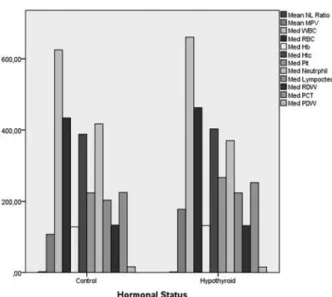
111 (35.46%) out of 313 cases were possesing the euthyroid state, whearas 202 (64.54%) had the hypo-thyroid condition. It had not been detected any statisti-cally significant difference between the cases with hypothyroidism, Group 1 and cases with the euthyroi-dism, Group 2, Control, in terms of the inflammatory hematological parameters, RBC, Hb, Htc, RDW, WBC, neutrophil, lymhocyte, N/L, Plt, MPV, PCT, PDW (p > 0.05) (Figure 1). Therefore, in accordance with the statistical test results, no any difference bet-ween the inflammatory hematological parameters and hypothyroidism was detected (Table 1).
DISCUSSION
Thyroid, a crucial endocrine organ, has a notable effect on the erythropoiesis by inducing EPO secretion and also the proliferation of erythroid progenitors (8, 11, 19). Hypothyrodism reported as cooccurence with some coagulation system abnormalities, the most
rele-Hematologic Parameters
Hormonal
Status n Min Max Mean SD p-value
N/L Control Hypothyroid 111 202 1,03 0,00 4,86 5,57 2,3022 1,8435 1,38137 0,86573 0,459 MPV Control Hypothyroid 111 202 101,00 8,00 122,00 1114,00 107,3333 177,8951 8,77876 240,82600 0,137 WBC Control Hypothyroid 111 202 157,00 6,00 762,00 1662,00 544,6667 621,9020 215,28090 267,47184 0,395 RBC Control Hypothyroid 111 202 412,00 146,00 492,00 692,00 442,3333 465,3333 31,92909 49,48820 0,155 Hb Control Hypothyroid 111 202 114,00 11,00 140,00 841,00 128,1667 135,6373 8,30462 59,18326 0,513 Htc Control Hypothyroid 111 202 343,00 35,00 438,00 551,00 388,5000 394,8693 30,87880 69,99557 0,302 Plt Control Hypothyroid 111 202 165,00 131,00 319,00 452,00 229,0000 272,3235 57,00175 54,12551 0,066 Neutrophil Control Hypothyroid 111 202 212,00 7,00 1209,00 1216,00 493,8333 381,6993 361,91956 153,05536 0,756 Lymphocyte Control Hypothyroid 111 202 172,00 2,00 266,00 434,00 211,0000 223,1373 43,27586 81,44708 0,623 RDW Control Hypothyroid 111 202 128,00 13,00 155,00 288,00 136,6667 131,0490 9,58471 33,26398 0,421 PCT Control Hypothyroid 111 202 202,00 12,00 321,00 741,00 243,3333 238,5869 46,16781 93,00368 0,61 PDW Control Hypothyroid 111 202 11,80 8,70 17,90 279,00 14,9500 15,9886 2,55402 21,16918 0,546
N/L; neutrophil lymphocyte ratio; MPV, mean platelet volume; RBC, red blood cell; Hb, Hemoglobin; Hct, Hemotocrit; Plt, platelet; RDW, red cell distribution width; PCT, plateletcrit; PDW, platelet distribution width
Table 1.The comparison of the inflammatuar hematological paramaters between the patients with hypothyroidism and euthyroidism, control
Figure 1.The comparison of the inflammatuar hematological paramaters with the hormonal status
whether any commitment between a kind of thyrocyte dysfunction, hypothyroidism, and inflammatory he-matologic parameters, consisting RBC, Hb, Htc, RDW, WBC, neutrophil, lymhocyte, N/L, Plt, MPV, PCT, PDW.
Geetha and Srikrishna (21) reported RBC indices, comparing in the cases with hypothyroidism and hyperthyroidism and the study revealed that RDW and MCV in these groups in comparison to the euthyroid individuals had statistically significant difference. Ho-vewer, the other RBC parameters, such as Hb and Hct, did not exhibit any significant difference in compari-son with the euthyroid hormonal status. Kawa et al (9) reported that RBC, Hb in the cases with hypothyroi-dism attenuated, while Hct was increased. They also showed that MCH, MCHC were lower and MCV was increased in hypothyroid group in comparison with the control group.
In the present study, the upper limit of the normal TSH reference range was determined as 4 mU/L and the comparision of the cases with hypothyroidism and the euthyroid ones had been performed for investigat-ing the prediction of the current hormonal status of the thyroid gland by means of their inflamatuar hematolo-gic parameters. However, it was not detected any signi-ficant difference between the inflammatory hematolo-gic parameters, RBC, Hb, Htc, RDW, WBC, neutrop-hil, lymhocyte, N/L, Plt, MPV, PCT, PDW and a type of thyroid hormone disturbance, hypothyroidism.
CONCLUSION
The present study investigated just hypothyroi-dism, performed on the cases that had benign thyroid nodular diseases in the duration of five years. The limi-tations of the present study may be the retrospective design, studying on the cases with the benign thyroid nodular diseases and not analyzing the thyroid antibo-dies, like antithyroid peroxidase Ab (anti-TPO Ab) or antithyroid microsomal Ab, antithyroglobulin anti-body (anti-Tg Ab), and thyroid stimulating immuno-globulin (TSI Ab).
In conclusion, the usage of the inflammatory he-matological parameters may not be beneficial for esti-mating the hypothyroid hormonal status of the thyroid gland in cases with the benign thyroid nodules that was verified with the cytological evaluation, TBSRTC.
has been declared.
Acknowledgements
It has not been used any funding for the present work. DS had contributed in constituting the notion and hypothesis, intellectual planning and management of the study, collecting the data, performing the statisti-cal analysis, writing the whole manuscript, its linguis-tic and academical revisions. IS had contributed in exa-mining and follow-up of the patients, collecting the da-ta, academical revision of the manuscript. All the aut-hors finally approved the submitted and proof versions without any conflict of interest.
We would like to thank the resident and students of Department of General Surgery and all the staff and personnel of Department ofPathology and Biochemis-try, Giresun University-Ministery of Health Prof. Dr. A. Ilhan Ozdemir Education and Research Hospital, Giresun, Turkey.
Abreviations
T4— L-thyroxin
T3— L-triidothyronin
EPO— Erythropoietin
MCV— Mean corpuscular volume
MCH— Mean corpuscular hemoglobin
US— Ultrasonography
FNA— Fine needle aspiration
US-g-FNA— US guided FNA
FNAC— FNA cytology
RDW— Red cell distribution width
PDW— Platelet distribution width
N/L— Neutrophil lymphocyte ratio
MPV— Mean platelet volüme
Plt— Platelet
TSH— Thyroid stimulating hormone
TBSRTC— The Bethesda System for Reporting Thyroid Cytopathology
ATA— American Thyroid Association
anti-TPO Ab— Antithyroid peroxidase antibody
anti-Tg Ab— Antithyroglobulin antibody
TSI Ab— Thyroid stimulating immunoglobulin
Licensing
REFERENCES
1. Pankow BG, Michalak J, McGee MK. Adult human thy-roid weight. Health Phys. 1985; 49(6): 1097-103.
2. Hegedüs L. Thyroid size determined by ultrasound. In-fluence of physiological factors and non-thyroidal disease. Dan Med Bull. 1990; 37(3): 249–63.
3. Wartofsky L. The thyroid gland. In: Becker KL (ed) Principles and practice of endocrinology and metabolism. Lip-pincott Williams & Wilkins, Philedelphia, pp 308-401.
4. Spencer CA, LoPresti JS, Patel A, Guttler RB, Eigen A, Shen D, et al. Applications of a new chemiluminometric thyro-tropin assay to subnormal measurement. J Clin Endocrinol Me-tab. 1990; 70(2): 453-60.
5. Kondalenko VF, Kalinin AP, Odinokova VA. ŠUltra-structure of the normal and pathologic human thyroid gland¹. Arkh Patol. 1970; 32(4): 25-34.
6. Nesland JM, Sobrinho-Simoes M, Johannessen JV. Scanning electron microscopy of the human thyroid gland and its disorders. Scanning Microsc. 1987; 1(4): 1797-810.
7. Drews RE. Critical issues in hematology: anemia, throm-bocytopenia, coagulopathy, and blood product transfusions in cri-tically ill patients. Clin Chest Med. 2003; 24(4): 607-22.
8. Golde DW, Bersch N, Chopra IJ, Cline MJ. Thyroid hor-mones stimulate erythropoiesis in vitro. Br J Haematol. 1977; 37(2): 173-7.
9. Kawa MP, Grymula K, Paczkowska E, Baskiewicz-Ma-siuk M, Dabkowska E, Koziolek M, et al. Clinical relevance of thy-roid dysfunction in human haematopoiesis: biochemical and mole-cular studies. Eur J Endocrinol. 2010; 162(2): 295-305.
10. Mackenzie GM. Anemia in hypothyroidism. JAMA. 1926; 86(7): 462-64.
11. Das KC, Mukherjee M, Sarkar TK, Dash RJ, Rastogi GK. Erythropoiesis and erythropoietin in hypo- and hyperthyro-idism. J Clin Endocrinol Metab. 1975; 40(2): 211–20.
12. Fein HG, Rivlin RS. Anemia in thyroid diseases. Med Clin North Am. 1975; 59(5): 1133-45.
13. Surks MI, Hollowell JG. Age-specific distribution of serum thyrotropin and antithyroid antibodies in the US popula-tion: implications for the prevalence of subclinical hypothyroi-dism. J Clin Endocrinol Metab. 2007; 92(12): 4575-82.
14. Valdés S, Maldonado-Araque C, Lago-Sampedro A, Lillo-MuZoz JA, Garcia-Fuentes E, Perez-Valero V, et al. Refer-ence values for TSH may be inadequate to define hypothyroi-dism in persons with morbid obesity: Diªbet.es study. Obesity (Silver Spring). 2017; 25(4): 788-93.
15. Cibas ES, Ali SZ. The Bethesda system for reporting thyroid cytopathology. Thyroid. 2009; 19(11): 1159-65.
16. Haugen BR, Alexander EK, Bible KC, Doherty GM, Mandel SJ, Nikiforov YE, et al. 2015 American Thyroid Associa-tion Management Guidelines for Adult Patients with Thyroid No-dules and Differentiated Thyroid Cancer: The American Thyroid
Sa`etak
DA LI POSTOJI VEZA IZME\U TIPA TIREOCITNE DISFUNKCIJE,
HIPOTIREOIDIZMAI ZAPALJENSKIH HEMATOLO[KIH PARAMETARA
U SLU^AJEVIMA SA BENIGNIM TIROIDNIM NODUSIMA?
5-GODI[NJE ISKUSTVO JEDNOG CENTRA
Sengul Demet,1Sengul Ilker2 1
Department of Pathology, Giresun University Faculty of Medicine, Giresun, Turkey 2
Division of Endocrine Surgery, Department of General Surgery, Giresun University Faculty of Medicine, Giresun, Turkey
Uvod: Mikroskopski posmatrano, tireoidna `le-zda je sastavljena od sferi~nih folikula i tireoidnog pa-renhima koji uklju~uje dva glavna }elijska tipa, tireoci-te koji osloba|aju hormone i C }elije koje sekretuju zreli kalcitonin. Poznato je da je hipotireoidizam pove-zan sa mnogim abnormalnostima sistema za koagula-ciju. U ovoj studiji, cilj je bio da se ispita povezanost izme|u hematolo{kih parametara, RBC, Hb, Htc, RDW, WBC, neutrofila, limfocita, N/L, Plt, MPV, PCT, PDWi hipotireoidnog hormonskog statusa kod pacijenata sa benignim tireoidnim nodulima.
Materijal i metode:Ukupno 313 slu~aja, 202 sa hi-potireoidizmom, 111 sa eutireodizmom i benignim tireo-idnim nodulima, koji su verifikovani citolo{kom evalua-cijom nakon {to je ura|ena ultrasonografijom (US) vo|e-na aspiracija tankom iglom (FNA) (US-g-FNA) od strane endokrinog hirurga na Odseku za endokrinu hirurgiju, odeljenja op{te hirurgije, Medicinskog fakulteta, Univer-ziteta Giresun u Turskoj je u~estvovalo u studiji u periodu
od aprila 2010-te godine do aprila 2015-te godine. Peto-godi{nji dokumenti su se sastojali od laboratorisjkih te-stova za slu~ajeve koji su uklju~ivali tireoidne hormone, slobodni T3, slobodni T4 i TSH i inflamatorne hematolo-{ke parametre koji su prikazani i analizirani retrospektiv-no. Gornja granica za normalni tireostimuliraju}i hormo-ne (TSH) bila je 4 mU/L za potrebe studije.
Rezultati:Nije pokazana statisti~ki zna~ajna raz-lika izme|u inflamatornih hematolo{kih parametara, RBC, Hb, Htc, RDW, WBC, neutrofila, limfocita, N/L, Plt, MPV, PCT, PDW i hipotireodizma (p > 0,05).
Zaklju~ak: Inflamatorni hematolo{ki parametri verovatno nisu dovoljno korisni za procenu hormon-skog statusa tireoidne `lezde u slu~ajevima sa benig-nim tireoidbenig-nim nodulima koji su verifikovani citolo-{kom evaluacijom, TBSRTC.
17. American Thyroid Association (ATA) Guidelines Task-force on Thyroid Nodules and Differentiated Thyroid Cancer, Coo-per DS, Doherty GM, Haugen BR, Kloos RT, Lee SL, Mandel SJ, Mazzaferri EL, McIver B, Pacini F, Schlumberger M, Sherman SI, Steward DL, Tuttle RM. Revised American Thyroid Association management guidelines for patients with thyroid nodules and diffe-rentiated thyroid cancer. Thyroid 2009; 19(11): 1167–214.
18. Cooper DS, Doherty GM, Haugen BR, Kloos RT, Lee SL, Mandel SJ, et al. Management guidelines for patients with
19. Fujita H. Fine structure of the thyroid gland. Int Rev Cytol. 1975; 40: 197-280.
20. Yango J, Alexopoulou O, Eeckhoudt S, Hermans C, Daumerie C. Evaluation of the respective influence of thyroid hormones and TSH on blood coagulation parameters after total thyroidectomy. Eur J Endocrinol. 2011; 164(4): 599-603.
21. Geetha J, Srikrishna R. Role of red blood cell distribu-tion width (rdw) in thyroid dysfuncdistribu-tion. Int J Biol Med Res. 2012; 3(2): 1476–8.
Correspondence to/Autor za korespondenciju
Ilker SENGUL, M. D. The Founder Vice Dean,
Associated Professor of General Surgery The Founder Chairman, Endocrine Surgery The Founder Chairman, General Surgery Vice Chair, Department of Surgical Sciences Giresun University Faculty of Medicine
The Administrative Manager,
The Founder Chairman, Program Manager of Education General and Endocrine Surgery Clinic
Ministry of Health Giresun University-Prof. Dr. A. Ilhan Ozdemir Education and Research Hospital
Nizamiye Compound Mumcular Avenue